Continuing Education Activity
Milia are benign and transient subepidermal keratin cysts. These white appearing bumps are common, present in many different ways, and can develop in any area of the skin, most commonly the face. The lesions are seen in about half of full-term newborns, and due to a lack of clinical relevance, they are paid little to no attention. The natural course of milia is self-limited and, in most cases, will resolve without leaving a scar by one month of life. This activity reviews the evaluation and management of milia and highlights the role of the interprofessional team in evaluating and treating patients with this condition.
Objectives:
- Identify the etiology of milia.
- Describe the evaluation of milia.
- Review the management options available for milia.
Introduction
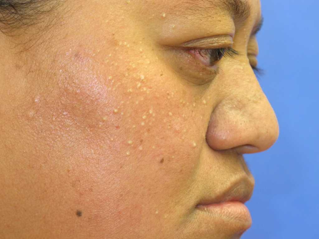
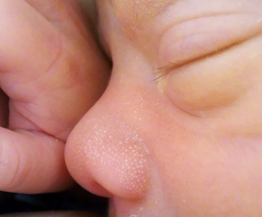
Milia (singular: milium) are benign and transient subepidermal keratin cysts that present as small firm white papules in various numbers most commonly distributed on the face, but they can also be present on other anatomical areas such as the upper trunk, extremities, and genital area (prepuce).
The classification of milia includes primary and secondary. The vast majority of primary milia accounts for congenital milia that occur spontaneously and are present at birth, mainly over the nose, scalp, eyelids, cheeks, gum border (Bohn nodules), and palate (Epstein pearls). Still, there is another percentage of primary milia that may occur in association with certain rare genodermatoses (inherited genetic skin disorders) in children and adults. Meanwhile, secondary milia manifest in association with underlying skin pathology, medications, or skin trauma.[1][2][3][4]
Etiology
Mainly, primary milia are considered to arise from the lower infundibular sebaceous collar of the vellus hair follicles; meanwhile, secondary milia are believed to derive more frequently from the eccrine ducts than from hair follicles, sebaceous ducts, or overlying epidermis.[5][6]
The presence of primary milia also is linked with specific inherited genetic skin disorders (genodermatoses), which include oro-facial digital syndrome type 1, congenital hereditary trichodysplasia (Marie-Unna hypotrichosis), Basex-Dupre-Christol syndrome, and some other ectodermal dysplasias. An uncommon presentation of primary milia occurring on an erythematous plaque is known as milia en plaque, which etiology is still not well understood.[1][2][6]
Secondary milia may arise after trauma, such as dermabrasion or radiotherapy. They also happen secondary to chronic topical steroid use with underlying atrophy and nonsteroidal anti-inflammatory drug use. Besides, they may manifest after the clearing of inflammatory skin diseases as sequelae of bullous disorders.[3][6]
Epidemiology
Primary congenital milia are so common that they could be considered as a typical variation, occurring in up to 40% to 50% of healthy full-term neonates. However, milia may be delayed in premature neonates. Primary and secondary milia occur without significant racial or sex differences; only milia en plaque lesions are more common in females. Moreover, milia can affect persons of any age but is most commonly seen in neonates with the congenital form.[2][7][8]
Histopathology
In 1956, Epstein and Kligman described that "milia are probably the commonest benign tumors of the skin." Histopathologic studies support the notion that milia are not retention cysts, but instead, they represent a simple keratinizing type of benign tumor. Histologically, primary congenital milia look like small cysts of the vellus hair follicles emerging at the level of the sebaceous duct and contain walls of several layers of thick stratified squamous epithelium with a granular cell layer and in the center contain keratinous material. On the other hand, secondary milia lesions present with the same histological characteristics as primary milia lesions. They may also derive from any epithelial structure, including parts of the pilosebaceous apparatus and eccrine glands, such as the epidermis, hair follicle, sebaceous duct, or sweat duct.[1][8][9]
History and Physical
Generally, the most common type (primary congenital milia) manifests at birth, but its onset in premature newborns could manifest weeks later. Milia lesions commonly are asymptomatic and will resolve spontaneously during the first months of life. However, clinical information may change according to the milia subtype.
When acquired (secondary milia), usually affect older children, and a previous history of trauma or bullous skin disease could be present. This type of milia may persist without treatment.
Classification of Milia
Primary Milia
- Congenital
- Benign primary milia of children and adults
- Milia en plaque
- Nodular grouped milia
- Multiple eruptive milia
- Nevus depigmentosus with milia
- Genodermatosis-associated
Secondary milia:
- Disease-associated
- Medication-associated
- Trauma-associated
Physical Examination
Typically, congenital milia lesions occur spontaneously. They consist of small white to yellow papules, less than 3 mm in diameter, and have a smooth dome shape. They could be associated with a faint blue hue in darkly pigmented skin. Milia vary from solitary to multiple-grouped lesions. Usually found on the face, characteristically, with the nose being involved most of the time.
Benign acquired milia of children and adults also develop spontaneously. The distribution in children and adults of this type of milia favors the eyelids, cheeks, forehead, and genitalia area.
Lesions of milia en plaque manifest as an erythematous plaque covered with multiple milia. They can reach several centimeters in size.
Another type includes the acquired and widespread multiple eruptive milia, which appears abruptly over weeks to months. Multiple eruptive milia also, in most cases, occur sporadically. Nonetheless, it may be inherited in an autosomal dominant fashion without other evident anomalies or be associated with certain genodermatosis.
Milia-related lesions to genodermatosis have been documented in association with Brooke-Spiegler syndrome, pachyonychia congenita type 2, and basal cell nevus syndrome, among others.
Disease-associated milia may occur with blistering skin diseases, such as porphyria cutanea tarda or epidermolysis bullosa.
Milia also happen secondary to chronic topical steroid use with underlying atrophy and nonsteroidal anti-inflammatory drug use.
Secondary traumatic milia most commonly arise after skin burns, skin grafting, dermabrasion, or radiotherapy.[1][2][5]
Evaluation
Milia lesions are diagnosed on clinical findings. Although seldom required, incision and drainage of the keratinous content of milia can corroborate the diagnosis. Additionally, persistent and widely distributed milia may require investigation for other causes of primary and secondary milia, such as an underlying genodermatosis, especially when other clinical findings are present.[5][8]
Treatment / Management
The lesions of congenital milia do not require any particular treatment since these lesions tend to resolve spontaneously. The other forms of primary milia, along with secondary milia, may not resolve on their own, and they could be treated with simple surgical interventions such as evacuation, with a tiny incision with a scalpel blade and tangential pressure applied with a comedone extractor or curette. Other treatment options for multiple milia include topical retinoids and electrodesiccation or electrocautery.[5][8]
Differential Diagnosis
The principal differential diagnosis is sebaceous hyperplasia, which manifests as white to yellow grouped papules around the upper lip and nose. Similarly to congenital milia, sebaceous hyperplasia is less frequent in premature newborns. Other differential diagnoses could include comedonal acne, flat warts, and milia-like idiopathic calcinosis cutie.[5]
Prognosis
Congenital milia tend to resolve spontaneously without scarring within a couple of weeks, though they may persist for several months, usually disappearing during the first month of life. Acquired milia may continue without treatment.[2][5][10]
Complications
No systemic complications have been documented. Milia are benign and asymptomatic lesions.[5]
Deterrence and Patient Education
Practitioners should educate and inform caregivers about the benign course of milia and the proneness to resolve without scarring spontaneously.
Enhancing Healthcare Team Outcomes
Healthcare workers need to be aware that milia are benign and transient dermal cysts of keratin affecting different areas of the skin, most commonly the face (nose, gums, and palate). Despite that these lesions are often seen in about half of full-term newborns, they receive little attention.
The natural course of milia is self-limited and, in most cases, will resolve without scaring at one month of life. However, if present in older children, they could be associated with an underlying inherited skin disorder or be secondary to skin trauma. Thus in older age groups, these possible etiologies may need to be considered by physicians and nurses based on other historical and clinical findings to improve outcomes. But when in doubt about the diagnosis, the patient should be referred to dermatology. Finally, neonatologists, primary care providers, and nursing staff working in nurseries should provide education to caregivers and their families about this skin condition to improve patient-centered care.[Level 5]