Continuing Education Activity
The term papilloma refers to a group of various benign epithelial proliferations that can affect the eyelid skin. These lesions are not necessarily associated with the papillomavirus. These lesions are not dangerous but can cause mild irritation or be cosmetically unfavorable for the patient. This activity describes the cause and pathophysiology of eyelid papilloma and highlghts the role of the interprofessional team in its management.
Objectives:
- Describe the presentation of an eyelid papilloma.
- Summarize the treatment of eyelid papilloma.
- Review the differential diagnosis of an eyelid mass.
- Outline the importance of improving care coordination among interprofessional team members to improve outcomes for patients affected by eyelid papilloma.
Introduction
Despite the small surface area of the eyelid, it serves an important role in protecting the eyeball. Skin on the eyelid has no subcutaneous fat and is the thinnest skin found in the body.[1] These characteristics, as well as the location of the eyelids on the body, make them particularly sensitive to irritants and UV damage. As such, eyelids are prone to developing benign and malignant eyelid tumors. One study reported that 5 to 10 percent of all skin cancers occur on the eyelids.[2]
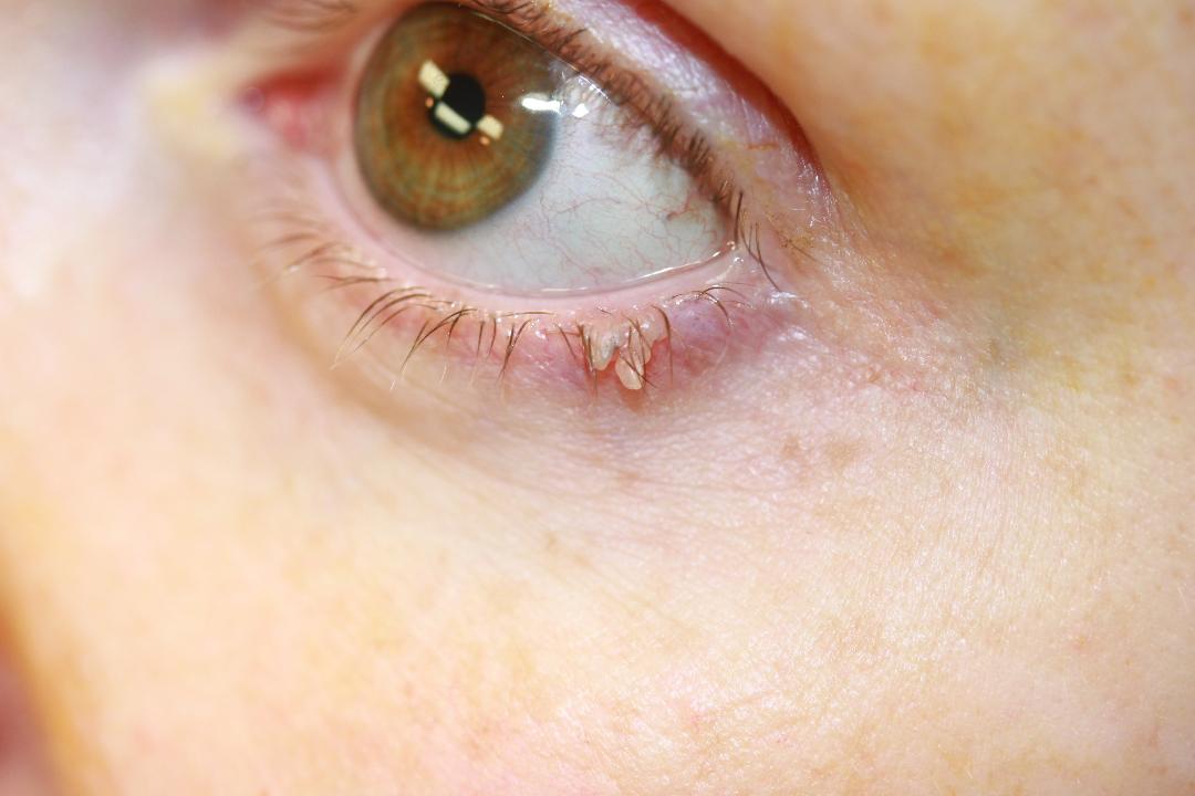
The term papilloma refers to a group of various benign epithelial proliferations that can affect the eyelid skin. These lesions are not necessarily associated with the papillomavirus. These lesions are not dangerous but can cause mild irritation or be cosmetically unfavorable for the patient. Also, it is important to be able to differentiate a benign lesion like a papilloma from a potentially malignant lesion on the eyelid. The lesions which fall into this classification include, but are not limited to seborrheic keratosis, pseudoepitheliomatous hyperplasia, inverted follicular keratosis, verruca vulgaris, squamous papilloma (acrochordon/skin tag), basosquamous acanthoma, and squamous acanthoma. Some types of papillomas are described below.
Squamous papilloma
Squamous papilloma is also known as acrochordon or skin tag. This is a soft, flesh-colored lesion that is smooth, round and/or pedunculated.
Seborrheic keratosis
This is a benign proliferation of cells. Classically has a “stuck on” appearance and can have varying degrees of pigmentation. Lesions tend to vary from a pink or flesh color to dark brown. These lesions are well circumscribed and are usually slightly elevated. Normally these lesions are benign, but the sudden appearance of multiple seborrheic keratoses in a region of the body could indicate a paraneoplastic process. These lesions often have an inflamed base. This is called Leser-Trelat sign.
Pseudoepitheliomatous Hyperplasia
These lesions are often reactive processes in response to trauma, wound, burn, pharmaceutical agents, etc. that can appear similar to basal cell or squamous cell carcinoma. These lesions are usually elevated on the skin with an irregular surface and occasionally ulceration or crusting.[1]
Inverted Follicular Keratosis
This is normally a solitary lesion affecting the eyelid margin. It may or may not be pigmented and has been described as either nodular or papillary in appearance.[1]
Verruca vulgaris
This is flesh-colored skin growth caused by human papillomavirus. This lesion is rare on the eyelid.
Etiology
Papilloma etiology depends on the type of epithelial proliferation. Squamous papillomas and seborrheic keratosis are idiopathic benign cellular proliferation. There is no known definitive cause of these lesions. However, malignant skin lesions that can look like papillomas are often associated with chronic ultraviolet (UV) exposure and sun-damaged skin. Verruca vulgaris is caused by human papillomavirus type 6 or 11.
Epidemiology
One study found that, overall, epidermal tumors make up the majority of eyelid tumors. Seborrheic keratosis was found by one study to be the most frequently encountered lesion, followed by squamous cell papilloma. [3] Another study, however, reported that squamous papillomas are the most common benign epithelial tumor of the eyelid.[1] Seborrheic keratosis is typically found in middle-aged or elderly patients. Squamous papillomas do not have a strong predilection for a particular race or sex. They tend to increase in frequency with older age but can occur at any age.
Histopathology
Seborrheic keratosis displays hyperkeratosis, acanthosis, and some degree of papillomatosis on histological preparation. By definition, the squamous cells that make up the lesion do not show dysplasia. One characteristic finding in some seborrheic keratosis are the pseudo-horn cysts. These are circular collections of surface keratin in the acanthotic epithelium of the seborrheic keratosis lesion.
Squamous papillomas display benign squamous epithelium consisting of variable levels of acanthosis and hyperkeratosis, and focal parakeratosis overlying a fibrovascular core. These have been shown to occasionally demonstrate features of chronic inflammation.[1]
Pseudoepitheliomatous hyperplasia demonstrates invasive elongated processes of hyperplastic epithelium and often has inflammatory cells; however, signs that indicate malignancy are absent such as dysplasia and atypical mitoses. [1]
Inverted follicular keratoses display endophytic proliferation of basal and squamous elements. Pigmentation, acantholysis and chronic inflammation can occur in these lesions. [1]
History and Physical
When evaluating a skin lesion on the eyelid, the following questions are important to ask the patient:
- How long has the lesion been present?
- Have you noticed the lesion changing color, size or shape?
- Does the lesion cause pain or irritation?
- Has the lesion bled or drained any fluid or purulent material?
- Do you have any other similar skin lesions on other parts of your body?
- Have you had a similar lesion in the past on the eyelid?
- Do you have a history of skin cancer?
- When patients present with eyelid papillomas, they typically have been present for months to years. These lesions do not grow rapidly, and patients do not notice them changing in character over time. Occasionally the patient can feel the papilloma from the weight of the eyelid, or the lesion can get mildly inflamed, but chronic or severe inflammation of the lesion is uncharacteristic of a papilloma. Papillomas do not bleed or drain purulent material unless the lesion was picked at by the patient and it subsequently became infected. A history of skin cancer in a patient should prompt the clinician to monitor for suspicious characteristics of the lesion more closely.
When performing the physical exam, it is important to perform the following evaluations:
- Examine the surrounding skin for additional skin lesions.
- Feel facial/neck lymph nodes to assess for lymphadenopathy.
- Using the slit lamp, examine lesion for the destruction of nearby tissues (skin ulceration, destruction of Meibomian glands, or eyelash loss (madarosis). Note any telangiectasia of the lesion, whitening of nearby eyelashes (poliosis), destruction of Meibomian glands. Evert eyelids to look for disruption of palpebral conjunctiva.
Physical exam finding for papillomas will vary slightly depending on the type.
Squamous papilloma: This is a soft, flesh-colored lesion that is generally smooth, round and/or pedunculated.
Seborrheic keratosis: Classically has a “stuck on” appearance and can have varying degrees of pigmentation. Lesions tend to vary from a pink or flesh color to dark brown. These lesions are well circumscribed and are usually slightly elevated.
Pseudoepitheliomatous Hyperplasia
These lesions are often reactive processes in response to trauma, wound, burn, pharmaceutical agents, etc. that can appear similar to basal cell or squamous cell carcinoma. These lesions are usually elevated on the skin with an irregular surface and occasionally ulceration or crusting.[1]
Inverted Follicular Keratosis
This is normally a solitary lesion affecting the eyelid margin. It may or may not be pigmented and has been described as either nodular or papillary in appearance.[1]
Verruca vulgaris: Flesh-colored skin growth. The superior portion of the lesion can have very tiny, fingerlike projections that are sometimes visible without magnification.
Evaluation
Consider documenting lesion with photo or drawing.
If lesion displays any characteristics that are concerning for malignancy, consider performing a biopsy. Typically an incisional biopsy is performed in these cases.
Treatment / Management
Lesions that are found to be eyelid papillomas can be observed. If the papilloma is causing irritation or is cosmetically unacceptable for the patient, it can be removed. Most papillomas can be removed with a bedside shave excision. See technique described below. Verruca vulgaris typically responds better to cryotherapy.
One study found intralesional interferon to treat a large eyelid papilloma successfully. A larger study involving 64 patients with eyelid papilloma-like lesions found that using a radiofrequency unit for lesion removal was safe and effective. In this study, 72% of the lesions treated were squamous papillomas.
Papilloma Excision Technique
Instill topical tetracaine drop in the ipsilateral eye to prevent eye irritation from the cleaning solution. Use the full strength povidone-iodine solution to clean papilloma area and surrounding eyelid tissue. Place small sterile drape with a hole cut out to isolate eyelid lesion.
Instill 1 mL to 2 mL of lidocaine with epinephrine directly underneath papilloma. Use the least amount of local anesthetic necessary to provide adequate patient comfort.
Using 0.5mm forceps, grasp tissue and elevate gently while using a 15 blade or iris scissor to remove the lesion. Be sure to start cutting at the base of the lesion to remove fully. There should be no need to enter into deeper tissues. A handheld cautery tool can be used to achieve hemostasis. Typically the remaining defect is small and does not require suturing to approximate the skin. Prescribed antibiotic (i.e., Erythromycin ophthalmic ointment) for the patient to place on healing incision three to four times daily for 1 to 2 weeks until healed to prevent infection.
Consider sending tissue specimen to pathology for evaluation if it has any suspicious characteristics. [4][5][6][5]
Differential Diagnosis
Differential diagnoses include: chalazion/hordeolum, epidermal inclusion cyst, molluscum contagiosum, xanthelasma, squamous cell carcinoma, nevus, actinic keratosis, basal cell carcinoma, sebaceous gland carcinoma.[7]
Enhancing Healthcare Team Outcomes
Lesions on the eyelid may be first encountered by the nurse practitioner, primary care provider, internist or the emergency department physician. It is important to refer these patients to an ophthalmologist because the general clinician is usually not familiar with the anatomy or surgical principles of eyelid surgery. Lesions on the eyelid may be benign or malignant and the type of treatment depends on the size of lesion, adjacent spread and involvement of other organs.