Continuing Education Activity
Disseminated superficial actinic porokeratosis (DSAP) is a disease of disordered keratinization. Disseminated superficial actinic porokeratosis is one of six variants of porokeratosis and is more extensive than most other variants. Risk factors for porokeratosis include genetic factors, immunosuppression, and exposure to ultraviolet light. The lesions seen in disseminated superficial actinic porokeratosis begin as pink to brown papules and macules with raised borders in sun-exposed areas and can be asymptomatic or slightly pruritic. There are many options for the treatment of disseminated superficial actinic porokeratosis. This activity describes the pathophysiology and presentation of superficial actinic porokeratosis and highlights the role of the interprofessional team in its management.
Objectives:
- Review the etiology of disseminated superficial actinic porokeratosis.
- Describe the presentation of disseminated superficial actinic porokeratosis.
- Outline the treatment options for disseminated superficial actinic porokeratosis.
- Explain why careful planning and discussion amongst interprofessional team members involved in the management of patients with disseminated superficial actinic porokeratosis will improve outcomes.
Introduction
Disseminated superficial actinic porokeratosis (DSAP) is a disease of disordered keratinization. Disseminated superficial actinic porokeratosis is one of six variants of porokeratosis. It has more extensive involvement than most other variants. These other variants include linear porokeratosis, porokeratosis of Mibelli, punctate porokeratosis, porokeratosis palmaris et plantaris disseminata, and disseminated superficial porokeratosis. Porokeratosis ptychotropica, facial porokeratosis, giant porokeratosis, hypertrophic verrucous porokeratosis, reticulate porokeratosis, and eruptive pruritic papular porokeratosis are other rare variants. The eruptive form of porokeratosis is associated with malignancy, immunosuppression, and a proinflammatory state. The lesions appear all over the body. A feature that is seen in all of these variants is the cornoid lamella. It is seen on histology as a column of parakeratotic cells and is characterized by a raised ridge circumscribing the porokeratotic lesions. Risk factors for porokeratosis include genetics, immunosuppression, and ultraviolet light. The lesions in disseminated superficial actinic porokeratosis start as pink to brown papules and macules with a raised border in sun-exposed areas that can be asymptomatic or slightly pruritic. There are many options for the treatment of disseminated superficial actinic porokeratosis, including topical diclofenac, photodynamic therapy (PDT), 5-fluorouracil (5-FU), imiquimod, vitamin D analogs, retinoids, and lasers. These lesions are considered precancerous. There is a 7.5 to 10% risk of malignant transformation to squamous cell carcinoma or basal cell carcinoma.[1][2]
Etiology
Genetics, ultraviolet radiation, trauma, infection, and immunosuppression (including post-transplant) are causes of porokeratosis. There is a form of disseminated superficial actinic porokeratosis that is familial. Familial DSAP has an autosomal dominant inheritance pattern with incomplete penetrance. Mutations in the mevalonate kinase gene (MVK) on chromosome 12q24 were seen in patients with disseminated superficial actinic porokeratosis. MVK gene codes for mevalonate kinase, an enzyme that is part of the cholesterol synthesis pathway that offers protection against ultraviolet light-induced cell death. Disseminated superficial actinic porokeratosis has a high incidence and is the most common form of porokeratosis. The possibility for malignant transformation is due to overexpression of p53 and is seen more commonly in lesions that have been present for a long time, larger lesions, lesions in the elderly, or lesions in patients that are immunocompromised.[3][4]
Epidemiology
There is a slight female predominance. It is seen more commonly in the 30s and 40s.
Histopathology
A skin biopsy should be done to include the border of the lesion. A column of parakeratotic cells is seen correlating to the raised border. This column is called the cornoid lamella. The granular layer underneath this column can be thin or not present. There is also dyskeratosis in the epidermis under the cornoid lamella. Spongiosis can be present.
History and Physical
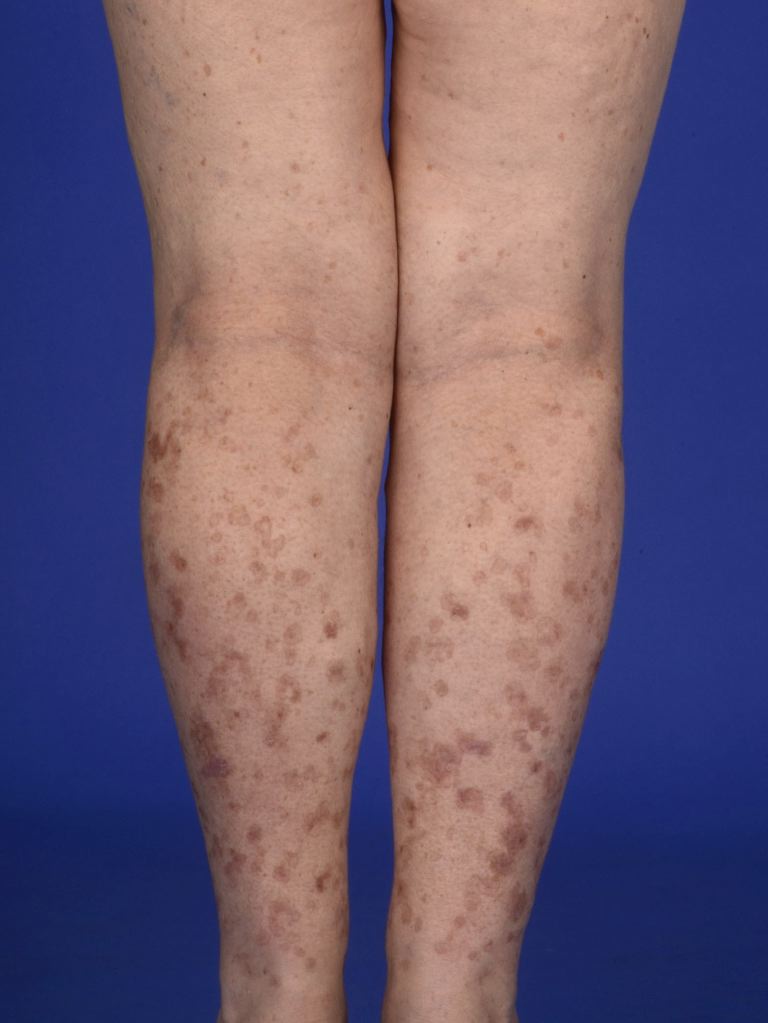
Lesions appear as asymptomatic or pruritic annular erythematous or brown circular macules, papules, or plaques with a raised hyperkeratotic border surrounding the lesions. It can occur bilaterally. Lesions are most commonly present in the third or fourth decade and occur in areas exposed to sunlight. The legs, forearms, shoulders, and back are the areas most commonly affected. The face can rarely be involved. The palms and soles are spared. Disseminated superficial actinic porokeratosis usually worsens when it is exposed to sunlight, and pruritus can intensify.[5]
Evaluation
Disseminated superficial actinic porokeratosis can be diagnosed clinically due to the characteristic appearance of the lesions. A skin biopsy can be performed if there is doubt. Dermoscopy can be a useful tool for evaluating disseminated superficial actinic porokeratosis.
Nicola et al. proposed dermoscopic features seen in porokeratotic lesions.
- A white border circumscribing the lesion
- Homogenous central white scar-like area
- Brownish globules or dots
- Vascular structures: pinpoint vessels or irregular linear vessels crossing the lesion[6]
Treatment / Management
Treatment options include the following.
Topical diclofenac
Diclofenac is an NSAID that inhibits COX-2. The medication was approved for actinic keratosis and has been used to treat disseminated superficial actinic porokeratosis with variable results. It has a good safety profile.
Ingenol mebutate
Ingenol mebutate is used in the treatment of actinic keratosis and can be used for disseminated superficial actinic porokeratosis lesions. It can help with hyperkeratosis but not atrophy or hypopigmentation.
Topical vitamin D analog
Vitamin D3 analogs have shown good responses after being used for 6 to 8 weeks. Vitamin D3 analogs are known to induce the transcription of genes that are responsible for the differentiation and proliferation of keratinocytes.
5-fluorouracil
5-FU inhibits thymidylate synthase and stops DNA synthesis, therefore, inhibiting fast proliferating cells. Its use results in a very severe and robust inflammatory reaction in some patients, including erythema, ulcerations, and dermatitis. The clinical response is usually temporary.
Imiquimod
Imiquimod works by recruiting the body's immune cells through the activation of toll-like receptors 7 and 8 and the induction of cytokines. It has been used mostly for porokeratosis of Mibelli and porokeratosis palmaris et plantaris. It can induce an inflammatory response similar to 5-FU.
Photodynamic therapy
Photodynamic therapy has been used to treat actinic keratosis, basal cell carcinoma, and squamous cell carcinoma in situ. A photosensitizer is applied that gets uptaken by atypical keratinocytes. The photosensitizers used are 5-aminolevulinic acid (ALA) and methyl aminolevulinate (MAL). The atypical keratinocytes are destroyed when light is applied because the photosensitizers generate reactive oxygen species. Some studies show that MAL might be better than ALA because it is more lipophilic.
Retinoids
Retinoids are vitamin A derivatives and are used in disorders where there is abnormal keratinocyte proliferation. Topical retinoids are preferred over systemic retinoids. Systemic retinoids have more side effects and are teratogenic. Relapse is common.
Cryotherapy and other
Cryotherapy, excision, curettage, and dermabrasion have been shown to have some good responses. It is, however, limited and is not used for extensive disease. Cryotherapy leaves a scar, and recurrence is common.
Lasers
Carbon dioxide (CO2), Q-switched ruby (QSRL), Neodymium:yttrium-aluminum-garnet (Nd:YAG), fractional photothermolysis (FP) lasers, and Grenz ray have been used to treat Disseminated superficial actinic porokeratosis. The carbon dioxide laser uses pulsed or scattered infrared light with wavelengths between 9.4 micrometers and 10.6 micrometers and works on the water inside cells. Vaporization of the liquid causes tissue destruction. Its use can leave hyperpigmentation. QSRL uses melanin as its target. It reduces hyperpigmentation but does not destroy the cornoid lamella. It has greater penetration than Nd:YAG. Nd:YAG laser is used at 532 nm for pigmented lesions. It removes the superficial papillary dermis. It was shown to decrease the hyperpigmentation and obliteration of the cornoid lamella. FP induces small zones of thermal necrosis that do not create too much damage, redness, or pain and allow for faster healing. Grenz ray uses electromagnetic radiation like x-rays. It inhibits proliferation by inhibiting DNA synthesis.[7]
Immunosuppressive agents
Medications that suppress the immune response, such as topical corticosteroids, are not usually effective because disseminated superficial actinic porokeratosis is not an inflammatory disease, but using these medications can help with the associated pruritus.[8][9][10][11][12]
Topical Statin-Cholesterol
The combination of 2% simvastatin or lovastatin and 2% cholesterol cream was demonstrated to reduce lesion number, scaling, and erythema.
Enhancing Healthcare Team Outcomes
An interprofessional team best manages superficial actinic porokeratosis. Primary care providers should refer patients to dermatologists if the diagnosis is in doubt or for some treatments. Oncologists and transplant physicians should be vigilant and have a high level of suspicion for this diagnosis. Clinicians should be aware that superficial actinic porokeratosis is a premalignant lesion, and patients should be advised to avoid the sun and tanning spas. Anytime there is a change in lesion characteristics, a biopsy is recommended. Pharmacists educate patients about medications, check for drug-drug interactions, and monitor for compliance. [Level 5]