Continuing Education Activity
Lunotriquetral joint instability is often misdiagnosed and if left untreated can result in lunotriquetral dissociation. Although patients may lack symptoms in low grade lunotriquetral injury, when symptomatic there is a typical pattern of pain and simple tests can be performed and obtained to more specifically identify injury. This activity outlines the identification and management of lunotriquetral joint instability for the clinician as well as the interprofessional team dealing with this pathology.
Objectives:
- Identify the classical presentation of lunotriquetral joint instability.
- Review the imaging modalities and imaging findings of lunotriquetral joint instability.
- Outline the treatment and management options for lunotriquetral joint instability.
- Explain the importance of improving care coordination amongst the interprofessional team to improve outcomes for patients with lunotriquetral joint instability type injuries.
Introduction
Lunotriquetral (LT) joint instability is an often missed diagnosis and can result in LT dissociation. The reasons for undiagnosed LT joint instability are in part due to multiple confounding injuries (usually associated with high energy/impact sports) and isolated injury to the LT ligament results in relatively normal-appearing radiographs. LT ligamentous injury is less common than other proximal, carpal row, ligamentous injury and thus, more poorly understood and diagnosed.
The earliest reported LT injury was in 1903, which was then followed by a description of carpal dissociation of the lunate and triquetrum without significant change in position in 1913. Later in the 1900s, volar intercalated segmental instability (VISI) began to be defined. In the mid to late 1900s, it was found that a cause of VISI was LT ligament dissociation.[1]
The wrist is comprised of numerous extrinsic and intrinsic ligaments that work together to provide stability. The complex, harmonious relationship between these ligaments indicates that compromise in the integrity of one of these parts will affect the overall position and strength of the wrist. The extrinsic ligaments that are most often involved in LT instability include the dorsal radiocarpal (providing dorsal support) and the radiolunate ligament (providing volar support). The interosseous LT ligament is the main intrinsic ligamentous supporter of the LT joint. It is described as “C” shaped and is divided into 3 parts. The volar portion is the strongest and thickest portion and provides the most stability and mainly prevents volar translation. The dorsal portion limits rotational movement, and the combination of all parts of the LT intrinsic ligament prevent dorsal translation. Both of these attach to bone, while the third, weaker portion, or membranous portion, connects to the articular cartilage. The dorsal radiocarpal ligament is an extrinsic ligament that when injured will cause further instability of the LT joint by allowing the lunate to flex more easily. The radiolunate ligament is extrinsic and can be involved in advanced cases of injury.
The lunate and triquetrum have been shown to have the same movements in flexion/extension motion as well as radial/ulnar deviation. This is likely why the lunotriquetral coalition is usually an asymptomatic variant.[2]
Etiology
LT joint instability is caused primarily by the disruption of the intrinsic LT ligament secondary to trauma to the wrist. Dorsal radiolunate and radiotriquetral ligament rupture are usually involved on presentation because isolated LT ligament injury seldom causes symptoms.[3]
Epidemiology
Young athletes who participate in high-energy sports are the group most commonly associated with LT instability. Injury to the ligaments that support the LT joint is the second most common cause of carpal instability which is reportedly one-sixth as common as scapholunate ligamentous injury.[3]
Pathophysiology
The mechanism of action for LT joint injury occurs with hyperextension of the wrist which can be caused by falling on an outstretched hand or a combination of radial deviation and extension motion. Lifting heavy objects with forced pronation has also been described as a mechanism of injury. A blow to the dorsum of the hand can provide this level of hyperextension, which is typical with sports injuries. Ulnar positive variance has also been associated with LT instability.
Isolated injury to the intrinsic LT ligament is usually asymptomatic initially. The extensive network of extrinsic ligaments within the wrist mostly maintain the LT joint, and only micromotion instability will occur, over time causing arthritis or synovitis. If a larger injury to the wrist occurs, which involves more than just the intrinsic LT ligament, VISI can occur.
In rare cases, VISI can be non-pathological due to ligamentous laxity. VISI can be seen with LT joint instability because the lunate tends to flex when there is no support from the triquetrum. As mentioned earlier, isolated injury to the LT ligament is often associated with other ligamentous injuries including dorsal radiotriquetral and volar radiolunate ligaments and the combination of these will cause a VISI deformity. VISI diagnosis will be discussed in the imaging section.[4][5]
History and Physical
LT joint injury can present asymptomatically or with gross deformity. An asymptomatic injury usually involves only an isolated assault to the LT ligament, which is often missed. The most common presentation is ulnar-sided pain in the proximal wrist with provocation during pronation and ulnar deviation. Patients may also present with decreased grip strength and a clicking noise with movement. A clue in history gathering can be participation in impact sports (football, hockey, rugby) or high energy injury. Often the history of the injury can occur more than a few weeks before presentation. The pain described is often constant with increased pain on provocation.
Upon examination, it is important to investigate the hand and forearm to identify other potential causes of the pain or co-injuries. In general, a physical exam that shows increased mobility of triquetrum in comparison to the lunate will likely be positive for LT joint instability. Almost always there will be point tenderness over the lunotriquetral joint. There are numerous described specific tests to evaluate this mobility more carefully. An important step in all of these tests is testing the contralateral wrist for comparison.
The LT shuck test or ballottement test is one of the most commonly used for diagnosis. It is performed by grasping the lunate and triquetrum between both thumbs and index fingers and applying alternating palmar and dorsal loads repeatedly. A positive exam will elicit pain, potentially a clicking noise, increased laxity, or crepitus.
Kleinman’s shear test is similar to the LT shuck test where the grasping locations are the same, but the test is performed by loading the triquetrum dorsally.
The LT compression test is performed by grasping the triquetrum and deviating it radially and ulnarly.
The click provocation test is performed by pronating the wrist after which ulnar deviation is added, and an axial compression load is applied.
The ulnar snuffbox test is a more specific localization method by palpation for tenderness which involves pressure between the extensor carpi ulnaris and flexor carpi ulnaris tendons.
Intra-articular steroid injection to the location has also been described as a test helpful for diagnosis if the injection relieved the pain patients are experiencing.[6]
Evaluation
Once the clinician performs the physical exam and obtains a thorough history and highly suspects lunotriquetral instability, he or she should order appropriate imaging which is greatly beneficial.
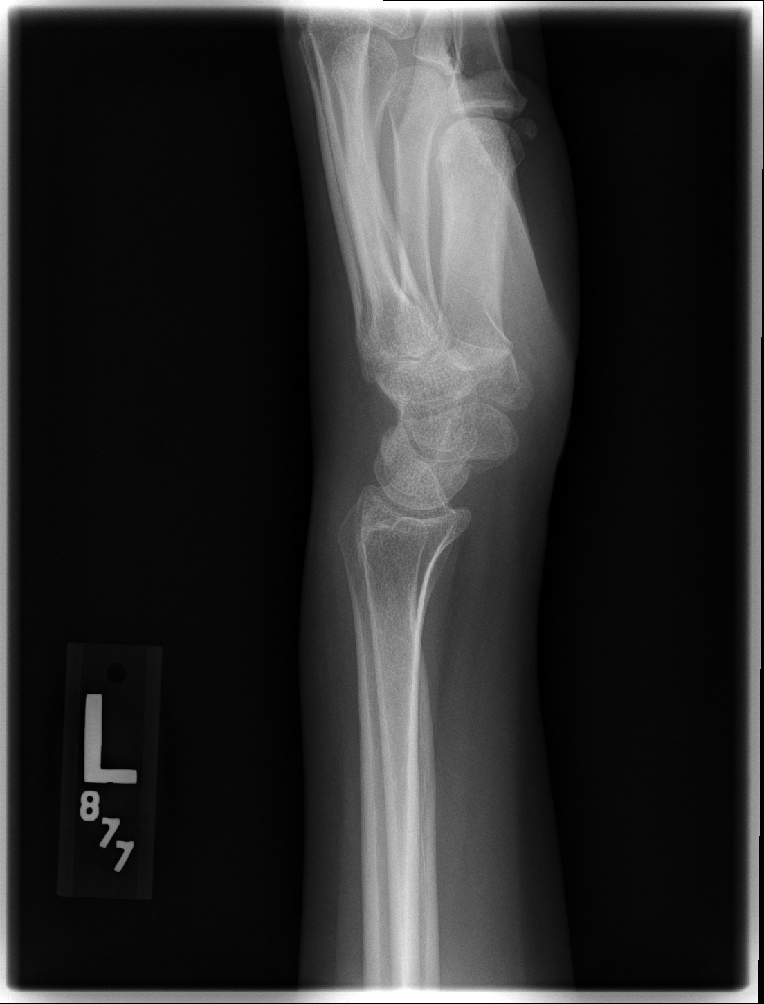
Plain radiography can be helpful if abnormal, but LT injury cannot be excluded if it is normal. A lateral and AP wrist radiograph of the injured wrist is the initial imaging to be obtained. On the lateral radiograph, a volar tilt of the lunate can be seen which causes the scapholunate angle to be less than 30 degrees, where a normal angle would be approximately 47 degrees. A scapholunate angle less than 30 degrees is consistent with VISI deformity. A capitolunate angle can also be seen where a normal capitolunate interaction is co-linear. On the AP radiograph, one can see a break in the proximal carpal arc and overlap of the lunate and triquetrum with proximal translation.
Historically, arthrography was beneficial in diagnosis as it demonstrated contrast interdigitating the lunotriquetral ligament; however, this finding was not specific as micro membrane perforations from a degenerative disease could be another cause. Magnetic resonance arthrography has since replaced arthrography.
If an audible clicking sound is associated with the pain, video fluoroscopy can be beneficial in localizing the joint in question.
MR imaging could potentially be very useful and specific in the diagnosis of LT instability, particularly when combined with arthrography. With MR arthrography, spillage of contrast from the radiocarpal to the intercarpal joints through the lunotriquetral interspace indicates disruption or injury to these intrinsic carpal ligaments. Occasionally this can be positive with age-related degenerative changes, similar to regular arthrography.
The most agreed upon and definitive way to diagnose LT joint instability is through direct visualization with arthroscopy.[6]
Treatment / Management
A nonoperative approach can initially be attempted with wrist immobilization and close observation. This allows for optimal healing and reduction of inflammation and will often be curative. If the pain is persistent, steroid injections can be considered. If these more conservative options for relatively stable LT injuries are unsuccessful, more aggressive surgical options are warranted if symptoms persist.
Surgeons may implement many operative paths, and each has slightly different indications. Arthroscopic intervention is considered the first line treatment, particularly in athletes with isolated LT ligament tears. The best view of the LT ligament in arthroscopy is the 4-5, 6R, and midcarpal portals. Closed reduction and percutaneous pinning with Kirschner wire fixation are indicated when there is acute instability. Direct, open primary repair of the LT ligament is indicated if there is gross instability of the ligament upon inspection during arthroscopy and occasionally capsulodesis can be considered. Another option is LT fusion which is indicated for chronic instability, but has fallen out of favor and nonunion is often a complication.
Post-operative management includes a short arm cast for up to 12 weeks with subsequent removal of K-wires when fully healed.[6]
Differential Diagnosis
The differential diagnosis list is extensive, and each option has distinct presenting symptoms on physical exam or imaging. Some osseous-related pain that can mimic LT instability includes fractures of the ulnar styloid, pisiform or hamate. Injury to the triangular fibrocartilage complex, extensor carpi ulnaris, and flexor carpi ulnaris are soft tissue injuries that can also mimic LT instability. Some vascular diseases that can mimic LT instability include Kienbock syndrome or hypothenar hammer syndrome. Neurological diseases that should be considered include ulnar nerve entrapment and ulnar dorsal sensory branch neuritis.[7]
Staging
A classification of LT ligament injury has been described by Viegas and colleagues which includes 3 levels of disease.
- Grade 1 is a partial or incomplete tear of the LT ligament without VISI deformity.
- Grade 2 is a complete tear of the LT ligament including a lesion within the palmar ligaments with a dynamic VISI deformity.
- Grade 3 is a complete tear of the LT ligament including a lesion within both the palmar and dorsal ligament with a static VISI deformity.
The Mayfield classification is commonly used as a grading system for arthroscopic assessment of carpal ligaments. Severity is described using a grading system of I through IV with an increasing number indicating worse injury. Grade I is attenuation/hemorrhage of ligament without malalignment. Grade II is attenuation/hemorrhage of the ligament with a small step-off of the carpal bones. Grade III is a step-off of the carpal bones with the ability to pass a probe through the gap between carpal bones. Grade IV is a step-off of the carpal bones with the ability to pass a probe through the gap between carpal bones and gross instability.[8][9]
Enhancing Healthcare Team Outcomes
Lunotriquetral instability frequently poses a diagnostic dilemma as many injuries could be a mimic of this pathology. Patients will present with general wrist pain to an urgent care facility or emergency department. There is usually no apparent deformity, and the pain is generalized over the ulnar region of the wrist. With non-specific signs such as these, an accurate and precise physical exam is vital in correctly identifying the abnormality. Often this will require a team of specialists which include an emergency physician, orthopedic surgeon, and radiologist for accurate diagnosis.
Imaging of the wrist will almost always be involved with generalized ulnar-sided wrist pain. A well-trained x-ray technician will aid by obtaining quality imaging for more accurate diagnoses by the radiologist. If no findings on the initial radiographs are present, MR imaging of the wrist can be obtained, and a well trained MRI technician is also utilized for a radiologist to make a diagnosis of LT ligamentous injury.
If intervention is indicated once the diagnosis of LT instability is made, an orthopedic surgeon and their staff including nurses, surgery technicians, and accompanying anesthesiologists will be involved for proper surgical fixation.
Almost always, even if surgery is not indicated, physical therapists will help with recovery of mobility, functioning and with pain relief. Physical therapists can also guide orthopedic surgeons in determining improvement or lack thereof for decisions on intervention with more invasive stabilization procedures.