Introduction
The head and neck, as a general anatomic region, are characterized by a large number of critical structures situated in a relatively small geographic area. It is inclusive of osseous, nervous, arterial, venous, muscular, and lymphatic structures. Lymphadenopathy is a significant clinical finding associated with acute infection, granulomatous disease, autoimmune disease, and malignancy. The involvement of specific nodal groups is an indicator of pathologically affected organs and tissues, especially in the context of malignancy. As such, intimate knowledge of the anatomic relationships of the lymphatic nodal levels and the structures they drain is critical in the delivery of appropriate therapy in many patients with cancers of the head and neck. This knowledge is especially crucial in guiding the approach to proper locoregional therapy, whether by surgery or irradiation. A detailed understanding of the principle lymphatic nodal levels of the neck is required, including their anatomical configuration and boundaries, patterns of drainage, and risk of metastatic involvement in the context of malignancy. See Image. Lymphatic System.
Blood Supply and Lymphatics
The head and neck contain a rich and elaborate lymphatic network of more than 300 nodes and their intermediate channels (see Image. Lymph Nodes of the Neck). Aponeuroses bind them together with the muscles, nerves, and vessels of the head and neck. These lymphatic chains are strongly lateralized and typically do not directly communicate between left and right in the absence of a pathologic process. This lymphatic drainage originates at the base of the skull, then proceeds to the jugular chain adjacent to the internal jugular vein. From there it moves into the spinal accessory chain adjacent to the spinal accessory nerve, or cranial nerve XI, and then meets the supraclavicular chain. The lymphatics then drain on both sides. On the left side, they drain either directly into the vasculature via the jugulo-subclavian venous confluence or directly into the thoracic duct. On the right side, they flow directly into the lymphatic duct. Conversely, most structures drain ipsilaterally, except in the case of structures situated at the anatomic midlines. These include the nasopharynx, pharyngeal wall, base of the tongue, soft palate, and larynx. The lymph nodes of the neck are further classified by level. These levels are Ia, Ib, II, III, IV, V, VI, VII, VIII, IX, X. [1][2][3][4][5]
Level Ia: Submental Group
- Anatomy
- Level I nodes are those bounded by the mandible superiorly and laterally and by the hyoid bone inferiorly. Level Ia contains the submental nodal group, bounded superiorly by the symphysis menti and inferiorly by the hyoid bone. It is bounded anteriorly by the platysma muscle, posteriorly by the mylohyoid muscles, laterally by the anterior belly of the digastric muscle, and medially by the virtual anatomic midline. These boundaries form a triangular region also termed the submental triangle.
- Drainage
- This group drains the skin of the mental region, or chin, the mid-lower lip, the anterior portion of the oral tongue, and the floor of the mouth.
- Associated primary malignancies
- These nodes most often contain metastatic deposits from malignancies of the floor of the mouth, anterior oral tongue, mandibular alveolar ridge, and lower lip.
Level Ib: Submandibular Group
- Anatomy
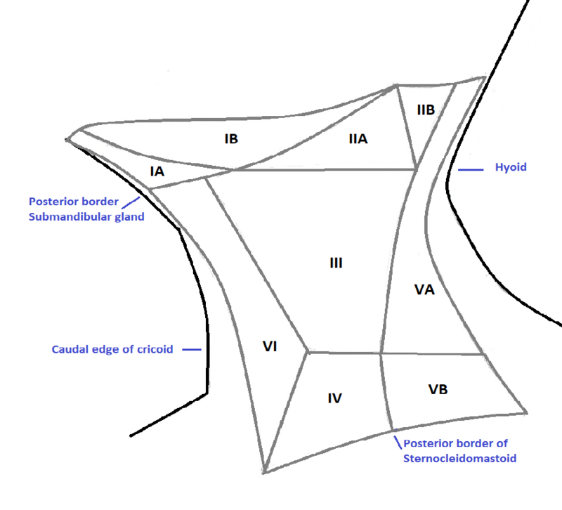
- Level Ib contains the submandibular nodal group, bounded superiorly by the mylohyoid muscle and inferiorly by the hyoid bone (see Diagram. Station for Cervical Lymph Nodes). It is bounded anteriorly by the symphysis menti, posteriorly by the posterior edge of the submandibular gland, laterally by the inner surface of the mandible, and medially by the digastric muscle. These boundaries form a triangular region also termed the submandibular triangle.
- Drainage
- They drain the efferent lymphatics from level Ia, the lower nasal cavity, both the hard and soft palates, and both maxillary and mandibular alveolar ridges. They also drain them from the skin and mucosa of the cheek, both upper and lower lips, the floor of the mouth, and the anterior oral tongue.
- Associated primary malignancies
- These nodes most often contain metastatic deposits from malignancies of the oral cavity, anterior nasal cavity, soft-tissues of the mid-face, and submandibular gland.
Level II: Upper Jugular Group
- Anatomy
- Level II represents the beginning of the jugular chain. It contains the upper jugular nodal group, adjacent to the top third of the internal jugular vein (IJV) and upper spinal accessory nerve. It is bounded superiorly by the insertion of the posterior belly of the digastric muscle into the mastoid process, and inferiorly by the caudal border of the hyoid bone or alternatively, as a surgical landmark, the carotid bifurcation. It is bounded anteriorly by the posterior edge of the submandibular gland, posteriorly by the posterior edge of the sternocleidomastoid muscle (SCM), laterally by the medial surface of the SCM, and medially by the internal carotid artery and scalenus muscle.
- Drainage
- This group drains the efferent lymphatics of the face, parotid gland, level Ia, level Ib, and retropharyngeal nodes. It receives direct drainage from the nasal cavity, the entire pharyngeal axis, larynx, external auditory canal, middle ear, and the sublingual and submandibular glands.
- Associated primary malignancies
- These nodes most often contain metastatic deposits from malignancies of the nasal and oral cavities, nasopharynx, oropharynx, hypopharynx, larynx, and major salivary glands. It is the most commonly involved nodal level. [6][7][8][9][10]
Level III: Middle Jugular Group
- Anatomy
- Level III contains the middle jugular nodal group, adjacent to the middle third of the IJV. It is bounded superiorly by the caudal border of the hyoid bone, and inferiorly by the caudal edge of the cricoid cartilage or alternatively, as a surgical landmark, the plan where the omohyoid muscle crosses the IJV. It is also bounded anteriorly by the anterior edge of the SCM, or the posterior third of the thyrohyoid muscle, and posteriorly by the posterior border of the SCM. Finally, it is bordered laterally by the medial surface of the SCM, and medially by the internal carotid artery and scalenus muscle.
- Drainage
- This group drains the efferent lymphatics from level II and level V, and partially from the retropharyngeal, pretracheal, and recurrent laryngeal nodes. It receives direct drainage from the base of the tongue, tonsils, larynx, hypopharynx, and thyroid gland.
- Associated primary malignancies
- These nodes most often contain metastatic deposits from malignancies of the oral cavity, nasopharynx, oropharynx, hypopharynx, and larynx. [11][12][13][14][15]
Level IVa: Lower Jugular Group
- Anatomy
- Level IVa contains the lower jugular nodal group adjacent to the inferior third of the IJV. It is bounded superiorly by the caudal border of the cricoid cartilage, and inferiorly by a virtual level two centimeters superior to the sternoclavicular joint, based off surgical conventions of level IVa dissection. It is bounded anteriorly by the anterior edge of the SCM (more superiorly) and the body of the SCM (more inferiorly), and posteriorly by the posterior edge of the SCM (more superiorly) and the SM(more inferiorly. This group is also laterally bound by the medial edge of the SCM (more superiorly) and the lateral edge of the SCM (more inferiorly). Finally, it is medially bordered by the medial edge of the common carotid artery, the medial edge of the thyroid gland and scalenus muscle (more superiorly), and the medial edge of the SCM (more inferiorly).
- Drainage
- This group drains the efferent lymphatics from levels III and V, and partially from the retropharyngeal, pretracheal, and recurrent laryngeal nodes. It receives direct drainage from the larynx, hypopharynx, and thyroid gland.
- Associated primary malignancies
- These nodes most often contain metastatic deposits from malignancies of the hypopharynx, larynx, thyroid, cervical esophagus, and rarely, the anterior oral cavity. Deposits from the anterior oral cavity can manifest without proximal nodal involvement.
Level IVb: Medial Supraclavicular Group
- Anatomy
- This nodal group is a continuation of level IVa to the superior edge of the sternal manubrium. It is bounded anteriorly by the deep surface of the SCM. Posteriorly, it is bound by the anterior edge of the scalenus muscle (more superiorly) and the lung apex, brachiocephalic vein, and artery on the right, as well as the common carotid and subclavian arteries on the left (more inferiorly). It is bounded laterally by the lateral edge of the scalenus muscle, and medially by the medial border of the common carotid artery which is also adjacent to level VI.
- Drainage
- This group drains the efferent lymphatics from levels IVa and Vc, and partially from the pretracheal and recurrent laryngeal nodes. It receives direct drainage from the larynx, trachea, hypopharynx, esophagus, and thyroid gland.
- Associated primary malignancies
- These nodes most often contain metastatic deposits from malignancies of the hypopharynx, subglottic larynx, trachea, thyroid, and cervical esophagus.
Level Va and Vb: Posterior Triangle Group
- Anatomy
- These nodal groups are contained with the posterior triangle. They are situated posteriorly to the SCM, and adjacent to the inferior portion of the spinal accessory nerve and transverse cervical vessels. It is bounded superiorly by the superior edge of the hyoid bone and inferiorly by a virtual plane crossing the transverse vessels. It is bound anteriorly by the posterior margin of the SCM, and posteriorly by the anterior border of the trapezius muscle. It is also bound by the platysma muscle and skin laterally, and by the levator scapulae (more superiorly) and scalenus muscle (more inferiorly) medially. A virtual plane at the inferior edge of the cricoid cartilage divides this group into upper, or Va, and lower, or Vb, posterior triangles.
- Drainage
- These nodal groups drain the efferent lymphatics from the occipital, retro-auricular, occipital, and parietal scalp nodes. It receives direct drainage from the skin of the lateral and posterior neck and shoulder, the nasopharynx, oropharynx, and thyroid gland.
- Associated primary malignancies
- These nodes most often contain metastatic deposits from malignancies of the nasopharynx, oropharynx, and thyroid.
Level Vc: Lateral Supraclavicular Group
- Anatomy
- This nodal group is a continuation of levels Va and Vb; it contains the lateral supraclavicular group. It is bounded superiorly by a virtual plan crossing the transverse vessels, and inferiorly by a virtual plan 2 cm superior to the sternoclavicular join. It is also bounded anteriorly by the skin and posteriorly by the anterior border of the trapezius muscles (more superiorly) and the serratus anterior (more inferiorly). Laterally, it is bounded by the trapezius muscle (more superiorly) and the clavicle (more inferiorly). Medially, it is bordered by the scalenus muscle and lateral edge of the SCM, and is directly adjacent to the lateral edge of level IVa.
- Drainage
- This group drains the efferent lymphatics from levels Va and Vb.
- Associated primary malignancies
- These nodes most often contain metastatic deposits from malignancies of the nasopharynx.
Level VI: Anterior Compartment Group
The anterior compartment contains this nodal group, which is symmetric about the anatomic midline. It is also further subdivided into the superficially-located anterior jugular nodes, or level VIa, and the deeper pre-laryngeal, pre-tracheal, and para-tracheal (recurrent laryngeal) nodes, or level VIb. Level VIa
- Anatomy
- Level VIa is bounded superiorly by the inferior edge of level Ib and inferiorly by the superior edge of the sternal manubrium. It is bounded anteriorly by the skin and platysma, posteriorly by the anterior surface of the infrahyoid muscles, and bilaterally by the anterior edges of the SCMs.
- Drainage
- Level VIa drains the integuments of the lower face and the anterior neck.
- Associated primary malignancies
- These nodes most often contain metastatic deposits from malignancies of the lower lip and soft tissues of the chin, such as advanced gingiva-mandibular carcinoma.
Level VIb
- Anatomy
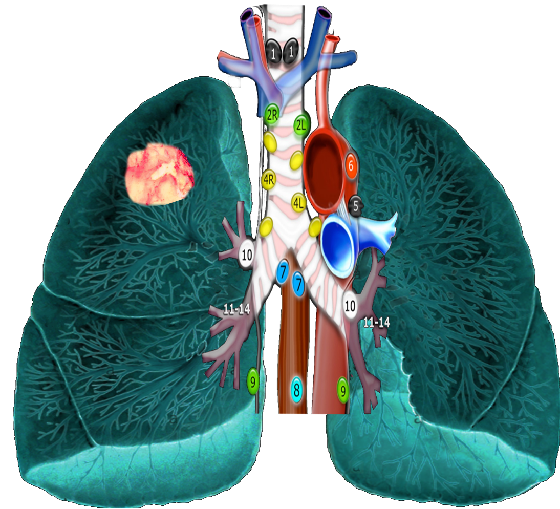
- Level VIb is bounded superiorly by the superior edge of the thyroid cartilage and inferiorly by the superior border of the sternal manubrium (see Image. Mediastinal Lymph Nodes). It is also bounded anteriorly by the posterior margin of the infrahyoid muscles, and posteriorly by the anterior larynx, thyroid gland, and trachea at the midline, the pre-vertebral muscles on the right, and the esophagus on the left. This group is bordered laterally by the common carotid artery and medially by the lateral aspects of the trachea and esophagus.
- Drainage
- Level VIb drains the efferent lymphatics from the anterior floor of the mouth, tip of the oral tongue, lower lip, thyroid gland, glottic and supraglottic larynx, hypopharynx, and cervical esophagus.
- Associated primary malignancies
- These nodes most often contain metastatic deposits from malignancies of the lower lip, oral cavity (floor of the mouth and anterior oral tongue), thyroid, glottic and subglottic larynx, the apex of the piriform sinus, and the cervical esophagus. [16][17]
Level VII: Prevertebral Compartment Group, including Levels VIIa and VIIbLevel VIIa: Retropharyngeal Nodes
- Anatomy
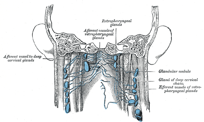
- These nodes are contained in the retropharyngeal space. They are divided into medial and lateral subgroups. The lateral groups are bounded superiorly by the superior edge of the C1 vertebral body, or the hard palate, and inferiorly by the superior edge of the body of the hyoid bone. Anteriorly, they are bounded by the posterior edge of the superior/middle pharyngeal constrictor muscles. They are bordered posteriorly by the longus capitis and longus colli muscles, laterally by the medial edge of the internal carotid artery, and medially by a virtual line parallel to the lateral edge of the longus capitis muscle. The medial groups are approximated at the midline and not well-defined.
- Drainage
- These nodes drain the efferent lymphatics from the nasopharynx, eustachian tube, and soft palate.
- Associated primary malignancies
- These nodes most often contain metastatic deposits from malignancies of the nasopharynx, pharyngeal wall, and oropharynx including tonsillar fossa and soft palate.
Level VIIb: Retrostyloid Nodes
- Anatomy
- These nodes are contained in the fatty space surrounding the large vessels of the neck leading to the jugular foramen. They are the superior continuation of level II. Level VIIb is bounded superiorly by the jugular foramen at the base of skull, and inferiorly by the inferior edge of the lateral process of the C1 vertebral body, the superior boundary of level II. These nodes are bounded anteriorly by the posterior edge of the prestyloid parapharyngeal space, and posteriorly by the C1 vertebral body and base of skull. Finally, they are bordered laterally by the styloid process and deep parotid lobe, and medially by the medial edge of the internal carotid artery.
- Drainage
- These nodes drain the efferent lymphatics from the nasopharynx.
- Associated primary malignancies
- These nodes most often contain metastatic deposits from malignancies of the nasopharynx and anywhere in the head and neck resulting in significant infiltration of upper-level II nodes causing via retrograde flow.
Level VIII: Parotid Group
- Anatomy
- This group includes the subcutaneous pre-auricular, superficial and deep intraparotid, and subparotid nodes. It is bounded superiorly by the zygomatic arch and external auditory canal, and inferiorly by the mandibular angle. This group is bounded anteriorly by the posterior edge of the mandibular ramus, the posterior edge of the masseter muscle (more laterally), and medial pterygoid muscle (medially). It is also bordered posteriorly by the anterior edge of the SCM (more laterally) and posterior belly of the digastric muscle (more medially). These nodes are bordered laterally by superficial muscular aponeurotic system (SMAS) layer within the subcutaneous tissues, and medially by the styloid process and muscle.
- Drainage
- These nodes drain the efferent lymphatics from the frontal and temporal skin, eyelids, conjunctivae, auricles, external acoustic meatus, tympanum, nasal cavities, the root of the nose, nasopharynx, and the eustachian tube.
- Associated primary malignancies
- These nodes most often contain metastatic deposits from malignancies of the previously named draining structures, as well as the orbit, external auditory canal, and parotid gland.[18]
Level IX: Buccofacial group
- Anatomy
- This group contains the malar and the buccofacial nodes. These are superficial nodes surrounding the facial vessels on the external surface of the buccinator muscle. It is bounded superiorly by the inferior edge of the orbit and inferiorly by the inferior border of the mandible. It is also bounded anteriorly by the SMAS layer within the subcutaneous tissue, and posteriorly by the anterior edge of the masseter muscle and the corpus adiposum buccae. The lateral border is the SMAS layer, and the medial border is the buccinator muscle.
- Drainage
- These nodes drain the efferent vessels of the nose, eyelids, and cheek.
- Associated primary malignancies
- These nodes most often contain metastatic deposits from malignancies of the facial skin, nose, and buccal mucosa, as well as the maxillary sinus if invading soft tissues of the cheek.
Level X: Posterior Skull Group, including Levels Xa and Xb
Level Xa: Retroauricular and Subauricular Nodes
- Anatomy
- This group includes superficial nodes on the mastoid process. It is bounded superiorly by the superior edge of the external auditory canal, and inferiorly by the mastoid tip. It is also bounded anteriorly by the anterior edge of the mastoid (inferiorly) and posterior edge of the external auditory canal (superiorly), and posteriorly by the posterior edge of the SCM. This group is bordered laterally by subcutaneous tissue, and medially by the splenius capitis muscles (inferiorly) and the temporal bone (superiorly).
- Drainage
- These nodes drain the efferent vessels from the posterior auricular surface, external auditory canal, and adjacent scalp.
- Associated primary malignancies
- These nodes most often contain metastatic deposits from malignancies of the retro-auricular skin.
Level Xb: Occipital Nodes
- Anatomy
- This group is the superior and superficial continuation of level Va. It is bounded superiorly by the external occipital protuberance, and inferiorly by the superior border of level V. It is also bounded anteriorly by the posterior edge of the SCM, which is the posterior border of level Xa, and posteriorly by the anterior/lateral side of the trapezius muscle. Finally, this group is bordered laterally by subcutaneous tissues, and medially by the splenius capitis muscle.
- Drainage
- These nodes drain efferent vessels from the posterior hairy scalp.
- Associated primary malignancies
- These nodes most often contain metastatic deposits from malignancies of the occipital skin. [5]
Clinical Significance
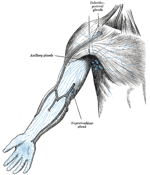
The lymphatic drainage of the head and neck is unique. Its remarkably well-delineated and characterized anatomic subgroups are closely associated with draining anatomic structure (see Image. Lymph Nodes of the Arm). These, in turn, are related to malignant neoplasms arising from specific anatomic structures. Intimate knowledge of this network allows the surgeon to complete an oncologically-appropriate dissection. It also helps the radiation oncologist appropriately treat elective nodal levels to reduce recurrence and the primary care physician to guide the path to the necessary workup.