Continuing Education Activity
Arnold-Chiari or Chiari malformations describe a group of deformities of the posterior fossa and hindbrain, which includes the cerebellum, pons, and medulla oblongata. These deformities lead to problems ranging from cerebellar tonsillar herniation through the foramen magnum to the absence of the cerebellum, with or without other associated intracranial or extracranial defects such as hydrocephalus, encephalocele, syrinx, or spinal dysraphism. This activity examines when this condition should be considered within a differential diagnosis and how to evaluate the patient for it properly. This activity highlights the role of the interprofessional team in caring for patients with this condition.
Objectives:
- Review the classification of Chiari malformations.
- Describe the presentation of a patient with a Chiari malformation.
- Outline the management options available for Chiari malformations.
- Summarize interprofessional team strategies for improving care coordination and outcomes in patients with Chiari malformations.
Introduction
Arnold-Chiari, or simply Chiari malformation, is the name given to a group of deformities of the posterior fossa and hindbrain (cerebellum, pons, and medulla oblongata). Issues range from cerebellar tonsillar herniation through the foramen magnum to the absence of the cerebellum with or without other associated intracranial or extracranial defects such as hydrocephalus, syrinx, encephalocele, or spinal dysraphism.[1][2][3]
Classification
Chiari malformations are classified based on their morphology and severity of anatomic defects, typically through imaging (or autopsy).
Chiari I is the least severe and often found incidentally. It is characterized by one or both pointed (not rounded) cerebellar tonsils that project 5 mm below the foramen magnum, measured by a line drawn from the basion to the opisthion (McRae Line).
Chiari II consists of brainstem herniation and a towering cerebellum in addition to the herniated cerebellar tonsils and vermis due to an open distal spinal dysraphism/myelomeningocele.
Chiari III involves herniation of the hindbrain (cerebellum with or without the brainstem) into a low occipital or high cervical meningoencephalocele.
Chiari IV is now considered obsolete.[4] Prior to becoming an obsolete diagnosis, it was already a more controversial and rare variant that demonstrated severe cerebellar hypoplasia, similar to primary cerebellar agenesis. Previously some stated that myelomeningocele could be present,[5] while others argued that the presence of myelomeningocele should then be classified as a Chiari II with a vanishing cerebellum.[6]
There are other controversial reported classifications, including Chiari 0, Chiari 1.5, and Chiari V. Chiari 0 is characterized by syringomyelia without hindbrain herniation while Chiari 1.5 is felt to be the progression of Chiari I with increased cerebellar tonsillar descent and some involvement of the brainstem.[7][8] Chiari V, the most severe variant, represents cerebellar agenesis with occipital lobe descent and herniation through the foramen magnum.[9]
Etiology
There are multiple proposed theories, including molecular, hydrodynamic, and mechanical, with the likelihood that different mechanisms can have the same resulting Chiari malformation.[10]
Reduced volume of the posterior fossa leads to displacement of the cerebellar tonsils through the foramen magnum in Chiari I malformations. Causes include primary congenital hypoplasia or secondarily from acquired morphologic changes, such as premature closure of sutures, calvarial dysplasia, or genetic/syndromic. Mutations on chromosomes 1 and 22 have been identified as possible causes for hereditary posterior fossa hypoplasia.[11]
McClone and Knepper proposed an open neural tube defect (myelomeningocele) is the underlying cause of Chiari II malformations.[12] This leads to leakage or redirected flow of cerebrospinal fluid resulting in a fourth ventricle that is unable to maintain distension. This continued collapse of the fourth ventricle in utero results in a hypoplastic posterior fossa and cerebellar tonsillar herniation. This is also the suspected cause of Chiari III as well, though in the setting of an encephalocele or high cervical myelomeningocele, as opposed to the lumbar or sacral myelomeningocele in Chiari II. Folate deficiency and methylenetetrahydrofolate reductase mutations increase the risk of neural tube defects and can thus be an underlying cause of Chiari II and III.
The etiology of the remaining Chiari variants is still debated and not clearly known. Trauma can be an etiology of cerebellar tonsillar herniation, though, in the setting of a normal-sized posterior fossa, a designation of Chiari is not appropriate.
Epidemiology
Chiari I malformation is the most common type and occurs in approximately 0.5 to 3.5% of the general population with a slight female predominance (1.3:1).[13][14]
Chiari II occurs in 0.44/1000 births without gender predominance but can have a decreased incidence with folate replacement therapy by the mother in utero.
The remaining Chiari malformations are much rarer. Chiari III is the most common of these other variants, consisting of 1-4.5% of all Chiari malformations.
Pathophysiology
Neurologic signs and symptoms can arise from 2 mechanisms:
- Direct compression of neurological structures against the surrounding foramen magnum and spinal canal
- Syringomyelia or syringobulbia development
- The obstruction of cerebrospinal fluid (CSF) outflow eventually results in syrinx development.
- Fluid-filled cavities (syrinx) develop within the spinal cord or brainstem, resulting in neurologic symptoms as the cavity expands.[15]
In patients with the Chiari I malformation, the bones of the skull base often are underdeveloped, which results in a reduced volume of the posterior fossa, the volume of which is inadequate to contain the entire cerebellum; thus, cerebellar tonsils are displaced through the foramen magnum.
The posterior fossa in Chiari type II malformation is even smaller than in Chiari I malformation. The cerebrospinal fluid (CSF) cisterns are poorly developed due to lack of fourth ventricular expansion as a consequence of in-utero derivation of CSF circulation to the neural tube defect, all of which results in hindbrain structures downward herniating with subsequent compression of these structures against the foramen magnum.[15]
In both type I and II, there is CSF-flow obstruction by the foramen magnum crowding, and consequently, hydrocephalus and/or syringomyelia formation are possible over time.
History and Physical
In Chiari I malformation, the most common presentation is suboccipital headaches and/or neck pain (80%). Symptoms are exacerbated when asked to perform the Valsalva maneuver. Other common presentations include ocular disturbances, otoneurologic symptoms (dizziness, hearing loss, vertigo), gait ataxia, and generalized fatigue. Although much less common, the literature reports multiple case studies in which patients have presented with isolated extremity pain or weakness, one such report including a presentation of unilateral shoulder pain with isolated muscle weakness presenting to a sports medicine clinic.[16]
Myelopathy classically presents with “dissociated sensory loss” (loss of pain and temperature sensation, preserved fine touch and proprioception) and motor weakness.[17][18]
Cerebellar signs, including ataxia, dysmetria, and nystagmus, and lower cranial nerve deficits (IX, X, XI, XII CN) result either from direct compression of the cerebellum or medulla at the foramen magnum or from syringomyelia or syringobulbia.
Sleep apnea can occur in a patient with Chiari malformation due to a weakness of pharyngeal muscles elicited by the brainstem, upper spinal cord, or lower cranial nerve compression.
It is not an uncommon scenario to find patients with radiological findings compatible with Chiari malformation with no clinical manifestations of the disease (incidental Chiari malformation). Therefore, nonspecific symptoms such as generalized fatigue or classic pattern migraines are not necessarily related to the Chiari malformation.
The remaining variants (with the exception of Chiari 0 and 1.5) are diagnosed often in utero or at birth.
Evaluation
Imaging evaluation of Chiari malformations varies, with Chiari I evaluated using magnetic resonance imaging (MRI) typically in a child or adult, whereas Chiari II to IV are often first evaluated by ultrasound in utero, with fetal MRI assessment performed for further characterization.
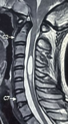
MRI of the head and cervical spine is the test of choice in evaluating Chiari I. This will demonstrate cerebellar tonsillar descent greater than 5 mm below the foramen magnum (McRae line). In addition, a decreased size of the posterior fossa and a syrinx may be seen.[19] Depending on the extent of the syrinx, the addition of a thoracic and/or lumbar spine MRI may be needed. In the setting of ventricular dilation, CSF flow (or cine) sequences may be performed to assess for CSF flow dynamics and evaluate for obstruction at the foramen magnum.
Other useful tests in the management of patients with Chiari I malformation include:
- Myelography: Of special value as an alternative in patients in who an MRI cannot be obtained.
- CT or x-rays of the neck and head: May reveal common associated bony defects, particularly of the craniocervical junction relevant for surgical planning, such as basilar invagination.
Fetal sonography, often during the second-trimester anatomy scan, demonstrates the typical imaging features of Chiari II and III malformations. One classic imaging finding on ultrasound is the lemon sign of the anterior frontal calvarium, with loss of the normal convex curvature and flattening or inward bowing/scalloping that results in a shape similar to a lemon.[20] The banana sign is another classic sign of Chiari II and distal neural tube defect, this time regarding the cerebellum, which demonstrates an abnormal morphology with anterior curvature of the cerebellum and obliteration of the cisterna magna.[21] Chiari III will demonstrate the occipital or high cervical meningoencephalocele when evaluating the posterior fossa during a prenatal exam.
Fetal MRI can demonstrate the cerebellar hypoplasia/aplasia of Chiari IV and V and further evaluate the neural tube defects and hindbrain herniation of Chiari II and III. MRI also better demonstrates the tectal beaking that occurs in Chiari II.
Laboratory studies are not of help in the evaluation of patients with Chiari malformation. However, laboratory studies are needed during planning for surgery. Routine studies like the complete blood count (CBC), coagulation profile, electrolyte levels, chest X-ray, and electrocardiogram (ECG) will suffice.
Treatment / Management
Medical Management
Patients with Chiari malformation and who have no symptoms can be managed medically. Headaches and neck pain can be treated with muscle relaxants, NSAIDs, and temporary use of a cervical collar. However, studies show that while a headache and nausea may improve, there will be no improvement in gait with medical management in many symptomatic patients. Close to 90% of patients with Chiari type I may remain asymptomatic even if they have syringomyelia.
Surgical Management
The main treatment for Chiari malformation is surgical with the goal of re-establishing the CSF flow across the craniovertebral junction and relieving pressure on the cerebellum and hindbrain by decompressing the posterior fossa.[22][10]
Surgery is recommended for persistently symptomatic patients and confirmed tonsillar herniation. In the setting of asymptomatic tonsillar herniation, with or without syrinx, observation is recommended unless symptoms develop.
Better surgical results are seen when surgery is performed within 2 years of symptoms onset.
Surgical Techniques
The standard surgical technique for Chiari I is a posterior fossa decompression.[23][10][24] This is obtained by a suboccipital craniectomy enlarging the foramen magnum, often in conjunction with C1, and possible C2, laminectomy. The dura may or may not be opened, with subsequent dissection of arachnoid adhesions if present. Depending on the available dural expansion and size of the posterior fossa, a duraplasty may need to be performed. The dural graft can be an autograft such as occipital fascia or tensor fascia lata (TFL) tendon, or artificial dura.[25] In the setting of a syrinx, a shunt can also be placed if decompression alone is not effective. Tonsillar cauterization may also be performed.
More recently, minimally invasive techniques have been described similar to those used in the spine. These allow for smaller incisions, less soft tissue damage, less dural manipulation, shorter hospital stays, faster recovery, and fewer complications.[26][27][28][29][30][31]
Initial surgical correction for Chiari II is the correction of the myelomeningocele, generally in the first 48 hours. This can also be done in utero through a hysterotomy.[32] Closure of the spinal dysraphism can be done in a variety of ways, with either primary skin closure, myocutaneous flap, or fasciocutaneous flap, depending on the severity, involved layers, and the available adjacent tissue. The vast majority will eventually need a ventricular shunt for CSF diversion in the setting of hydrocephalus.[33] If needed, a posterior decompression is performed later to allow suboccipital expansion.
Chiari III follows a similar course to Chiari II. The occipital/high cervical encephalocele is corrected first, with resection of herniated contents, as these are typically non-viable, followed by dural closure and a cranioplasty.[34] If the amount of herniated tissue is greater than intracranial contents, the patient is deemed a nonsurgical candidate. A ventricular shunt is placed if the patient has concomitant hydrocephalus.
Contraindications to Surgery
Suboccipital decompression is contraindicated when the tonsillar herniation is due to a pathology other than Chiari malformation. Some examples of this are intracranial hypotension or mass effect in the posterior fossa due to a mass.
Differential Diagnosis
- Intracranial hypotension – sagging midbrain may mimic tonsillar or hindbrain herniation.
- Normal variant cerebellar tonsillar ectopia – does not meet the criteria for Chiari malformation and an incidental finding in an asymptomatic patient.
- Tonsillar herniation from increased intracranial pressure (ICP) – assess for causes of ICP such as mass effect from neoplasm, hydrocephalus, trauma, or hemorrhage.
Prognosis
Chiari I has a good prognosis, but it also depends on the presence of preexisting neurological deficits. Most patients who have no neurological deficits have an excellent outcome.[35][14] Chiari II has a 3% neonatal in-hospital mortality and 15% 3-year mortality rate. Those that survive can have increasing motor dysfunction over time. Continued follow-up for shunt placement evaluation or shunt failure is recommended. Prognosis in the more severe Chiari variants is poor and often dismal, with early death.
Individuals who have chronic weakness or gait problems usually do not improve, and their prognosis is guarded.[36]
Complications
- Pseudomeningocele
- CSF leak
- Meningitis
- Wound infection
- Lower brainstem malfunction
- Epidural hematoma
- Apnea
- Vertebral artery injury
Postoperative and Rehabilitation Care
In the postoperative period, monitoring for CSF leak is vital. Some patients may develop a pseudomeningocele, which may require drainage.
Patients can also be evaluated postoperatively with the Chicago Chiari Outcome Scale for a more subjective evaluation of postoperative improvement.[37][38][39] This scoring system factors pain, non-pain symptoms, functionality, and postoperative complications into a 1-4 point scale for each component. This results in a total score ranging from 4-16, with 4 representing an incapacitated outcome, while 16 represents an excellent outcome.
Exercise and heavy lifting should not be done for at least 3 to 4 weeks after the procedure.
Most patients require 6 to 8 weeks to recover from surgery and reverse any major neurological deficit fully. Patients should consider refraining from contact sports following surgery, even after the surgical site is well healed.
Repeat MRI is necessary to ensure that the syrinx has responded to the treatment.
Deterrence and Patient Education
If the Chiari I is being managed conservatively, the patient should be instructed to come back if he/she develops cough, headaches, or progressive limb weakness. In the postoperative period, the patient should be conscious of the risk of developing pseudomeningocele, CSF leak, and meningitis.
Pearls and Other Issues
Arnold-Chiari, also known as Chiari malformation, is the name given to a group of deformities of the hindbrain (cerebellum, pons, and medulla oblongata).
Issues range from herniation of the posterior fossa contents outside of the cranial cavity to the absence of the cerebellum with or without other associated intracranial or extracranial defects such as hydrocephalus, syrinx, encephalocele, or spinal dysraphism.
For Chiari I malformation, the prognosis is good, but it also depends on the presence of preexisting neurological deficits. Most patients who have no neurological deficits have an excellent outcome.
A controversial subject in clinical management entails the diagnosis in the setting of possible sport-specific participation. Historically, symptomatic and postsurgical Chiari I patients were advised not to return to contact sports, but recent studies have shown that the risk of catastrophic injury in this population is low.[40][41] Regardless, a decision should be made on a case-by-case basis.
Individuals who have chronic weakness or gait problems usually do not improve, and their prognosis is guarded.
Enhancing Healthcare Team Outcomes
Arnold Chiari malformations are relatively common and represent a spectrum of hindbrain anomalies. The diagnosis and treatment of this condition require an interprofessional team consisting of primary care, neurologists, radiologists, neurosurgeons, and specialty trained nurses. [Level 5] Depending on the severity of the malformation, the individual may be asymptomatic or have severe neurological symptoms. While the patients are often managed with decompressive surgery, the nurses are responsible for looking after these individuals. Hence, the nurse must be aware of the potential post-surgical complications and their presentation. The prognosis for most patients with a Chiari I malformation is good, but it also depends on the initial neurological presentation. Those patients with mild neurological deficits tend to have good outcomes, but those with moderate to severe symptoms tend to have a guarded prognosis. The surgery is also associated with several complications, of which the most common are CSF leak and pseudomeningocele. A few individuals may have a persistent syrinx and may require a shunt.[42][Level 3]

