Continuing Education Activity
Laparoscopic cholecystectomy is performed over 750,000 times in the United States annually. As laparoscopic cholecystectomy has been increasingly used to treat symptomatic cholelithiasis, the number of bile duct injuries (BDI) has also increased. The biliary tree and relationship of the cystic duct and its insertion onto the common hepatic duct is noted to have variable and anomalous anatomy. The most common reason for injuring the bile duct is due to the misidentification of normal biliary anatomy. Once the bile duct is injured, early recognition is crucial to facilitate appropriate treatment. This activity outlines the evaluation and management of iatrogenic and traumatic injury to the biliary system and highlights the role of the interprofessional team in evaluating and managing patients with this condition.
Objectives:
- Describe the etiology of bile duct injury.
- Outline the methods and classification systems for evaluating any injury to the biliary system.
- Explain the management considerations for patients with bile duct injury based on the etiology.
- Review the importance of collaboration and coordination amongst the interprofessional team members to improve outcomes of patients with injury to their biliary system.
Introduction
Injury to the biliary tree poses a unique challenge for the surgeon due to the variable anatomy, limited working space, and morbidity of its complications.
Minimally invasive cholecystectomy is performed over 750,000 times in the United States annually. As laparoscopic cholecystectomy has been increasingly used to treat symptomatic cholelithiasis, the number of bile duct injuries (BDI) has also increased. The biliary tree and relationship of the cystic duct and its insertion onto the common hepatic duct is noted to have variable and anomalous anatomy. The most common reason for injuring the bile duct is due to the misidentification of normal biliary anatomy.[1] Once the bile duct is injured, early recognition is crucial to facilitate appropriate treatment.
Etiology
Iatrogenic biliary injury most commonly occurs by misidentifying the common bile duct for the cystic duct during laparoscopic cholecystectomy[2], with an incidence of 0.3 to 0.7%, which is historically three times higher than in open cholecystectomy.[3] The variable biliary anatomy is one of the factors in the causation of this injury. Injury to the biliary tree rarely occurs in penetrating or blunt abdominal trauma, with an incidence of 0.1% of hospital admissions for trauma. Depending on the location and time of diagnosis, the management ranges from cholecystectomy, drainage, reconstruction to restore the flow of bile into the intestine, or hepatic resection.[4]
Epidemiology
The incidence of bile duct injury increased with increasing adoption of the laparoscopic technique for cholecystectomy and treatment of other biliary pathologies. Bile duct injury occurs in 0.3 to 0.7% of the approximately 750000 laparoscopic cholecystectomies performed in the United States every year.[1] Iatrogenic bile duct injury is a cause of significant morbidity and mortality, especially when not recognized intraoperatively, which only occurs 25% to 32.4% of the time.[5] This frequently results in a reduction in quality of life for the patient and litigation for the surgeon. When the bile duct is injured laparoscopically, the injuries tend to be more complex, due to the limited visualization and adoption of electrosurgery.[6]
Risk factors of bile duct injury include anatomic variants, patient condition, gallbladder pathology, and surgeon related factors. A short cystic duct or cystic duct that runs parallel to the common bile duct can lead to the misidentification of the cystic duct. Other factors, such as variation of the cystic duct and common hepatic duct junction, a cystic duct that inserts onto the right hepatic duct, an accessory cystic duct, or presence of ducts of Luschka also contribute to injury or leak. Patients who have severe obesity, prior hepatobiliary surgery, or underlying liver disease can impair visualization and increase the rate of injury, though 80% of injuries occur in the absence of any risk factors. Acute cholecystitis increased the rate of bile duct injuries due to the associated inflammation, adhesions, gallbladder wall thickening, and increased bleeding.[7] As surgeons perform more laparoscopic cholecystectomies, the rate of bile duct injury decreases. Routine intraoperative cholangiography does not decrease the incidence of bile duct injuries.[8] However in cases of uncertain anatomy or suspicion of BDI, an intraoperative cholangiogram(IOC) or another alternative method to delineate biliary anatomy has been recommended.[9]
Histopathology
Histologically, the bile duct injury may be inconspicuous, consisting of reactive and focal degenerative changes in bile duct epithelium. These cells become ballooned and vacuolated. The pathologist may note pyknotic nuclei and mitotic figures.
In the most advanced stage, there is often portal edema with polymorphic inflammatory cells containing neutrophils. Frank cholestasis may present with the presence of bile pigment within Kupffer cells and hepatocytes and bile duct proliferation with canalicular bile plugs.
At late stages, portal tract fibrosis may be prominent, and appropriate treatment must precede this irreversible stage.
History and Physical
Only approximately 25 to 40% of bile duct injuries are recognized intraoperatively. The injury manifests as biliary obstruction, biliary leak, or biliary stricture. History of gallbladder empyema[10], gangrenous cholecystitis[11] as indications for cholecystectomy should raise higher suspicion for BDI. If the bile duct injury is not noted immediately, the patient may present with bile in the drain if one was left in place. Otherwise, the incision may drain bile. Fever, vague abdominal pain, nausea, pruritis, and inability to tolerate diet are postoperative findings that can indicate a BDI.
If there is a large leak or bile collection, the patient can present with an acute abdomen[12] or sepsis.
In cases of biliary obstruction, the patient will have features of obstructive jaundice. Early recognition of the injury is essential to minimize the morbidity associated with untreated bile duct injuries, such as cholangitis, portal hypertension, or cirrhosis.
Evaluation
If there is a bile duct injury or leak noted intraoperatively, the surgeon must decide whether he has adequate training, staff, and resources to evaluate and treat the injury appropriately. If the surgeon decides to proceed, cholangiography must be performed to delineate anatomy and plan treatment.[13] If the surgeon feels he cannot safely repair the injury, no further dissection or conversion to laparotomy should be performed, and the patient should have a drain placed and transferred to an institution with experienced surgeons. If a cholangiogram catheter can easily be placed into the injury, this can help the next team identify the injury and perform prompt cholangiogram.
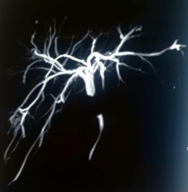
In patients presenting postoperatively, an abdominal ultrasound can identify the presence of a fluid collection in the gallbladder fossa or ductal dilation, which can pair with clinical findings of abdominal pain and hyperbilirubinemia to indicate the presence of a bile leak. CT scan is very sensitive to evaluate free fluid within the abdomen. A HIDA scan[14] can distinguish the normal postoperative fluid collections from irrigation fluid or bile spillage intraoperatively from an active bile leak, but it is difficult to ascertain the level of the leak from HIDA scan. A final delayed image of 3 hours post radiotracer injection is necessary if no major leak is initially noted to confirm the absence of a leak. Endoscopic retrograde cholangiography is used to evaluate the level of the leak and provide therapeutic intervention through stenting. Magnetic resonance cholangiopancreatography can help diagnose a biliary leak, especially the level of the leak.
There are many proposed classification systems of injuries to the bile duct.
The Strasberg-Bismuth classification defines injuries and biliary strictures based on their anatomic location within the biliary system relating to the biliary confluence.[15] Type A, the most common, is a leak from the cystic duct or small ducts in the gallbladder or liver bed. Type B is an occlusion of an aberrant right hepatic duct, while type C is transection of this aberrant right hepatic duct with subsequent leakage. These injuries often occur in conjunction with a cystic duct that drains into an aberrant right hepatic duct. At the level of the aberrant right hepatic duct draining into the common hepatic or common bile duct, it may be misidentified as the cystic duct and transected. Type D is an injury to the lateral bile duct involving less than 50% of the duct circumference. Type E injuries are defined as strictures to the hepatic ducts and are further classified by the proximal extent. Type E1 strictures have a common duct stump of greater than 2 cm. Type E2 strictures have less than 2 cm of common duct available for anastomosis. Type E3 strictures occur at the confluence. Type E4 strictures indicate a separation of the right and left hepatic ducts due to the destruction of the confluence. Type E5 stricture is due to the injury of an aberrant right hepatic duct with concomitant stricture of the common hepatic duct.[16]
The McMahon classification system classifies injuries after laparoscopic cholecystectomy as either major or minor based on the depth of injury. A laceration of less than 25% of the circumference of the common bile duct or cystic-CBD confluence ranks as a minor injury. A complete transection or laceration greater than 25% to the CBD or any postoperative bile duct stricture is a major injury.[17]
The Stewart-Way classification system stratifies injuries based on mechanism after examining surgeon operative reports and the described process of injury.[18] Class I injuries occur with a prevalence of 7% when there is partial transection of the common duct due to being mistaken for the cystic duct causing minimal loss of tissue. Most often this is due to the misidentification of the cystic duct with the common hepatic duct. Less commonly, the extension of the cystic duct opening during cholangiography can involve the common hepatic or common bile duct. Class II injuries occur with a prevalence of 2% from thermal injury or inadvertent clipping of the lateral common hepatic duct, causing stricture or leak due to limited visibility or an attempt to control bleeding. The common hepatic duct is only partially damaged, and there is an associated hepatic artery injury in 18% of cases, especially in cases of anomalous origin of the right hepatic artery from the superior mesenteric artery. Class III injuries involve complete transection of the common hepatic-common bile duct with excision of a variable portion of duct proximal to this junction and occurs when mistaking the common duct for the cystic duct with subsequent partial excision of the duct as the gallbladder gets removed. Class III injuries are the most common type, occurring in 60% of cases. Subdivision class IIIa injuries have a remnant of common hepatic duct, while class IIIb involves transection at the junction of the cystic duct-CHD. In class IIIc, the bifurcation has been excised with complete loss of the confluence. In class IIId, the transection is above the level of a lobar duct or secondary bile duct. Class IV injuries involve trauma to the right hepatic or right segmental hepatic duct, 60% of the time with additional injury to the right hepatic artery. Again, misidentification of the right hepatic duct as the cystic duct is the most common mechanism or inadvertent injury to the lateral wall of an inferior lying right hepatic duct.
Treatment / Management
Depending on both the level and complexity of the injury, the treatment can range from simple drainage procedures to reconstruction of the biliary system.[19] With all injuries, noted either at the time of injury or in a delayed fashion, the patient can always transfer to a specialized center with clinicians and resources better suited to treat bile duct injuries.
The Strasberg classification can help guide treatment. In Strasberg type A injuries, the biliary system is in continuity with a leak from the cystic duct or minor hepatic duct through the liver bed. If there is a drain already in place, the output can be monitored to evaluate for spontaneous closure of the leak. Endoscopic stenting across the lesion can help occlude the leak and facilitate drainage through the biliary system by decreasing the pressure in the proximal biliary system. Sphincterotomy may be necessary in cases of retained choledocholithiasis. If the patient has peritonitis or worsening intraabdominal sepsis, exploration may be necessary for washout. For Type A injuries involving leakage from the cystic duct stump, coil embolization the cystic duct by interventional radiology has been described in a few cases.[20]
Strasberg type B injuries with only minimal pain and mild elevation in liver function tests can be treated conservatively and monitored. The occlusion may go unnoticed and cause segmental cholestasis, intrahepatic stones, and atrophy of the right lobe of the liver. If there is evidence of cholangitis due to the occlusion, the patient will require drainage of the affected segment via percutaneous transhepatic cholangiogram with biliary drainage tube placement or hepaticojejunostomy. Segmental resection of the involved segments may be necessary if the atrophy is significant. Endoscopic management of occlusive injuries is typically unsuccessful as the occlusion puts the proximal biliary segment in discontinuity with the distal biliary tree.
Strasberg type C injuries are similar to type B, but the injured accessory duct is leaking instead of occluded. Both types B and C injuries are not amenable to endoscopic intervention as the segment proximal to the injury is not in continuity with the biliary system. Percutaneous drainage of the subhepatic biliary leak can result in spontaneous closure and prevention of biliary peritonitis. In rare instances, the patient may require hepaticojejunostomy or hepatectomy.
Strasberg type D injuries involve a medial partial injury to the common bile duct. If the injury is small with no evidence of devascularization, the defect can be closed using interrupted 5-0 absorbable monofilament suture with a drain left in place, endoscopic sphincterotomy, and stent placement. If there is devascularization, the surgeon should still repair the injury with a drain left in place in anticipation of an expected bile leak. Endoscopic stent placement with interventional radiologic drain placement is an alternative, especially for injuries noted in the postoperative period. A multidisciplinary approach is necessary to ensure proper treatment and avoid further injury or complications. A HIDA scan is obtained two to four weeks after the insertion of the endoscopic stent to evaluate for a continued leak. In the absence of a leak on HIDA, the stent is removed endoscopically, and cholangiogram is performed. A leak noted on cholangiogram is treated with sphincterotomy or stent replacement for an additional four weeks. The clinician should follow these patients with repeat ERCP or MRCP to evaluate for the development of stricture, leak, or progression to a Strasberg type E injury.
Strasberg type E injuries that are noted at the time of injury can be repaired primarily in an end to end fashion if there is no tension on the anastomosis. This repair should be done over a T-tube that provides external drainage or a Y tube that drains into the duodenum. If the anastomosis is not performable in a tension-free fashion, a Roux-en-Y hepaticojejunostomy is the preferred reconstruction option. In cases of friable tissue or very dense adhesions where hepaticojejunostomy is not feasible, a pedicled omental patch can be used as a temporary repair to control bile leak until definitive reconstruction.[21]
Vascular injuries associated with bile duct injuries can present with hemobilia, abscess, or ischemia, for which the management is angioembolization, percutaneous drainage, or liver resection, respectively.[22]
Differential Diagnosis
Patients who continue to have symptoms of nausea, vomiting, jaundice, or abdominal pain after cholecystectomy may be suffering from post-cholecystectomy syndrome (PCS).[23] The reported incidence varies from 5 to 60%, and symptoms can result from a bile duct injury, stricture, retained stones, biliary dyskinesia, or sphincter of Oddi dysfunction.
Patients may also have symptoms of PCS caused by extra-biliary disorders, such as peptic ulcer disease, pancreas divisum, pancreatitis, pancreatic masses, mesenteric ischemia, diverticulitis, or intestinal motility disorders.[24] Rarely, patients can have biliary like symptoms due to extraintestinal disorders, such as a psychosomatic manifestation of psychiatric disorder or coronary artery disease.
Prognosis
Bile duct injury can cause serious complications, such as strictures, ascending cholangitis, cirrhosis, or portal hypertension, if unrecognized or improperly managed. Overall, there is a reduction in long term survival and quality of life with high rates of litigation.[25]
The surgeon should defer management of the injury to an experienced center with trained hepatobiliary experts, as the reconstruction performed by the surgeon causing the injury is only successful in treating the injury 21% of the time.[26]
The factors associated with successful repair include control of intraabdominal infection, intraoperative cholangiography, using the correct surgical reconstructive technique, and a repair performed by a hepatobiliary surgeon. The timing of repair has not been shown to influence the success of the repair as long as there is control of intraabdominal sepsis. Studies have demonstrated worse outcomes with early repair in the presence of intraabdominal infection.[27]
Complications
Complications of bile duct injury vary in their potential morbidity. A biliary leak can cause biloma, abscess, wound infection, intraabdominal infection, and sepsis. Almost all bile duct leaks are successfully treatable with endoscopic stent (96%). Bile duct reconstruction with hepaticojejunostomy for transection or stricture can result in wound infection, bile leak, a migration of stents in approximately 11% of patients.[28] Overall morbidity after hepaticojejunostomy reconstruction is 36% and mortality approximately 2%. More serious complications after reconstruction include stricture (30%) which can be treated conservatively, with a percutaneous stent, or with a redo hepaticojejunostomy. If this fails or the complication is recognized too late, the patient may develop secondary biliary cirrhosis and require liver resection or transplant. Other surgical related complications include dehiscence of the anastomosis, pulmonary embolism, bleeding, or uncontrolled sepsis. About 50% of patients have no complications after hepaticojejunostomy.
Postoperative and Rehabilitation Care
Most patients will do well after the endoscopic management of the bile duct injury. They should not require a prolonged in-hospital stay. For patients requiring complex repairs of the BDI will frequently have a longer inpatient stay and are candidates for post-operative rehabilitation. In the elderly population, it may take even longer to get back to the baseline functional level.
Consultations
Bile duct injury is a complex and challenging problem for the surgeon. It frequently requires expertise from the endoscopist, gastroenterologists, and interventional radiologists. In more complicated cases hepatobiliary or even transplant surgeons may need to intervene. Infectious disease physicians will need to provide input in the patient presenting with uncontrolled sepsis. Intensivists play a vital role in the management of these patients in the immediate postoperative period after complex reconstructions or for patients presenting with severe sepsis and multiorgan failure.
Deterrence and Patient Education
There are different proposed techniques to prevent bile duct injury during laparoscopic cholecystectomy. The critical view of safety(CVS) is one such technique to ensure proper identification of the cystic duct and artery to prevent common bile duct or hepatic duct injury. The technique requires the separation of the lower third of the gallbladder from the cystic plate, clearance of fat and fibrous tissue from the hepatocystic triangle, and identifying two and only two structures entering the gallbladder, which are the cystic duct and cystic artery.[29] Local inflammation can cause scarring and distorted anatomy resulting in the appearance of the common bile duct arising from the gallbladder infundibulum.[30] Difficulty cholecystectomies have higher rates of bile duct injury and conversion to open, so if the CVS is not obtainable, the surgeon should consider a bailout cholecystostomy, subtotal cholecystectomy, or referral to a tertiary care center.
The rate of bile duct injury in laparoscopic cholecystectomy is less than 1%, which limits the ability to collect enough data through randomized controlled trials to demonstrate statistical significance, though smaller studies do support the use of CVS. SAGES created a Safe Cholecystectomy program online to teach and promote the adoption of attaining the CVS and avoid inadvertent injury, especially in the difficult gallbladder.
Pearls and Other Issues
The protocol includes the achievement of the CVS, recognition of aberrant anatomy, the performance of intra-operative time out before clipping or cutting the cystic duct or artery, use of intraoperative cholangiogram to define unusual anatomy, knowing the appropriate bailout procedures, and possible referral to a tertiary care center. Infrared fluorescent cholangiography can aid in defining anatomy and conversion to open can sometimes enable safer dissection.
Techniques during the procedure that can facilitate visualization of the anatomy include retraction of the gallbladder infundibulum towards the umbilical fissure to open the hepatocystic triangle. Another method for avoiding injury is the identification of anatomical fixed landmarks with the pneumonic B-SAFE, which stands for the bile duct and the base of segment 4 (B), Rouviere's sulcus (S), hepatic artery (A), umbilical fissure or the fissure between the left lateral and left medial segments and is a continuation of the falciform ligament (F), and enteric viscera such as the duodenum and pylorus (E).[31] As with all surgery, the judicious use of electrosurgery and proper tissue handling will avoid inadvertent tissue injury due to thermal spread or excessive traction.[32]
Enhancing Healthcare Team Outcomes
Bile duct injury is a rare surgical complication, but it accounts for a major burden of morbidity and litigation. Bile duct injury can be the result of significant inflammation around the gallbladder or due to the variable anatomy of the biliary system. Using techniques outlined above combined with thorough documentation and even intraoperative photography can ensure that structures were adequately identified according to the standard of care. If there is a bile duct injury, the surgeon should evaluate the injury and his or her ability to manage the injury safely. Bailout procedures or simply aborting the procedure can prevent further damage, especially when the cause of the injury is severe inflammation and scarring. Bile duct injuries repaired by hepatobiliary specialists have higher success rates and lower rates of complications, so referral to a tertiary center is always an option if the surgeon is not well trained in bile duct reconstruction.
The surgeon is in charge of facilitating the appropriate diagnostic and therapeutic options in bile duct injury. The role of an interprofessional team effort is at the heart of the management of patients with biliary ductal system injuries. The nurses are a valuable part of this interprofessional group as they help monitor patient's vital signs for timely recognition of complications such as anastomotic leaks and sepsis. The diagnostic and interventional radiologist plays a crucial role in the evaluation of bile leaks suspected postoperatively using ultrasound, MRCP, or HIDA scan. The surgeon should communicate with the radiologist and discuss the patient's history and presumptive diagnosis to focus on the evaluation. Nursing will again be heavily involved post-operatively with medication administration, monitoring the patient's condition, and updating the team on progress or issues that may arise. The nurse should also assist with family and patient education. That pharmacist will work with the nursing staff and surgeons on pain control and medication reconciliation. The patient may be on antibiotics to treat potential intraabdominal infection, and appropriate consultation to the pharmacist and potentially infectious disease physicians ensures satisfactory serum levels and proper antimicrobial coverage. Such a high level of communication and coordination ensures patient safety and improves outcomes. Physicians, specialists, nursing, and pharmacy must all collaborate as an interprofessional team to ensure optimal results and patient progress in bile duct injury cases. [Level V]