Continuing Education Activity
Tinea pedis, also known as athlete's foot, results from dermatophytes infecting the skin of the feet. Patients contract the infection by directly contacting the organism while walking barefoot. Symptoms typically develop in the interdigital clefts of the toes but can also affect the soles and medial and lateral edges. Generally, physicians treat tinea pedis with topical antifungals and advise maintaining proper foot hygiene for this benign infection. If not treated appropriately, tinea pedis can lead to significant morbidity, including cellulitis, osteomyelitis, and lymphangitis.
A practice gap exists among clinicians in recognizing the risk factors and potential complications associated with tinea pedis. Although it's a common condition, healthcare providers may not be fully aware of the heightened risks in individuals with diabetes and those wearing occlusive shoes. Additionally, there is a need for improved awareness about the progression of untreated tinea pedis, which can lead to severe complications such as cellulitis, pyoderma, and osteomyelitis, especially in immunocompromised patients. Bridging this gap is crucial to ensure timely diagnosis, effective management, and the prevention of complications in patients with this fungal skin infection.
Objectives:
Identify the clinical manifestations and characteristic features of tinea pedis accurately.
Differentiate tinea pedis from other dermatological conditions with similar presentations.
Implement evidence-based treatment strategies, including the appropriate use of topical and oral antifungals, tailored to the severity of the infection.
Coordinate care with other healthcare team members to provide well-rounded support and education for patients at risk of tinea pedis infection, thereby promoting optimal patient outcomes.
Introduction
Tinea pedis, commonly known as athlete's foot, results from fungal infections on the skin of the feet [1][2][3] caused by dermatophytes, including Trichophyton rubrum, T mentagrophytes, T interdigitale, and Epidermophyton floccosum. This infection typically occurs through direct contact with the organism while walking barefoot in locker rooms, showers, and swimming complexes.[4] Individuals with diabetes and those who wear occlusive shoes are at an increased risk of developing tinea pedis.
Tinea pedis typically presents with pruritic scales and erosions between the toes. Some patients may experience areas of hyperkeratosis with underlying erythema on the medial and lateral aspects and soles of the feet. Occasionally, patients with this condition may present with painful bullous lesions concurrently develop tinea corporis, onychomycosis, and tinea manuum.
Untreated tinea pedis can lead to cellulitis, pyoderma, and osteomyelitis, especially in patients with immunocompromised conditions, diabetes, or peripheral vascular disease. This topic explores the etiology and pathophysiology of tinea pedis, as well as highlights the critical roles of the interprofessional healthcare team in evaluating, managing, and preventing recurrence and complications of the condition.
Etiology
T rubrum accounts for approximately 70% of tinea pedis cases,[5][6] whereas T interdigitale and E floccosum are responsible for the remainder. Occasionally, Tricholosporum violaceum can also cause tinea pedis. The United States of America has seen a few cases of T indotineae recently—a dermatophyte commonly found in India, Canada, and the Middle East. Notably, T indotineaee exhibits resistance to traditional antifungal treatments. Dermatophytes typically thrive in keratinized tissues such as hair, nails, and the outer layer of the skin.
The following factors contribute to the growth of dermatophytes:
- A hot and humid environment
- Prolonged use of tightly fitting footwear
- Excessive sweating
- Prolonged exposure to water
Epidemiology
Tinea pedis may affect approximately 10% of the total population. Adult males have a higher prevalence of tinea pedis than females. Wearing occlusive shoes for extended periods predisposes patients to dermatophyte infections. Community facilities involving water are likely to increase the chances of infection, as tinea pedis rates are higher among those who use community baths, showers, and pools.[7] One study reported an average onset age of 15 years.[5][8]
Pathophysiology
The occlusion of toe clefts, maceration, and wet conditions, along with a simultaneous increase in bacterial flora, contribute to tinea pedis infection. Furthermore, skin breakdown, humidity, and temperature also affect this infection. Once the dermatophyte infiltrates the patient's skin, it adheres through adhesins in the fungal cell wall. Proteases, such as serine subtilisin and fungalysin, digest keratin and trigger an immune response. In response, keratinocytes produce cytokines to combat the dermatophyte. In addition, molecules called "mannans" in the dermatophyte cell wall suppress the body's immune response, making the person more susceptible to infection. Exposure to ultraviolet light, humidity deficiency, and temperature contribute to the body's defense against a dermatophyte infection.
Histopathology
Histologically, tinea pedis presents with acanthosis, hyperkeratosis, and a sparse, superficial, perivascular infiltrate in the dermis. The vesiculobullous form exhibits spongiosis and parakeratosis. Fungal filaments can be demonstrated through Periodic acid-Schiff staining or methenamine silver stain.
History and Physical
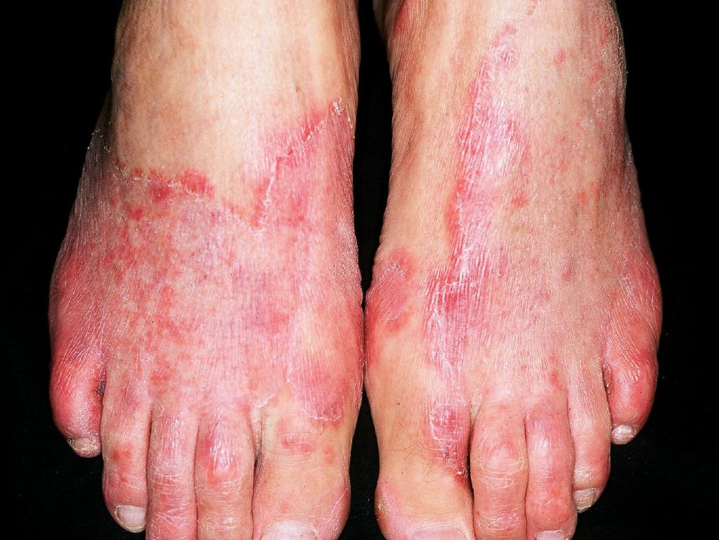
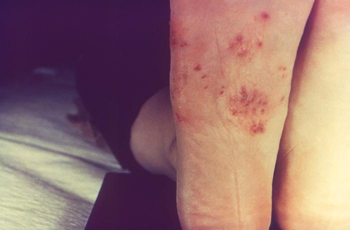
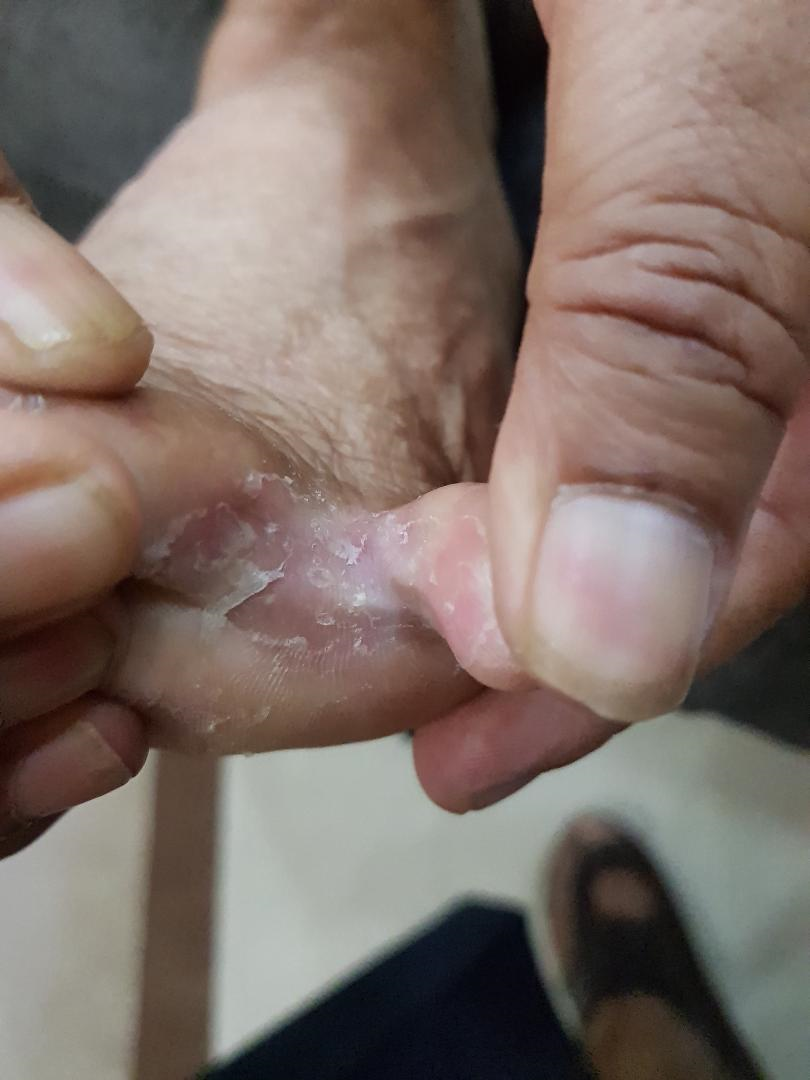
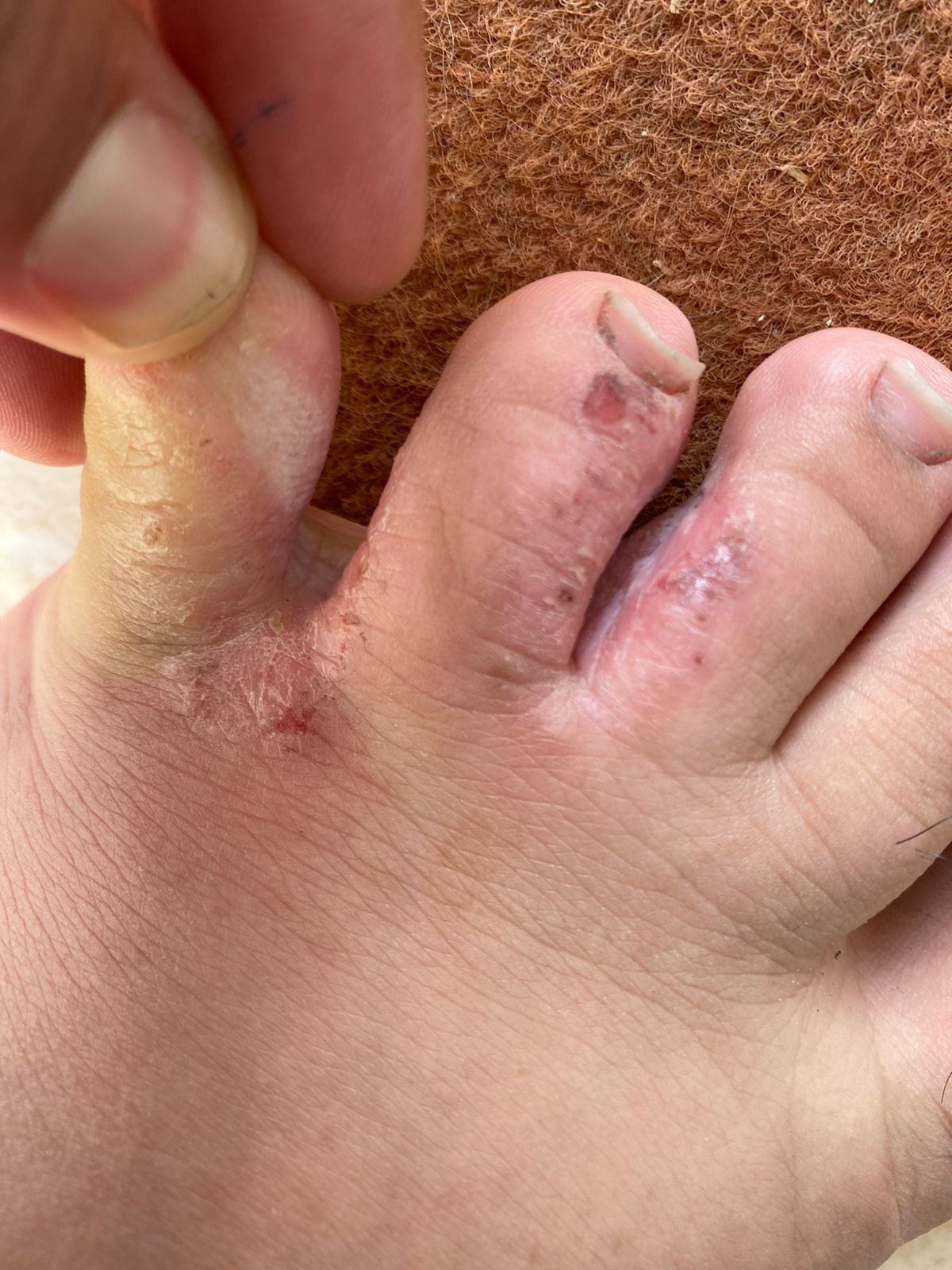
Patients with tinea pedis typically present with long-standing, itchy, intertriginous dermatitis of the toes characterized by peeling, maceration, and fissuring (see Image. Tinea Pedis). The undersurface of the toes primarily affects lateral toe clefts, exhibiting erythema and fine, silvery-white scales. Occasionally, a vesicular eruption resembling pompholyx can appear on the sole, but the most common presentation is the chronic intertriginous type.
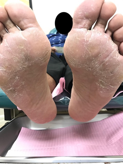
Chronic hyperkeratotic or moccasin-type tinea pedis typically presents with patchy or diffuse scaling on the bottom, medial, and lateral sides of the feet. The vesiculobullous form, often caused by T mentagrophytes, manifests as tense vesicles or bullae on the soles. The burning and itching resulting from bullae can cause significant discomfort. An associated bacterial infection can lead to an acute ulcerative form of tinea pedis.
Evaluation
Although the physical examination may strongly indicate a dermatophyte infection, tinea can overlap with other skin conditions. Confirmation through microscopy and culture may be necessary. A thorough skin examination is warranted as patients can experience dermatophyte infections in multiple body areas.
Microscopic examination of scrapings from the affected site can confirm the diagnosis by demonstrating segmented hyphae. A glass microscope slide should be used to scrape the foot's instep, heel, and sides to collect dry scales from the foot. Bullae should be unroofed, and either the entire roof or scrapings made from the underside of the roof should be mounted.
To perform the procedure, one should place a few drops of a 10% to 20% potassium hydroxide (KOH) solution on the glass slide, followed by a coverslip, and then observe the sample under the microscope. Adding 20% to 40% dimethyl sulfoxide (DMSO) accelerates the clearing of keratin without requiring heating. A staining method that involves mixing 100 mg of chlorazol black E dye with 10 mL of DMSO and adding it to a 5% aqueous solution of KOH can be beneficial. Toluidine blue can also be applied to thin specimens at a concentration of 0.1%. Although mycelia may be visible at low power magnification, using the 10× objective on a microscope is advisable to observe both hyphae and spores better.[2][9][10]
Treatment / Management
Improving hygiene in swimming pools and bathing areas and frequently washing and cleaning changing room floors and walkways may help control the spread of tinea pedis. Most patients can usually manage their condition effectively with topical treatment. Magenta paint or Castellani paint may be beneficial in certain cases of inflammatory tinea pedis, especially when a coexisting bacterial infection is present. Topical imidazoles such as clotrimazole, econazole, ketoconazole, miconazole, isoconazole, tioconazole, and sulconazole offer effective remedies with a very low incidence of adverse effects.
Topical application of terbinafine and amorolfine yields faster results in treating tinea pedis than clotrimazole treatment. Moreover, adding a topical keratolytic, such as salicylic acid, can be beneficial in patients with hyperkeratosis. Using prophylactic tolnaftate powder after swimming and showering in community settings reduces the levels of toe cleft tinea pedis caused by T interdigitale. Generally, topical treatment lasts for 4 weeks, although some patients may experience symptom resolution sooner. Terbinafine 1% can provide effective results for interdigital tinea pedis after 1 week. Repeated KOH scrapings and cultures should yield negative results.[11][12][13]
Patients who do not respond to topical treatment require systemic therapy.[12] Acceptable systemic treatment options include:
- Terbinafine at a daily dosage of 250 mg for 2 weeks.
- Itraconazole at a twice-daily dosage of 200 mg for 1 week.
- Fluconazole at 150 mg per week for 2 to 6 weeks.
- Griseofulvin, for adults, at a daily dosage of 1000 mg of griseofulvin microsize for 4 to 8 weeks or 750 mg of griseofulvin ultramicro size for 4 to 8 weeks. However, the latter option may be less effective than the other options.
- Griseofulvin, for children, at a recommended daily dosage of 10 to 20 mg/kg of griseofulvin microsize, 5 to 15 mg/kg of griseofulvin ultramicrosize, or itraconazole at 3 to 5 mg/kg.[14][12][15]
Although rare, gastrointestinal adverse effects may occur from taking fluconazole. Itraconazole can lead to gastrointestinal upsets, diarrhea, and peripheral edema, particularly when combined with calcium channel blockers. Fluconazole and itraconazole have a significantly lower rate of hepatotoxicity compared to ketoconazole. Terbinafine can also result in gastrointestinal disorders and, in rare cases, hepatitis.[16]
Differential Diagnosis
The hyperkeratotic lesions of tinea pedis can resemble various conditions, including:
- Psoriasis
- Hereditary or acquired keratodermas
- Atopic or contact dermatitis
- Palmopustular eczema
- Pitted keratolysis
- Juvenile plantar dermatosis
- Keratolysis exfoliativa
Interdigital lesions may appear similar to the following conditions:
- Psoriasis
- Erythrasma
- Candida infections
Vesiculobullous lesions may resemble to the following conditions:
- Pustular psoriasis
- Dyshidrosis
- Acute contact dermatitis
- Scabies
Reiter syndrome also exhibits lesions that bear some resemblance to those seen in tinea pedis.
Prognosis
As long as individuals take preventive measures, the outlook for tinea pedis is favorable. The risk of tinea infections is elevated with excessive sweating, known as hyperhidrosis. To prevent the disease, patients should thoroughly dry their toes after bathing. Maintaining dryness is crucial to prevent reinfection. Individuals susceptible to tinea pedis should wear moisture-wicking socks, apply high-quality antifungal powder inside their shoes, and use drying foot powders after bathing, focusing on the spaces between the toes. Tolnaftate or clotrimazole powders serve as excellent options to maintain dry feet. Regularly applying a topical antifungal cream may be required in certain instances, particularly when wearing hot, enclosed footwear.[17]
Complications
The majority of patients will not experience significant issues related to tinea pedis. However, complications can arise due to inadequate treatment and persistent skin breakdown.
Secondary Bacterial Infection
Prolonged or severe cases of tinea pedis may result in a secondary bacterial infection. The compromised skin barrier enables bacteria to enter, potentially leading to conditions such as abscess formation.
Impetigo
Tinea pedis can make the affected areas more susceptible to impetigo—a highly contagious infection that manifests as honey-colored, crusted lesions capable of spreading to other body parts or individuals through direct contact.
Fungal Nail Infection
Untreated or inadequately managed tinea pedis can progress to toenail involvement or onychomycosis. Fungal organisms can infiltrate and cause onychomycosis, which is characterized by thickened, discolored, and brittle nails.
Cellulitis
In severe cases, tinea pedis can advance to cellulitis of the foot and leg. Cellulitis manifests as redness, warmth, swelling, and pain in the affected area, often accompanied by systemic symptoms.[18][19]
Lymphangitis
Chronic or severe tinea pedis can result in lymphangitis, characterized by the appearance of red streaks extending from the affected area and swelling and tenderness of the lymphatic vessels.
Allergic Contact Dermatitis
Some individuals may experience a local allergic reaction to antifungal medications or other topical treatments for tinea pedis. This can lead to allergic contact dermatitis, characterized by redness, itching, and a rash at the application site.
Chronic or Recurrent Infection
Occasionally, tinea pedis may develop into a chronic or recurrent condition, necessitating long-term management and preventive measures. The persistent nature of the infection can substantially impact the individual's quality of life and foot health.
Although tinea pedis can be associated with these complications, not all patients will experience them. Prompt and appropriate treatment and adherence to preventive measures can help minimize the risk of complications and contribute to the successful management of tinea pedis.
Deterrence and Patient Education
Tinea pedis is a fungal infection that affects the skin of the feet. The individuals at highest risk of developing tinea pedis are those with persistently wet feet, diabetes, immunocompromised conditions, and occlusive shoe wear. The most common places to contract tinea pedis are community showers, swimming pools, and bathing facilities or locker rooms.
The most prevalent symptoms of tinea pedis are itching, scaling, and developing skin cracks between the toes. In some cases, patients may develop thickening of the skin on the edges and underside of the foot, often accompanied by underlying redness. Most patients apply a topical cream for a duration of 4 weeks, with some experiencing a quicker response. Individuals who do not respond to topical treatment can consider oral therapy. To prevent a recurrence, patients should adhere to several key recommendations.
The following recommendations can aid in resolving a current infection and preventing future ones:
- Washing feet daily with mild soap and warm water.
- Drying the feet thoroughly after washing them, especially between the toes, after activities that involve water exposure.
- Using a separate towel for the feet and avoid sharing it with others.
- Wearing breathable shoes made of natural materials such as leather or canvas.
- Avoiding tight-fitting shoes that may create a warm and moist environment promoting fungal growth.[20]
- Choosing moisture-wicking socks to keep the feet dry.
- Wearing flip-flops or sandals in public places such as pools, gyms, and communal showers.
- Avoiding sharing shoes, socks, towels, or any other personal items with others.
- Inspecting feet for signs of infection, such as redness, itching, or scaling, regularly.
- Addressing any cuts, blisters, or breaks in the skin promptly to prevent the entry of fungal organisms.
- Wearing breathable shoes and moisture-wicking socks to minimize the risk of reinfection.
- Keeping the feet clean, dry, and well-maintained.
- Informing family members and close contacts about the tinea pedis infection and educating them on preventive measures to avoid its spread.
- Encouraging family members to follow similar hygiene practices to minimize the risk of spreading the infection.
Pearls and Other Issues
Regularly hosing the floors of shower rooms and the sides of swimming pools decreases the presence of dermatophytes on these surfaces. The use of topical antifungal powder in shoes, such as tolnaftate, can also help reduce the risk of tinea pedis infection. Patients with diabetes with tinea pedis are at a higher risk of developing onychomycosis. Furthermore, the presence of interdigital tinea pedis is a risk factor for cellulitis in patients with lymphoedema.
Enhancing Healthcare Team Outcomes
Tinea pedis is a widespread infection frequently encountered by healthcare professionals. Each member of the interprofessional healthcare team can contribute to preventing future infections and reducing morbidity associated with the infection. Pharmacists can guide patients to the appropriate over-the-counter medication, ensuring they do not use a treatment better suited for a Candida infection. Clinicians caring for patients with diabetes can regularly inspect patients' feet and consistently educate them on self-foot care and examinations. As over-the-counter medications do not appear on shared medication lists, open communication among team members is crucial to prevent the morbidity associated with tinea pedis infection.[4][21]