Continuing Education Activity
This activity outlines the evaluation and management of koilonychia and highlights the role of the interprofessional team in evaluating, treating and managing patients with this condition. Koilonychia represents an abnormal nail curvature that can be a sign of an underlying systemic disease mandating thorough systemic evaluation of patients with koilonychia before the commencement of any therapy.
Objectives:
- Identify the etiology and epidemiology of koilonychia, and the common medical conditions associated with it.
- Recall and analyze appropriate history, physical examination and laboratory evaluation of patients with koilonychia.
- Outline the approach to a patient with koilonychia and the treatment and management options as per the underlying systemic disease
- Discuss inter-professional team strategies and coordination for improving the outcomes and care of patients with koilonychia.
Introduction
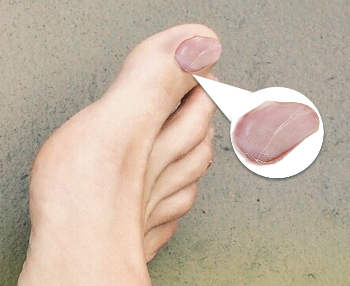
The term “koilonychia” is derived from the Greek word ‘koilos’ which means ‘hollow’ and ‘onikh’ means ‘nail’. This abnormality of the nails is also called as spoon-shaped (concave) nails. The nail abnormality is characterized by thin, brittle, concave nail dystrophy either placed horizontally or vertically, where the finger or toenail plate seems to be centrally depressed and everted laterally. It can be found in newborns, children, and adults. When a patient presents with this symptom, it is imperative to look into further to see if there are more serious health concerns that may be present.
Etiology
Although the aetiologies of koilonychia are myriad, to simplify they may be divided into hereditary or congenital, acquired and idiopathic causes. It is primarily considered to be a manifestation of chronic iron deficiency anemia due to malnutrition, worms, celiac disease, gastrointestinal blood loss, and malignancy.
At times, koilonychia may also be a manifestation of inflammatory skin diseases such as lichen planus or psoriasis, or maybe secondary to systemic alterations such as endocrine disorders (hypo or hyperthyroidism and diabetes mellitus), Plummer-Vinson syndrome, alopecia areata, onychomycosis , trauma, occupational exposure to petroleum products, high altitude, vascular disorders, musculoskeletal disorders, genetic or autoimmune disorders etc.[1] However, the common setting in which koilonychia is observed include, 5.4% of the patients with iron deficiency anemia, inflammatory skin disease, and onychomycosis.
Nail diseases in the pediatric population differ from those in adults with respect to the diagnostic and management approaches; however, few of them are exclusively manifested only in children. Pediatric patients with underlying systemic disorders are more likely to manifest acquired disorders of the nails. In the newborns, koilonychia is frequently found to be idiopathic and presents as a normal variant in 33% of cases, especially affecting the big toe, which regresses spontaneously after the age of 9 years once the nail plate thickens and becomes hard.
Epidemiology
Considering the numerous underlying etiologies, it is difficult to estimate the incidence of koilonychia and currently, there is no information stating the prevalence of koilonychia within different age groups and gender. In addition, not everyone with an underlying disease may present with the clinical manifestation of koilonychia. Similarly, the epidemiological data about the incidence and prevalence of other associated conditions like the Plummer-Vinson syndrome are not widely available. Iron deficiency is by far the commonest nutritional cause of anemia and thus koilonychia and is considerably more prevalent in the developing rather than in the developed countries (36% or 1.4 billion out of 3.8 billion estimated population in developing countries, versus 8% or just under 100 million out of 1.2 billion estimated population in developed countries). Given its impact on psychological and physical development, behavior and work performance, iron deficiency anemia remains a serious problem of public health significance to be tackled.
Pathophysiology
The pathophysiology of koilonychia is poorly understood; however, several mechanisms have been contemplated, including primary dermatoses, deficiency in metalloenzymes or sulfur-containing amino acids, nail matrix changes due to blood flow abnormalities (age, nervous or vascular changes), endocrinopathies and trauma.[2] It may also be related to the poor digital blood flow with successive weakening and disruption of subungual connective tissue, culminating in the depression of the distal matrix or reduced iron in iron-containing enzymes of the epithelial cells.[3][4] Another hypothesis highlights mechanical pressure causing the upward deformation of lateral and distal flexible iron-deficient nail plates.
Histopathology
Since it is a clinical diagnosis, there is no histopathology of koilonychia available.
History and Physical
The history given by a patient with koilonychia can vary greatly. It can either be a congenital defect that is present at birth or it can be a defect that has gradually become apparent. It can be present at any age. Patients or their family members may complain of a nail that appears scooped out. The abnormality is more clearly appreciated from the lateral side of the nail plate. The findings on history and physical examination shall further assist in narrowing the differential diagnosis.
Subtle koilonychia may be difficult to grasp only on visual examination and in the clinical setting, it is not practical to take appropriate measurements of the nail plate convexity as complex mathematics is required. So, to diagnose koilonychia, a clinical test has been classically described called, the “Water-drop test”.[5] Using a 1 ml syringe, few water drops are poured over the nail plate in this test. The water droplets pool over the concave nail plates in koilonychia, unlike the normal convex nail plates. Sometimes, in individuals with normal smooth convex nail plates, a false-positive water-drop test may be observed. To sidestep this, spherical plastic beads have been used on the nail plates in a study of 5 patients with koilonychia and the authors concluded that this test not only circumvents false-positive interpretation but also gives an excellent visual impression to both the clinicians and patients.[6] However, this test needs further validation in larger studies.
Evaluation
Appropriate workup is important to exclude an underlying cutaneous or systemic disorder and the workup should be based on patient profile, clinical history, and physical examination for a better understanding of the underlying condition.
Thus, the numerous etiologies of koilonychia warrant proper workup including a thorough history regarding any other underlying medical issues, medications, surgery, family history, diet and lifestyle choices. After getting a detailed history, a review of systems and physical examination is a good start. Depending on the findings on physical examination, pertinent blood work can be ordered. For example, a CBC can tell you if the patient is anemic and give you information about the size and chromaticity of the red blood cells. If the patient is anemic, an iron panel can be ordered for further evaluation. If the CBC is negative, other blood workups can be ordered at the physician's discretion keeping in mind the underlying systemic condition.
Patients of PVS presenting with dysphagia and long-standing iron deficiency anemia should be evaluated with an esophagogram to rule out esophageal rings and webs as they may be sometimes missed on esophagogastroduodenoscopy.
In cases in which the cause is unknown even after blood investigations, the possibility of hereditary/familial koilonychia should be considered.
Treatment / Management
The treatment of koilonychia generally includes treatment of the underlying disease. Although the aetiologies of koilonychia are numerous, the underlying diagnosis can be narrowed down based on the age, personal, family and occupational history, review of systems and a thorough physical examination. Koilonychia can be an important clue for either the dermatologic or systemic condition.
Appropriate workup is important not only to exclude an underlying cutaneous or systemic disorder but also to distinguish between the different aetiologies to provide appropriate management of koilonychia. Most of the cases with acquired causes are reversible.
The nails in newborns are usually thin and soft and present frequently with physiological alteration (koilonychia), which are better left untreated and in addition need a wait-and-watch policy and reassurance of the parents. However, it is important to follow up these patients to verify that this physiological condition does not turn into a pathological one.
Differential Diagnosis
Dermatologic conditions: Koilonychia is seen more commonly with the dermatologic conditions directly affecting the nail bed and therefore is observed frequently in lichen planus (LP) of the nail,[7] nail psoriasis,[8] and 20-nail dystrophy, another term for trachyonychia.[1] However, in onychomycosis, the mechanical subungual pressure on the nail bed is the reason for the development of spoon nails.[1]
Systemic conditions:
Nutritional deficiencies: Koilonychia occurs in approximately 5.4% of iron deficiency patients and it the classic manifestation of the iron store abnormalities.[9] As per some reports, in children, iron deficiency remains the most common cause of koilonychia and it may precede laboratory iron abnormalities.[10], [11] The spoon-shaped deformity is usually associated with nail thinning and brittleness.5 Although nail iron levels increase with oral supplementation, the severity of iron deficiency does not necessarily correlate with the development of koilonychia.[9]
Patients with poor nutrition as a result of deficiencies in vitamin C, zinc, copper, selenium, cysteine and other amino acids may present with koilonychia.[12], [13] Few studies have documented koilonychia in 5.5–18% of the populations subjected to poor nutrition such as rural villagers, alcoholics, child laborers, and patients with chronic kidney disease.[12], [14], [15]
Plummer–Vinson syndrome (PVS): Also known as Paterson– Kelly syndrome, is a rare entity characterized by the triad of iron deficiency anemia, dysphagia, and oesophageal webs. Koilonychia may be the presenting sign and is nearly seen in 37–50% of the cases.[16]
Hemochromatosis: Koilonychia has been also noticed in approximately 49% of hemochromatosis patients, an autosomal recessive disorder of iron accumulation (rather than deficiency). Nail findings may not only occur at any time during the disease course but also are the presenting sign of the disease. Phlebotomy has been tried as a treatment option, however, the nail plate deformity does not seem to be rectified by it.[17]
Endocrine disorders: Koilonychia has been observed in hypothyroidism, where the spoon-shaped deformity is accompanied by slow growth and brittleness of the nail plate.[18] The koilonychia in diabetes may be a result of nutrient deficiencies or related to microvascular dysfunction.[19]
Autoimmune conditions: Also rarely observed in systemic lupus erythematosus and Raynaud’s disease. spooning is hypothesized to be secondary to vasculopathy-induced hypoxia of the nail matrix.[20]
Congenital and Early Childhood Koilonychia: Transient acquired koilonychia, especially of the great toe has been observed in at least 5% of children generally in the first two years of life and 15 is rarely present after the age of nine.[21] In children, isolated koilonychia of the toenails is usually idiopathic and remains a diagnosis of exclusion, hence nutritional deficiencies and anemia should be considered.[22]
Familial koilonychia: Familial koilonychia, is a rare condition, usually transmitted through autosomal dominant inheritance and has been appreciated in several pedigrees with a greater degree of penetrance without any predilection for sex.[23],[24] It usually presents at birth or within the initial few years of life. The nails in these patients are typically thin and flat and develop various degrees of concavity over time. However, within the same family members, the variable expression may be noticed with only affection of fingernails or selected fingers or toes only or fingernails on one hand or with accentuation of the great toe and/ or thumb.[25] At times, koilonychia may be rarely seen as a part of a genodermatosis.
Trauma: Is a common cause of koilonychia in children, often due to thumb/finger sucking habit and use of tightly fitting shoes. However, with behavior modification, the nail plate growth usually normalizes.[1]
Occupational Koilonychia: Koilonychia may be seen in certain occupations, generally where the individual is exposed to mineral oil (petroleum), organic solvents and chemicals causing contact dermatitis and/or repetitive trauma and mechanical stress on the fingers. But with time, these changes become irreversible.[26], [27] Nail spooning over time has also been observed in hairdressers dealing with ammonium thioglycolate.[28]
Regional Koilonychia: The spoon-shaped deformity has been observed in the 4 or 5 decades of certain populations in India after several years of high-altitude habitation (>3000 meters above sea level).[29], [30] Other reasons speculated to contribute to this deformity in these communities include vegetarian diets with low iron content, manual labor (trauma), high altitude-associated haematologic changes and the high silica content of the soil.[31]
Pertinent Studies and Ongoing Trials
There are no ongoing studies regarding koilonychia.
Treatment Planning
The spoon shape abnormality observed in severe chronic iron deficiency anemia is usually reversible with replenishment of iron stores through food sources containing high iron content or medical management by taking iron supplements. However, any underlying malignancies, occult or overt blood loss or iron malabsorption should be ruled out before the commencement of therapy. For correction of iron deficiency anemia, 150-200 mg of elemental iron is generally required.[32] The best sources of dietary iron for non-vegetarians include meat and animal products while for vegetarians, beans, leafy greens, and fortified grain products are considered good sources of iron. Combining these iron sources with citrus fruits and vitamin C rich food sources of iron help to increase iron absorption. The nail abnormality usually returns to normal within 4–6 months of iron repletion.
Patients with Plummer–Vinson syndrome (PVS) are generally better treated with iron supplements i.e. 150-200 mg of elemental iron and endoscopic mechanical dilatation (balloon dilatation or Savary-Gilliard dilators) of the oesophageal webs.[33] Within weeks of iron therapy, rapid improvement in the symptoms of fatigue and dysphagia may occur.[34]
If the cause of the koilonychia happens to be either idiopathic or hereditary, there is no effective treatment available.
Staging
There is no staging for koilonychia.
Prognosis
Once the koilonychia has been diagnosed, the nail can become more blanched and spoon-shaped if the underlying disease is not being treated. How quickly the spoon nails return to normalcy, usually depends on the underlying cause. If they are related to anemia, the nails seem normal within 4–6 months of increasing the iron intake. Patients with PVS have an excellent outcome but are at an increased risk of developing squamous cell carcinoma of the hypopharynx or upper esophagus. Hence, patients with PVS should be screened for aerodigestive tract carcinoma. The toenails grow out much slower than the fingernails and may take a year and a half to regrow compared to half a year by the fingernail.
Complications
There are no complications due to koilonychia per se because it is merely a symptom of an underlying disease, however, its cause or other dermatological or systemic associations may lead to some serious consequences.
Chronic iron deficiency may cause irreversible mucosal changes which potentially lead to malignant degeneration. Also, if PVS is left untreated, there are high chances of progression of the esophagus to squamous cell carcinoma and there may be a significant luminal obstruction and persistent dysphagia if the esophageal webs are left untreated.[34]
Consultations
No consultations are necessary after diagnosing koilonychia unless there are hematologic issues or any underlying systemic involvement, which may further need detailed evaluation by the respective specialist doctor.
Deterrence and Patient Education
Koilonychia is a deformity of the nails where the central portion of the nail is depressed and the lateral aspects of the nail are elevated. This symptom can be a sign of an underlying disease process or a congenital process. Patients should be advised to trim their nails short and clean to prevent spoon nails, and also to prevent infection in the damaged areas. Further, regular use of a moisturizer or oil on the nails post showering or bathing helps to keep the nails in good condition.
Educational efforts towards the health and agricultural organizations of the country need to be undertaken in the long-term considering iron deficiency anemia which remains the common cause of koilonychia. Adequate educational national programs with changes in the economic and socio-cultural may aid in alleviating iron deficiency anemia and malnutrition among the low socio-economic classes. For such programmes to be effective, attention should be paid to the nutritional properties of foods, nutrient needs, environmental sanitation, personal hygiene and health and cost values of various foodstuffs. Other programs providing iron supplementation and parasitic control may also serve as motivational stimuli for millions towards nutrition and health education.
Moreover, to combat iron deficiency anemia and any other nutritional problem, adequate intake of affordable and acceptable nutritious foods needs to be encouraged. This necessitates to improve the availability of foods and upgrade the economic level of the population to purchase them in adequate quantity.
Similarly, the benefits of birth spacing and education of parents regarding the special needs of young mothers may additionally reduce the problems of anemia. In addition, the use of more complex laboratory methods in institutions should be encouraged to assist further more research groups with more resources and capabilities in developing countries.
PVS is an extremely rare condition, hence, deterrence of this condition is difficult. Further discussions should be held with the physician for any other questions or concerns.
Pearls and Other Issues
Due to the many possible causes of koilonychia there are no major prevention methods that are widely recommended. The cause of koilonychia can be a congenital defect, a presenting symptom of an autoimmune condition, or associated with other disease processes.
Enhancing Healthcare Team Outcomes
When a patient presents with koilonychia, it takes multiple types of healthcare professionals to get to the root of the cause. Realistically, a patient will research his or her condition and then present their concerns to the physician. While the primary care physician can start preliminary testing, it usually requires the help of multiple specialists to determine the cause of koilonychia. There have been no randomized controlled trials involving koilonychia so there are no levels of evidence regarding prevention or treatment of the same.
Patients with PVS are best managed by an interprofessional approach involving a hematologist, gastroenterologist, and a thoracic surgeon. These patients should be followed as outpatients to rule out the complications associated with this entity and prompt treatment should be given whenever needed.