Continuing Education Activity
A hallux valgus is a complex deformity of the first ray of the forefoot. This deformity is, at times, red and painful and can disrupt daily activities. If correctly identified and treated, the symptoms will vastly improve. Hallux valgus deformity is typically diagnosable through a physical exam, and imaging is important as it can evaluate whether there is damage to the first metatarsophalangeal (MTP) joint. Treatment centers upon first trialing non-surgical approaches such as wider shoes, orthotics, and night splinting. If conservative treatment proves ineffective, surgical management is the next recommended course of action. Patients tolerate the surgery well, with bony union usually occurring around 6 to 7 weeks postoperatively. This article highlights the role of the multidisciplinary team in evaluating and managing patients with this condition.
Objectives:
- Describe the etiology of hallux valgus.
- Identify the components of an appropriate history, physical, and evaluation of hallux valgus.
- Outline the treatment and management options available for hallux valgus.
- Review the importance of the interprofessional team to improve outcomes for patients affected by hallux valgus.
Introduction
Hallux valgus (HV), also known as a bunion, is one of the most common forefoot deformities. HV manifests with the proximal phalanx deviating laterally and the first metatarsal head deviating medially and due to the adduction of the first metatarsus, called metatarsus primus varus. However, the precise etiology is not fully understood. HV tends to occur more commonly in women than in men, with a ratio as high as 15:1 in one study, and occurs more in those who wear tight shoes or heels.[1]
Hallux valgus deformity is typically diagnosable through a physical exam, and imaging is important as it can evaluate whether there is damage to the first metatarsophalangeal (MTP) joint. Treatment centers upon first trialing non-surgical approaches such as wider shoes, use of orthotics, and night splinting. If conservative treatment proves ineffective, surgical management is the next recommended course of action. Patients tolerate the surgery well, with bony union usually occurring around 6 to 7 weeks postoperatively.[1]
Etiology
The precise etiology is not fully understood but are many proposed theories. HV deformity is most likely a result of multiple contributing factors, including genetics, short first metatarsal, dorsiflexed first metatarsal, flexible or rigid forefoot varus, rigid or flexible pes planovalgus, gastrocnemius equinus, abnormal foot mechanics, and joint hypermobility. Interestingly, certain arthritic conditions such as gouty arthritis, psoriatic arthritis, and research show that rheumatoid arthritis predisposes patients to HV deformity. Furthermore, HV deformity is more commonly seen in connective tissue disorders such as Marfan syndrome and Ehlers-Danlos syndrome, as well as in Down syndrome.[2]
Any muscle imbalance in the foot due to conditions such as a stroke, cerebral palsy, or myelomeningocele can also cause an HV deformity.
HV deformity is common in people who wear tight shoes and heels, which is regularly cited as the cause. However, men who wear sensible footwear often have marked HV deformity, while women who wear footwear that significantly compresses their feet have no deformity. This fact has given rise to the thought that footwear exacerbates an underlying bony abnormality rather than acting as the primary cause.
Epidemiology
HV deformity is a relatively common condition. It occurs in approximately 23% of adults aged 18 to 65 years and up 36% of adults older than 65 years. When looking at adult females, HV deformity occurs as high as 30%.[3] The prevalence is higher in those who wear shoes or heels when compared to the barefoot population. Interestingly, when comparing women and men in barefoot populations, women are found to have HV deformity twice as often.
Pathophysiology
The pathophysiology of HV is complex, but the general assumption is that an imbalance exists between the extrinsic and intrinsic muscles of the foot with the involvement of the ligaments also. Maintenance of the first metatarsal alignment is by the tension created by the peroneus longus laterally and the abductor hallucis muscle medially. Collateral ligaments prevent movement along the transverse plane at the first MTP joint. If there is increased pressure at the head of the first metatarsal, the metatarsal will begin to move medial-dorsally. This force increases the hallux angle, which is also worsened by muscle stabilization while walking. As these forces push the first metatarsal medially and the hallux laterally, the medial collateral ligament and the medial capsule become strained, eventually rupturing. Without stabilizing structures medially, the lateral structures (adductor hallucis muscle and collateral/lateral joint capsule ligaments) exacerbate this deformity.[4]
History and Physical
History
HV deformity commonly presents with a chronic progressive onset. The proximal phalanx pronates and deviates laterally, while the first metatarsal head deviates medially, often becoming red and painful. Patients typically present with a chronic onset of sharp or deep pain at the MTP joint that is exacerbated by ambulation. The patient occasionally describes an aching pain at the head of the second metatarsal. As HV deformity progresses, the frequency, duration, and severity of the pain gradually increase. Frequently patients describe an associated increase in the size of the deformity. Another relatively common presentation is tingling or burning pain at the dorsal part of the deformity. This symptomatology is indicative of a medial dorsal cutaneous nerve neuritis likely caused by compression from the deformity. These symptoms are primarily a result of three different processes:
- The bunion itself centers upon the medial aspect of the first metatarsal.
- Pressure against the toes that are superiorly displaced
- Increased pressure on second through fifth metatarsal bones
Other symptoms that affect patients include blisters, ulcerations, interdigital keratosis, and irritated skin adjacent to the deformity.[5] These symptoms can cause significant morbidity, often limiting physical activities.
Physical
For the physical exam, a biomechanical exam will look for possible causes of HV deformity. Common items to evaluate include forefoot/rearfoot varus or valgus, first ray hypermobility, subtalar joint stiffness, subtalar joint stiffness, midtarsal joint stiffness, resting calcaneal stance position, tibial torsion, and neutral calcaneal stance position. Assessment of the pathology is often divided into a non-weight bearing and weight bearing evaluation, as illustrated below.
Nonweightbearing
The position of the hallux, with respect to the second digit, should be assessed in the transverse plane. The hallux can be under-riding, overriding, or without contact. The lateral deviation of the MTP may represent subluxation at the MTP joint. The medial prominence also requires close evaluation.
The first MTP joint range of motion should undergo assessment for the maximum available motion (normal - plantar flexion less than 15 degrees and dorsiflexion 65 to 75 degrees). Next, the quality of the first MTP joint is assessed (pain, crepitation). Lastly, the MTP is evaluated for its axis of motion.
Weightbearing
HV deformity severity tends to be more obvious when the patient is weight-bearing. When weight-bearing, patients should be evaluated for increased hallux abduction, increase in the medial prominence, first MTP joint dorsiflexion, hallux purchase, and metatarsus varus.
Evaluation
Typically, routine assessment laboratory studies are not required. However, certain laboratory studies can be considered if there is a suspicion of metabolic or systemic disease. These include rheumatoid factor, antinuclear antibody, c-reactive protein, erythrocyte sedimentation rate, uric acid, and complete blood count. If there is a high degree of suspicion of osteomyelitis, the clinician can consider MRI and radionuclide imaging.
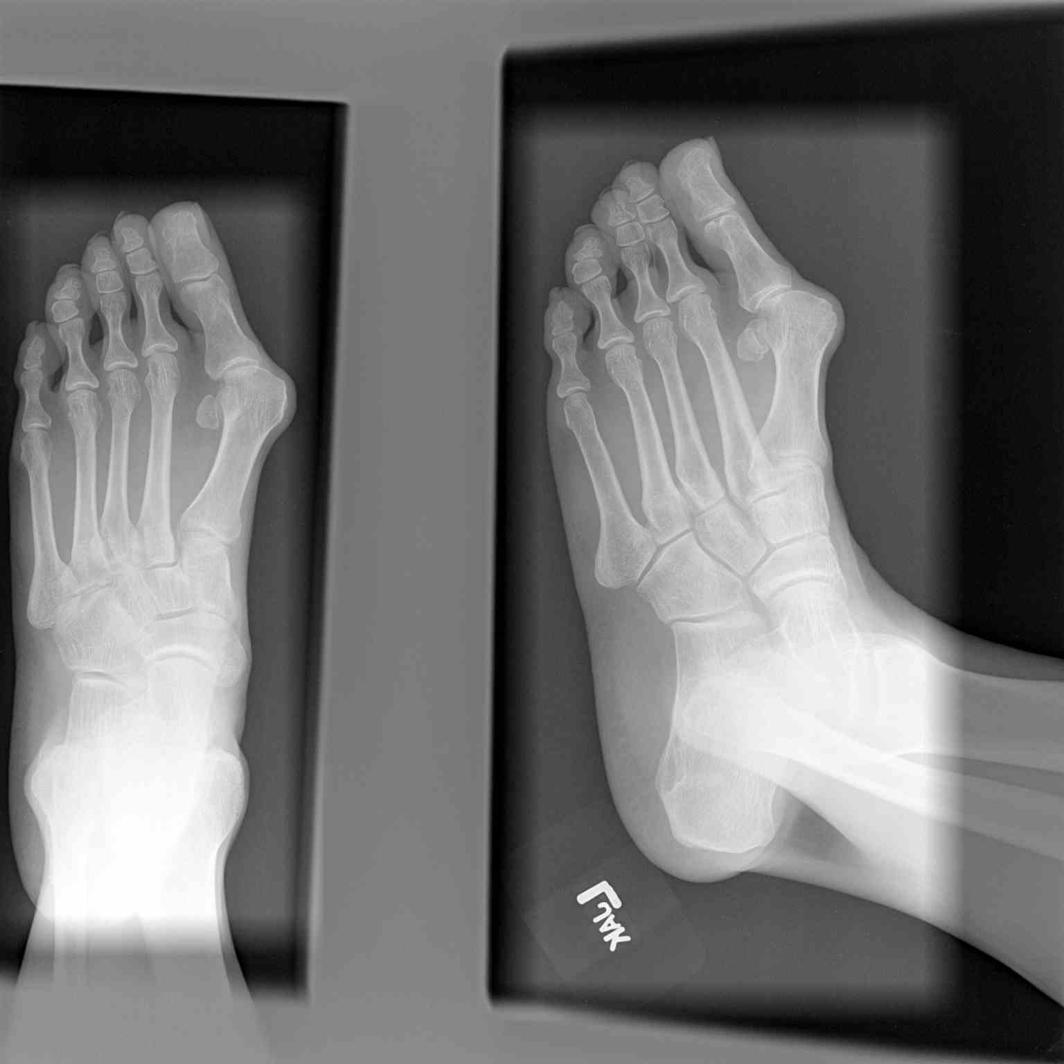
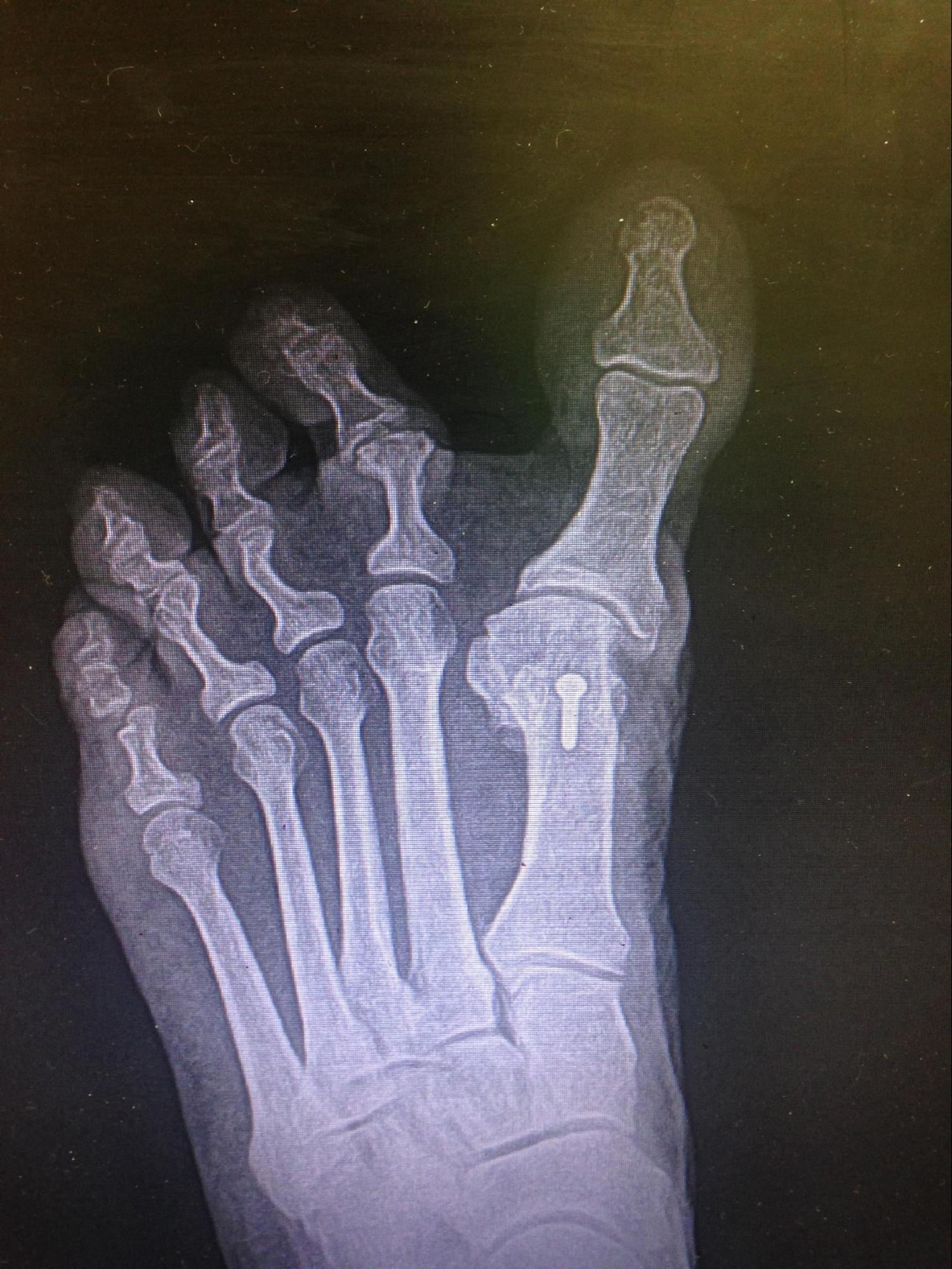
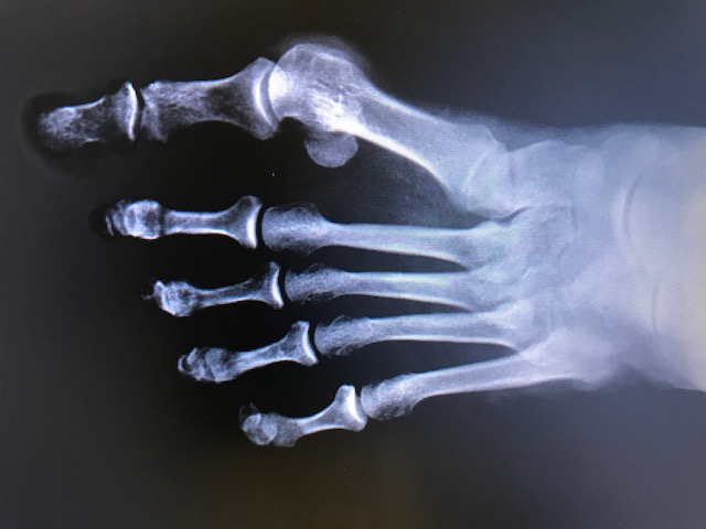
Establishing a diagnosis can be typically done through a physical exam. Imaging can help clinicians determine the extent to which the first MTP joint has suffered damage. Evaluation is primarily with plain radiography, with X-rays (AP and lateral weight-bearing) of the foot. Imaging demonstrates a lateral hallux deviation at the first metatarsal (normal hallux valgus angle is less than 15 degrees, and the intermetatarsal angle is less than 9 degrees). Typically, the deviation is in the transverse plane. However, HV deformity can cause rotation of the hallux resulting in the nail facing medially (pronation of the frontal plane). Following the determination of the severity of the deformity has been determined, the clinician can perform the most appropriate procedure.
The classification is mild, moderate, and severe based on the weight-bearing anteroposterior, lateral oblique, lateral, and sesamoid axial views. This imaging helps evaluate the structural status of the foot. The AP projection helps assess the intermetatarsal angle, hallux abductus angle, metatarsus adductus angle, hallux abductus interphalangeal, hallux rotation, and the condition of the first MTP joint. The lateral projection primarily serves to assess the first metatarsal position (elevated or plantar-flexed) and dorsal exostosis/osteophytes. The lateral oblique projection helps evaluate the density, uniformity, and trabeculation of the bone (bone stock). The sesamoid axial view looks primarily for sesamoid subluxations and degenerative joint changes to the cristae.
Degree: Hallux valgus angle (HVA) / Intermetatarsal angle (IMA)
- Normal: less than15 degrees / 9 degrees
- Mild: 15 to 30 degrees / 9 to 13 degrees
- Moderate: 30 to 40 degrees / 13 to 20degrees
- Severe: over 40 degrees / over 20 degrees
Treatment / Management
Treatment of patients with HV deformity revolves around non-surgical and surgical treatments. Usually, non-surgical treatments are attempted first. If there is treatment failure, surgical repair should be considered. Interestingly, there is no definitive evidence that conservative treatment is effective; however, the American College of Foot and Ankle Surgeons still recommends using conservative therapy before considering a surgical solution. It is essential for patients to try wide shoes and orthotics before considering more invasive options.
The goal of conservative treatment is to manage the symptoms without correcting the anatomical deformity. Non-surgical treatments options include:
- Shoe modification: Low-heeled, wide shoes.
- Orthoses: Improves alignment and support.
- Analgesics: Acetaminophen and NSAIDs.
- Ice: Icing the inflamed deformity to reduce inflammation.
- Medial bunion pads: Prevents irritation of HV deformity.
- Stretching: Helps maintain joint mobility in the affected joint.
If non-surgical treatments are unable to control the pain, the treatment is deemed to have failed. At that point, surgical management should be considered. Indication for surgery is primarily based on symptoms (difficulty with ambulation, pain). Interestingly, radiographic appearance does not play a significant role. The presence of arthritis and the severity of the deformity helps guide surgeons in performing the most appropriate procedure. Over 150 surgical procedures have been described for the correction of HV deformity. Although there are many described procedures, they all involve one of the basic approaches outlined below:
- Osteotomy - A cut is made in the first metatarsal bone and put into a less adducted position. The cut varies in position and shape depending on the surgical strategy. For example, A Wilson osteotomy utilizes a straight cut, while a chevron osteotomy uses a wedge-shaped cut. The location of the cut may occur near the base of the metatarsal (proximal osteotomy), in the shaft (scarf osteotomy), or the neck (distal osteotomy). The chevron osteotomy has been looked at most closely. A randomized control trial comparing chevron osteotomy to no treatment or orthosis found chevron osteotomy outperformed the other treatment strategies. At 12 months, the hallux abductus angle was normal in the osteotomy group, with an 80% satisfaction rate. However, approximately 61% of the patients in the osteotomy group had moderate footwear problems. The surgery group also required the greatest number of sick days and had higher foot care costs.[6]Most commonly, a distal metatarsal open approach is taken, resulting in a 3 to 5 cm scar. However, there are newer minimally invasive techniques that are increasing in popularity. A study comparing open osteotomy to minimally invasive surgery revealed no significant difference in the success of the surgery, but the surgical time was reduced, and the scar was smaller in the minimally invasive surgery. In addition, a randomized control trial comparing open vs. minimally invasive chevron osteotomies found clinical and radiographic outcomes to be similar between the two treatment groups.[7]
- Arthroplasty – The mobility of the first MTP joint is maintained while relieving the pain (replacing the joint with an implant or removing the joint). Both hemiarthroplasty and total joint arthroplasty have been developed. Hemiarthroplasty maintains toe length and requires less bone resection. An interpositional arthroplasty is performed in patients with severe hallux rigidus and helps maintain joint range of motion. The most common arthroplasty is the Keller resection. In this procedure, as much as 50% of the proximal phalanx is resected to increase dorsiflexion and decompress the joint. Postoperatively from a Keller’s arthroplasty, 75% of patients were satisfied with the results, and 88% had complete pain relief. However, around 12% of patients experienced increased pain. [8]
- Arthrodesis- Fusing the MTP joint into the correct position. This is only performed when the joint is severely degenerated and regaining functionality is unlikely.[9] The procedure is typically reserved for older patients. Postoperatively, up to 81% of patients had pain relief and improved ambulation. However, nonunion complications have been found to be up to 20%. Patients often complain of limited footwear options, metatarsalgia, and joint stiffness.Another arthrodesis option is the fusion of the 1st metatarsal-cuneiform joint, which is often reserved for cases of hypermobility of the 1st ray and severely increased hallux valgus and 1st intermetatarsal angles. More recently, the arthrodesis at this level has been modified to address the tri-planar deformity of HV and has shown positive results when assessing functional outcomes, diminished recurrence rates, and early return to weight-bearing activities.[10]
- Soft tissue procedures – The procedure that primarily involves soft tissue is the McBride procedure. In this procedure, the fibular sesamoid is excised, causing Interphalangeal joint flexion, MTP joint hyperextension, and medial deviation of the hallux. Interestingly, no reports have studied soft tissue procedures for HV deformity correction independently. One study compared the chevron plus adductor tenotomy to the chevron osteotomy alone. They found little difference in mechanical correction and no difference in patient satisfaction.[11]
There are very few randomized trials evaluating the efficacy of these procedures. However, as surgical techniques have improved, patient satisfaction has also improved, ranging from 50 to 90%. Interestingly, surgical outcomes do not appear to correlate with patient satisfaction. This is thought to be related to the unmet postsurgical expectations of the patient.
Postoperative care is primarily determined by the type of procedure performed. However, dressing is typically applied at the time of surgery to supply corrective forces. The dressing also compresses the surgical wound to help minimize postoperative edema. The weight-bearing status is procedure dependent but is commonly limited for the first two weeks. Once the sutures are removed, the patient can begin range of motion exercises and increasing weight-bearing activities. Postsurgical imaging is typically obtained as well when the patient becomes more active.
Long-term monitoring is directed to understanding the exact etiology to ensure HV deformity does not recur. If identified, clinicians should treat the causes. Postsurgical patients may still benefit from orthotic devices, notably in diseases that accelerate joint degeneration, such as rheumatoid arthritis. By better controlling these factors, long-term postsurgical results can be obtained.
Differential Diagnosis
Conditions to consider when evaluating patients with a possible HV deformity:
- Osteoarthrosis
- Freiberg disease
- Hallux rigidus
- Morton neuroma
- Turf toe
- Gout
- Septic joint
Staging
Root et al. divided HV deformity into four stages, as outlined below:
- Stage 1: Lateral displacement of the hallux at the MTP joint
- Stage 2: Progression of the hallux abduction (hallux pressing against the second toe)
- Stage 3: Increased intermetatarsal angle, possible associated second hammertoe deformity
- Stage 4: Partial/Complete hallux dislocation at the MTP joint[12]
Prognosis
The overall prognosis of HV deformity is good. Patients should first have a trial of conservative treatments. If the pain and functionality of the patient do not improve, surgery should then be a consideration. The postoperative recovery is dependent on the procedure. With any bone procedure, such as an osteotomy, healing takes around 6 to 7 weeks (corresponding to the complete bony union). If the patient is a smoker, the healing may take longer.[13] Patients typically return to work approximately 6 to 12 weeks post-surgery. Improvement has shown to take up to 1-year post-surgery.[14]
Postsurgical complications vary depending on the surgical procedure and technique. The more common complications include osseous non-union, hematoma, numbness, hardware failure, osteomyelitis, cellulitis, avascular necrosis, hallux varus, limited joint range of motion, and recurrence.[15]
Recurrence rates differ based on procedure, ranging from 10 to 47%. The etiology is typically multifactorial but typically includes anatomic predisposition, compliance with postsurgical instructions, medical comorbidities, and poor surgical technique.[16]
Complications
HV deformity can result in several complications, including:
- Bursitis (most common)
- Second toe hammertoe deformity
- Degenerative disease of the metatarsal head
- Central metatarsalgia
- Medial dorsal cutaneous nerve entrapment
- MTP joint synovitis[17]
Deterrence and Patient Education
HV deformity, also known as a bunion, is one of the common forefoot deformities. It manifests as a deformity in the big toe, causing it to bend towards the other toes, and the joint tends to become red and painful. The exact cause is not well understood. However, it tends to occur more often in women and people who wear tight shoes or heels. The diagnosis is typically through a physical exam. However, imaging is essential because it helps assess the severity of the deformity. Treatment centers upon first trialing non-surgical approaches such as wider shoes, use of orthotics, and night splinting. If this is ineffective, surgical management is the recommendation. Patients typically tolerate the surgery well, with bony union usually occurring around 6 to 7 weeks postoperatively.[18]
If the patient is a smoker, the healing may take longer, and there is a higher risk of complications such as non-union or wound infection.[13] Patients typically return to work approximately 6 to 12 weeks post-surgery. Improvement has shown to occur as far out as one-year post-surgery.[14]
Enhancing Healthcare Team Outcomes
Managing patients with HV deformity is best with an interprofessional team that includes primary care doctors, radiologists, physical therapists, pharmacists, pain specialists, and surgeons. The patient may present to their primary care doctor for swelling or pain. The diagnosis is typically made through a physical exam. Further imaging can help assess the severity of the deformity and guide the treatment. Conservative treatment should always be the first step in the management of this condition. If this approach is ineffective, the patient should receive a referral to a surgeon for evaluation for surgical management. If surgery is performed, rehabilitation should follow to maximize post-surgery functionality. A pain specialist and a pharmacist can be used to help manage post-surgical pain, with a focus on minimizing the use of opioids. Orthopedic nurses can assist at every step of the process, from conservative treatment to helping during surgery and post-op, as well as coordinating with therapists for post-surgical rehab. Patients should have long-term follow up to ensure they are meeting their recovery milestones. Open communication between members of the interprofessional treatment team is invaluable for improved outcomes.
This research was supported (in whole or part) by HCA Healthcare and/or an HCA Healthcare-affiliated entity. The views expressed in this publication represent those of the author and do not necessarily represent the official view of HCA healthcare or any of its affiliated entities.