Introduction
Hand motion results from a variety of muscle contractions. The contraction of muscles manifest as movement of the hand. These contractions may come from the muscles both within the hand (intrinsic hand muscles) and the muscles that originate from the forearm with tendon attachments to the hand (extrinsic hand muscles).
The action of making a fist and palmar flexion of the hand involves the muscles that originate in the forearm. These muscles are called the long flexor muscles of the hand.
Structure and Function
Six long flexor muscles originate in the forearm but have tendon attachments in the hand. The six muscles are the flex carpal radialis muscle, the flexor carpal ulnaris muscle, the flexor pollicis longus muscle, the flexor digitorum profundus muscle, the flexor digitorum superficialis muscle, and the palmaris longus muscle. These long flexor muscles either flex the fingers or flex the hand at the wrist joint.
Attachments and Origins
All of the long flexor muscles of the hand will have their muscle bellies in the forearm while their tendon attaches somewhere in the hand. The flexor carpal radialis muscle originates from the medial epicondyle of the humerus. The muscle and tendon of the flexor carpal radialis muscle will travel diagonally down the arm to attach to the base of the second and third metacarpal bone and the tuberosity of the trapezium bone. The flexor carpal ulnaris muscle originates from the medial epicondyle and the medial border of the olecranon. The flexor carpal ulnaris muscle attaches to the base of the fifth metacarpal bone, the pisiform bone, and the hamate bone. The flexor pollicis longus has its origin from the anterior surface of the radius. The flexor pollicis longus muscle inserts on the thumb at the distal phalanx. The flexor digitorum profundus muscle originates from the proximal end of the radius and ulnar. The flexor digitorum profundus muscle descends to attach at the base of distal phalanx of the second, third, fourth, and fifth digits. The flexor digitorum superificialis muscle originates from the medial epicondyle of the humerus with some attachments to the radius and ulnar. The flexor digitorum superifcialis muscle attaches to the middle phalanx of the second, third, fourth, and fifth digits. The flexor palmaris longus muscle originates from the medial epicondyle of the humerus. The palmaris longus attaches to the palmar aponeurosis and the transverse carpal ligament.
Motions
The long flexor muscles contract and manifest flexion in the hand. Due to the different location of the tendon attachments, flexion will occur at the joint where the tendon is attached closest. The contraction of the flexor carpal radialis muscle causes the hand to flex laterally. When the flexor carpal ulnaris muscle contracts it will cause the hand to flex medially. The motions of the flex carpal radialis muscle and the flexor carpal ulnaris muscle will equilibrate and manifest as palmar flexion. The action created by the flexor pollicis longus muscle is entirely on the thumb. When the flexor pollicis muscle contracts it will cause the thumb to flex toward the palm. When the thumb is isolated, the flexor pollicis longus muscle will assist in palmar flexion of the hand. The contraction of the flexor digitorum profundus muscle allows the distal phalanges to flex. The actions created by the flexor digitorum superifcialis muscle will only enable the flexion of the fingers up to the middle phalangeal joint. The sync contraction of the flexor digitorum profundus muscle and the flexor digitorum superficialis muscle allows for complete flexion of the second, third, fourth, and fifth digits. The muscle that provides the least amount of flexion to the hand and wrist is the palmaris longus muscle. The palmaris longus muscle only supports further flexion of the wrist, but its contribution is virtually unnoticeable.
Tendon Sheaths
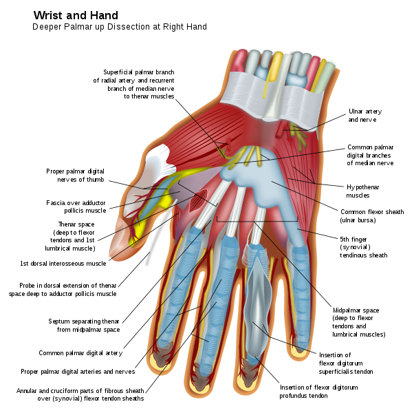
As the long flexor muscles descend into the hand, they are cover by a synovial sheath. This synovial sheath acts as a bursa for the tendons by reducing friction. The tendon sheath is similar to a sac of synovial fluid that allows the muscle tendons to move and stretch; this protects the muscles from adhering to each other or directly rubbing against each other. There is one tendon sheath that occupies the most space in the hand. The common flexor sheath is the largest tendon sheath in the hand and covers the tendons of the flexor digitorum profundus muscle and the flexor digitorum superficialis muscle as they pass under the transverse carpal ligament. Once the tendons from the flexor digitorum profundus muscle and the flexor digitorum superficialis muscle pass the transverse carpal ligament. The individual tendons will divide with their separate tendons sheaths towards the digits. The flexor pollicis longus, the flexor carpal radialis, and the flexor carpal ulnaris muscle have their own separate tendon sheaths. The palmaris longus muscle usually does not have a tendon sheath.
Embryology
Muscles, tendons, vessels, and connective tissue derive from the mesodermal germ layer. The mesodermal germ layer has mesenchymal tissue. The mesenchyme tissue will differentiate into the structures formed by the mesoderm layer. The muscles and tendons will grow as the limbs elongate. Fibroblast growth factor (FGF) induces elongation of the limbs.
Blood Supply and Lymphatics
The blood supply for the long flexors of the hand derives from the ulnar artery, the radial artery, and the interosseous arteries. The ulnar artery will provide blood to the flexor digitorum superficialis muscle, the palmaris longus muscle, the flexor carpal radialis muscle, and the flexor carpal ulnaris muscle. The anterior interosseus artery will perfuse the flexor pollicis longus muscle and the flexor digitorum profundus muscle. The ulnar artery will form the superficial palmar arch. The radial artery will form the deep palmar arch. These arterial arches will form anastomoses in the hand. These anastomoses provide collateral perfuse to the long flexors tendons.[1][2]
The lymphatic drainage of the long flexor of the hands will drain proximally to the cubital fossa. Once the lymph fluid is at the cubital fossa, it will drain back to the axillary lymph nodes. The lymph from the hand will eventually return to circulation via the right lymphatic duct or the thoracic duct. The right hand will drain into the right lymphatic duct, and the left hand will drain into the thoracic duct.
Nerves
The nerve innervation of the long flexor muscles of the hand subdivides between the median nerve and the ulnar nerve. The ulnar nerve (C8, T1) innervates the muscles on the ulnar side of the forearm and hand. The ulnar nerve only innervates one of the long flexor muscles, the flexor carpal ulnaris muscle.[3] The flexor pollicis longus muscle, the flexor digitorum superficialis muscle, the flexor digitorum profundus muscle, flexor carpal radialis muscle, and the palmaris longus muscle are all innervated by the median nerve (C5-T1).[4]
Muscles
There are six long flexor muscles of the hand:
- Flexor carpal radialis muscle
- Flexor carpal ulnaris muscle
- Flexor digitorum profundus muscle
- Flexor digitorum superficialis muscle
- Flexor pollicis longus muscle
- Palmaris longus muscle
All these flexor muscles will contribute to flexion of the hand, wrist, and fingers.
Physiologic Variants
The presence of the palmaris longus muscle is variable from individual to individual. The action of the palmaris longus muscle is minor, so the lack of this muscle will result in no deficits.[5] The attachments of the long flexor muscles to the carpal bones may vary slightly. Some of the long flexors will have small attachments to the carpal bones are they descend into the hand.[6][7][8]
Surgical Considerations
In hand surgery, the knowledge of the long flexor muscles of the hand can aid in successful surgeries by preventing avoidable damage to the muscles, nerves, or blood supply. In carpal tunnel syndrome repair, the transverse carpal ligament is split to allow more room for the structures that descend underneath the transverse carpal ligament. When dividing the ligament, it is crucial to avoid damage to the muscle tendons, vessels, and median nerve.
Surgeons commonly use the tendon of the palmaris longus muscle as a graft. When a tendon ruptures in the hand, the palmaris longus tendon may be used as a graft to repair that damaged tendon.[6][8]
Clinical Significance
The muscle and tendons of the long flexors of the hand may become inflamed, hypertrophied, or affected by fibrosis.
A condition called "Golfer's elbow" (medial epicondylitis) is a condition where the origins of the hand and wrist flexors are affected by inflammation. The origin of these flexors muscles is irritated by repetitive flexion of the hand and wrist. There will be localized pain upon palpation of the medial epicondyle. This condition commonly gets treated with nonsteroidal anti-inflammatories and rest.[9]
The tendons of the long flexor muscles of the hand can become thicken or affected by fibrosis. These alterations in the tendons tend to remain localized in the palmar region of the hand. A condition called "Dupuytren contracture" is caused by progressive fibrosis of the flexor tendons in the hand. The tendons are affected by fibrosis resulting in flexion of the digits. The pathological flexion of the fingers will be irreversible. This condition typically affects the third and fourth digits.[10][11]
The tendon sheath can also be affected by fibrosis or inflammation. When fibrosis and inflammation of the tendons occur to the flexors in the hand. Individuals will present with "trigger finger." Trigger finger is a result of fibrosis or inflammation limiting the movement of the fingers. The fingers will classically present in rigid flexion at rest. The fingers stay in flexion when the person tries to extend the fingers.
Other Issues
Since the nerve innervation of the long flexors of the hand splits between the ulnar nerve and the median nerve. If one of the nerves were to be compromised, it would manifest as radial or ulnar flexion when the individual attempts palmar flexion. If radial flexion predominates, this means there is damage to the ulnar nerve. If ulnar flexion predominates, this means the median nerve is damaged, since palmar flexion is a result of radial and ulnar flexion counteracting each other.