Introduction
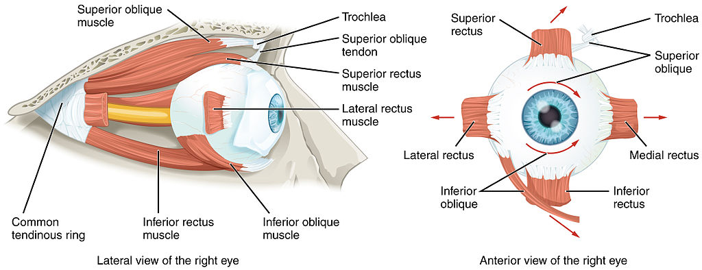
There are seven extraocular muscles. There are a total of four rectus muscles, two oblique muscles, and the standalone levator palpebrae superioris. The four rectus muscles are the medial rectus, lateral rectus, superior rectus, and inferior rectus. The oblique muscles are the superior and inferior obliques. The levator palpebrae superioris is primarily responsible for eyelid elevation.[1] See Image. The Extraocular Muscles of the Orbit.
Structure and Function
With the head facing straight and the eyes facing straight ahead, the eyes are said to be in primary gaze. From this position, an action from an extraocular muscle produces a secondary or tertiary action. Although the globe can be moved about 50 degrees from the primary position, usually during normal eye movement only 15 degrees of extraocular muscle movement occurs before head movement begins. The rectus and oblique muscles are involved in the different gaze positions of the eye.
Each of the rectus and oblique muscles have a functional insertion point, which is at the closest point where the muscle first contacts the globe. This point forms a tangential line from the globe to the muscle origin and is known as the arc of contact. The common site of origin for the rectus muscles in the Annulus of Zinn is located at the orbital apex.
The levator palpebrae superioris does not contact the globe directly but instead elevates the eyelid.[1]
Embryology
The mesenchyme of the head, including the orbit and its structures, are primarily embryologic derivatives of mesoderm and neural crest cells. The extraocular muscles originate from the mesoderm, but the satellite and connective tissue of the muscle arises from neural crest cells. Most of the remaining connective tissue of the orbit also is derived from neural crest cells.[2][3]
Blood Supply and Lymphatics
The primary blood supply for all of the extraocular muscles is the muscular branches of the ophthalmic artery, the lacrimal artery, and the infraorbital artery. The ophthalmic artery has two muscular branches, which are the superior and inferior muscular branches. The lateral rectus receives blood from a branch of the lacrimal artery, and the other rectus muscles receive blood via two anterior ciliary arteries that communicate with a structure called the anterior circle of the ciliary body.
Venous drainage is similar to the arterial system and empties into the superior and inferior orbital veins. Usually, there are a total of four vortex veins, and these are found at the lateral and medial sides of the superior and inferior rectus muscles. These vortex veins drain into the orbital venous system.[4]
Nerves
Cranial nerve III is divided into upper and lower divisions, with the upper division innervating the superior rectus as well as the levator palpebrae superioris, and the lower division innervating the medial rectus, inferior rectus, and inferior oblique. The superior oblique is innervated by cranial nerve IV (trochlear), while the lateral rectus is innervated by cranial nerve VI (abducens).[5]
Muscles
Each of the rectus muscles originates posteriorly at the Annulus of Zinn and courses anteriorly along the orbital walls until it reaches its insertion point. These muscles insert on the globe at varying distances from the limbus, and the curved line drawn along the insertion points makes a spiral that is known as the Spiral of Tillaux. Starting at the medial aspect of the globe, the medial rectus inserts at 5.5 mm from the limbus, the inferior rectus inserts at 6.5 mm from the limbus, the lateral rectus inserts at 6.9 mm from the limbus, and the superior rectus at 7.7 mm from the limbus.
The superior oblique originates medial to the optic foramen and travels through the trochlea, a pulley at the superonasal portion of the orbital rim. From here the muscle travels under the superior rectus and inserts slightly posterior to the insertion of the superior rectus. The inferior oblique originates from a depression on the orbital floor near the orbital rim, travels posteriorly and inferiorly, and inserts near the macula.
The levator palpebrae superioris originates from the lesser wing of the sphenoid and courses anteriorly. The body of the muscle travels over the superior rectus toward the eyelid. Where the connective tissue from the levator palpebrae superioris connects with similar tissue from the superior rectus, the Whitnall ligament is formed. Near this ligament, the levator palpebrae superioris fibers change to become more vertical, and they divide to the aponeurosis anteriorly and the superior tarsal muscle superiorly.[1]
Extraocular muscles have a large ratio of nerve fibers to skeletal muscle fibers. The ratio is 1:3 to 1:5, compared to other skeletal muscles which are 1:50 to 1:125. Extraocular muscles are a specialized form of skeletal muscle with a variety of fiber types, including both slow tonic types which resist fatigue, and also saccadic (rapid) type muscle fibers.[6][7]
Physiologic Variants
The size of the extraocular muscles, as well as their insertion point on the globe from the limbus and other anatomical measurements, may vary widely from one individual to the next. The numbers described in this article reflect average distances.
Occasionally, accessory extraocular muscles have been reported originating from the Annulus of Zinn and they insert in various locations. Both supernumerary and accessory extraocular muscles have been reported.
Congenital differences in extraocular muscles can cause ocular misalignment. See the Clinical Significance section for more details regarding strabismus.[1][8]
Surgical Considerations
The nerves to the rectus muscles and superior oblique muscles insert into the muscles at one-third the distance from the origin to the insertion. This makes damage to these nerves during anterior segment surgery difficult, but not impossible. Additionally, instruments that are advanced 26 mm posterior to the rectus muscle insertions can cause injury to the nerve.[9]
Blood vessels may be compromised during surgery of the inferior rectus muscle. The vessels which supply blood to the extraocular muscles also supply nearly all the temporal half of the anterior segment of the eye. Most of the nasal half of the anterior segment circulation is also derived from blood vessels that supply the extraocular muscles. Therefore, care must be taken during surgery of the medial rectus or other extraocular muscles to avoid disrupting this blood supply. [4],[10]
There are other complications that may result from strabismus surgery. Unsatisfactory alignment is the most common complication and may require additional surgery to correct this. Refractive changes may occur when two rectus muscles of one eye are operated on, and this may resolve over months. Other possible surgical complications include diplopia, perforation of the sclera, and postoperative infections. Although uncommon, serious infections may result after strabismus surgery, including pre-septal or orbital cellulitis and endophthalmitis.[11][12][13]
Clinical Significance
The function of the extraocular muscles can be assessed along with the other extraocular muscles during the clinical exam. The movement of the extraocular muscles can be assessed by having a patient look in nine directions. Starting with the primary gaze, followed by the secondary positions (up, down, left, and right) and the tertiary positions (up and right, up and left, down and right, down and left). The clinician can test these positions by having the patient follow the clinician's finger and trace a wide letter "H" in the air.
Further tests of ocular alignment can be tested further by several methods, including cover-uncover tests, corneal light reflex, dissimilar image tests, and dissimilar target tests. Since many patients with extraocular muscle abnormalities are young children, the clinician may need to employ various clever means such as using toys or other objects to elicit the cooperation of the child.
Strabismus, or ocular misalignment, can be caused by abnormalities in binocular vision or abnormalities of neuromuscular control. Weakness, injury, or paralysis that involves the inferior rectus muscle can be involved in strabismus.
Nerve palsies of the cranial nerves which innervate the extraocular muscles produce characteristic patterns which must be identified by the clinician.[14][15]
Other Issues
There is a capsule of connective tissue known as the Tenon capsule which is an envelope that fuses with the optic sheath posteriorly and comes to meet the intermuscular septum anteriorly. The Tenon capsule helps the globe to stay positioned in orbit along with the extraocular muscles.[16]