Continuing Education Activity
Bowel perforation results from insult or injury to the mucosa of the bowel wall caused due to a violation of the closed system. Bowel perforation can be secondary to many factors, including inflammation, infection, obstruction, trauma, or invasive procedure. This activity describes the clinical presentation, evaluation, and management of bowel perforation and the importance of interprofessional teams in the effective management of affected patients.
Objectives:
- Describe the epidemiology of bowel perforation.
- Outline the clinical presentation of bowel perforation.
- Explain the management and rehabilitation strategies in patients with bowel perforation.
- Describe how an interprofessional team can collaborate to improve the rapid diagnosis, resuscitation, evaluation, and management of patients with bowel perforation.
Introduction
Bowel perforation results from insult or injury to the mucosa of the bowel wall resulting from a violation of the closed system. This exposes the structures within the peritoneal cavity to gastrointestinal contents. Bowel perforation can be secondary to many factors, most commonly inflammation, infection, obstruction, trauma, or invasive procedure. Patients presenting with abdominal pain and distension, especially in the appropriate historical setting, must be evaluated for this entity as delayed diagnosis can be life-threatening due to the risk of developing infections such as peritonitis. Management includes stabilizing the patient while making surgical consultation. Even appropriately managed, bowel perforation can lead to increased morbidity and mortality from post-repair complications such as adhesions and fistula formation.[1]
Etiology
Bowel perforations can be separated based on their anatomic locations, but many causes are overlapping.
Small bowel: Erosion from duodenal ulcerations, tumor, infection or abscess, Meckel diverticulum, hernia with strangulation, inflammatory bowel disease/colitis, mesenteric ischemia, foreign body, obstruction, medication/radiation-related, iatrogenic, blunt or penetrating abdominal trauma
Large bowel: Tumor, diverticulitis, infection or abscess, colitis, foreign body, obstruction, volvulus, iatrogenic, blunt or penetrating abdominal trauma[2]
Epidemiology
In children, bowel perforation is most likely to follow abdominal trauma. The incidence of bowel perforation is 1% to 7% in pediatric trauma patients.
In adults, ulcerative disease represents the most common etiology of bowel perforation, with duodenal ulcers causing 2- to 3-times the rate of perforation than gastric ulcers do. Perforation secondary to diverticular disease represents up to 15% of cases. In the geriatric population, perforated appendicitis represents the most common cause of perforation.
The overall incidence of perforation from colonoscopy has been reported, on average, around 2% with higher rates during colonoscopy requiring therapeutic interventions.[3]
Pathophysiology
Bowel perforation results from a violation of the mucosal layers of the intestinal tract resulting in the spilling of air and digestive contents into the peritoneal cavity. These contents can range from highly acidic gastric contents in more proximal bowel perforation, to fecal material from a more distal area of perforation. Areas of partial erosion can become full-thickness tears over time, or a specific insult can cause spontaneous rupture. This means that pain may be gradual or sudden, but it usually progresses in severity. Feelings of abdominal distension and signs of abdominal muscular rigidity representing peritonitis may develop.[4]
History and Physical
Historical factors are exceptionally useful when making the diagnosis of bowel perforation. Lower chest or abdominal pain after recent instrumentation from endoscopy/colonoscopy or laparoscopic/open surgical procedure should raise high suspicion for this diagnosis. Previous medical, surgical, and social history to include a history of a prior hernia, bowel obstruction, known or suspected malignancy, foreign body insertion or ingestion, abdominal trauma, and regular medication use (NSAIDs, corticosteroids, and chemotherapy are the most common) should be elicited. The patient is likely to complain of pain and distension in the abdomen that is worsening. Patients may describe a pain-free period before the worsening of their pain, and this can represent decompression of an inflamed or injured area directly following the perforation.
Diffuse spillage of air and intestinal contents may make the pain difficult to localize during focused abdominal examination, but palpation is likely to identify tenderness. Patients usually have nausea and vomiting. As the situation progresses, peritoneal signs such as guarding and rigidity may develop. Vital signs may be normal, especially in early presentation; however, tachycardia, tachypnea, fever, and other signs of sepsis are likely to develop.[5]
Evaluation
Evaluation of a patient with suspected bowel perforation must focus on a thorough history and physical exam. Laboratory and radiographic workup should mimic that of any patient presenting with abdominal pain. Labs may include a complete blood count, basic metabolic panel, liver function tests, lipase, amylase, and inflammatory markers such as C-reactive protein. However, common findings such as a leukocytosis or elevated amylase or elevated CRP level are non-specific for diagnostic purposes.
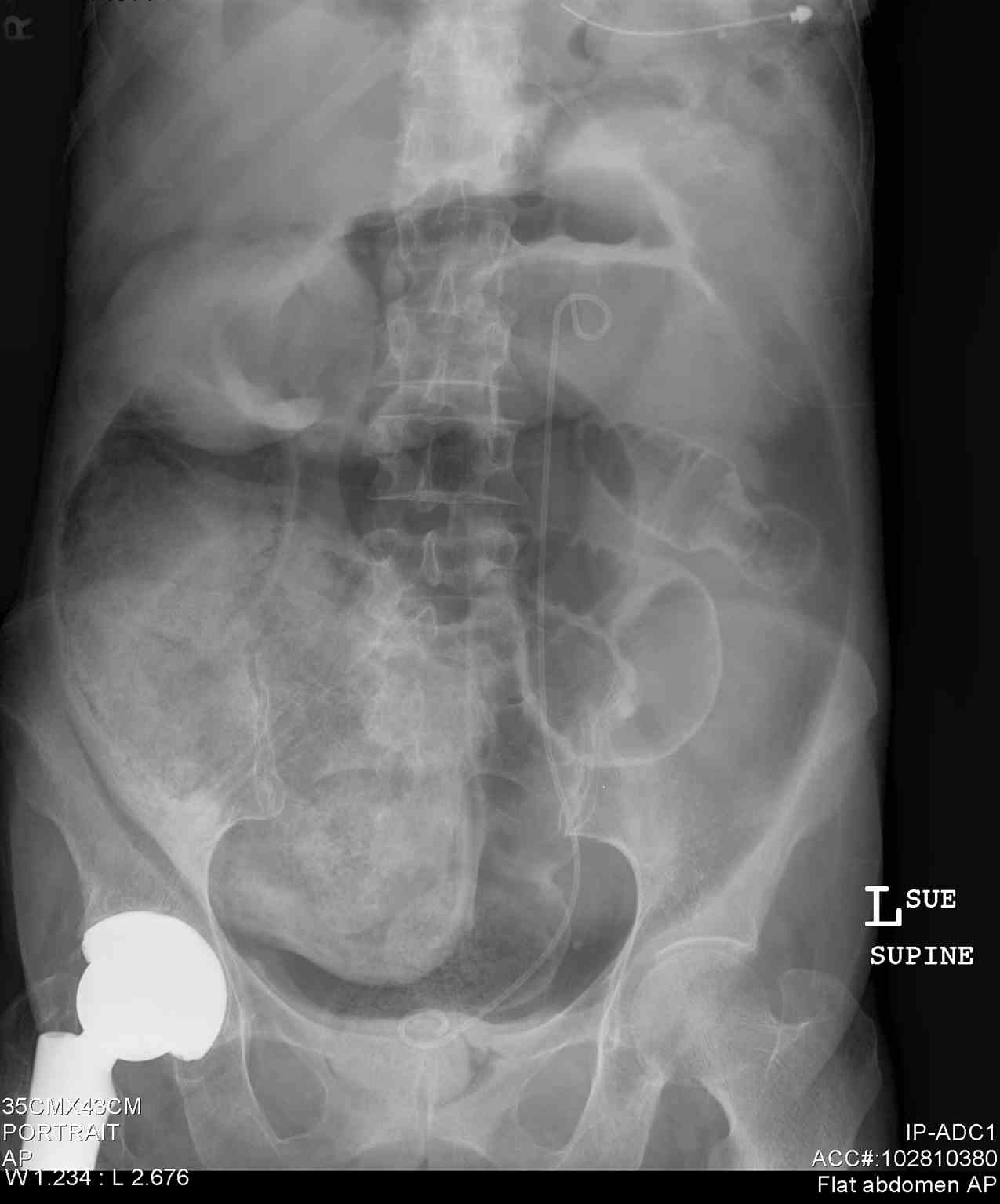
Upright chest radiography (50% to 70% sensitive) can help detect free intraperitoneal air below the diaphragm suggestive of bowel perforation. The likelihood of diagnosing free air is higher if the patient has been sitting upright for at least 15 minutes prior to the film. Lateral decubitus films can be used if the patient is unable to sit upright.
Up to 40 percent of patients will demonstrate greater than 2 cm of free gas at 24 hours post laparoscopy, without any clinical evidence of bowel perforation. After laparotomy, however, free intra-abdominal gas often may be present for a longer duration postoperatively (up to one week), but the volume should be gradually decreased.
Computer tomography (CT) scan is the modality of choice for not only diagnosing free air but also for localization of the site of perforation. A CT scan can also help to determine if the area has spontaneously walled off if there has been a progression to abscess formation or if there is an inflammatory involvement of surrounding structures.
Ultrasound has been used to localize gas collections suggestive of perforation. However, this modality is especially dependent on the experience level of the operator.
Surgical consultation should be pursued early in any patient with abdominal pain and signs of clinical deterioration.[6]
Treatment / Management
Treatment of a patient with suspected bowel perforation involves establishing intravenous (IV) access and initial hemodynamic management, especially if the patient presents with any signs or symptoms concerning sepsis or shock. Initiation of broad-spectrum antibiotics aimed at gram-negative and anaerobic organisms is essential and should be initiated early. For suspected distal perforation of the intestinal tract, nasogastric decompression should be performed, and the patient made NPO (nothing by mouth). Perforated appendix or appendicular abscess is categorized in the low-risk community-acquired intra-abdominal infections group. However, risk factors for antibiotic resistance or treatment failure should be excluded. Such risk factors include recent travel history to the high rates of antibiotic-resistant organisms’ sites, known colonization with antibiotic-resistant organisms, advanced age, immunocompromised patients.
If the patient is hemodynamically stable and there is no concern for peritonitis, such as in the case of a spontaneously contained perforation, the option for non-surgical management with antibiotics and observation may be pursued in consultation with the surgical team.
Localized abscess formation may be amenable to drainage by interventional radiology.
Most cases will progress to require direct investigation via laparoscopic or open (laparotomy) exploration. This allows for primary repair as well as infection control measures. Minimally invasive exploration and repair are often successfully performed. However, it should be noted that exploratory laparotomy is the surgical intervention of choice for any signs of clinical deterioration or hemodynamic instability.[7]
Differential Diagnosis
- Acute Cholecystitis and Biliary colic
- Inflammatory bowel disease
- Meckel diverticulum surgery
Prognosis
The patient’s medical state before the perforation occurs best predicts general prognosis. In patients without multiple comorbidities, the outcomes are more favorable. Management of the underlying etiology is key to preventing further episodes.[8]
Complications
Early
Hemodynamic instability leading to hypoperfusion, shock, and multi-organ system failure, infection (whether local abscess formation, peritonitis, or systemic bacteremia)
Late
Delayed wound healing, postoperative adhesions leading to bowel obstruction, fistula formation, and hernias [6]
Postoperative and Rehabilitation Care
Postoperative care should include a continued focus on hemodynamic resuscitation with monitoring of expected urine output, electrolyte repletion, infection monitoring and control measures, pain management, and nutritional considerations. [9]
Consultations
Emergency room consultation may include radiology, internal medicine, and, most importantly, surgery. In the long term, consultation should be aimed at the specialist team that will assist in the management of the underlying pathology that leads to perforation. This may include gastroenterology to assist in long-term surveillance and chronic disease management strategies in cases of inflammatory bowel disease or diverticulitis, oncology for the management of malignancy, or the primary care team for assistance with chronic medication management.[10]
Enhancing Healthcare Team Outcomes
Early and correct diagnosis is essential. This involves the recognition of a patient with an acute abdomen, proper resuscitation with antibiotics, crystalloid fluids, pain control and correction of other concurrent conditions. The interprofessional team should include emergency room physicians, general surgeons, specialty trained nurses, and pharmacists. The nursing staff must closely monitor patients for clinical deterioration, including hypotension. Pharmacists evaluate medical treatment and asses for drug-drug interactions. Communication between team members will improve outcomes. Proper treatment must involve the surgical team at presentation to ensure expedient surgical intervention which is the best predictor of improving morbidity and mortality.[11]