Continuing Education Activity
Blastomycosis is a fungal infection caused by Blastomyces dermatitidis. It presents as a pulmonary infection after the inhalation of spores, and it may be either asymptomatic or have severe life-threatening complications like acute respiratory distress syndrome. This activity reviews the evaluation and treatment of blastomycosis and explains the role of the interprofessional team in managing patients with this condition.
Objectives:
- Identify Blastomyces dermatitidis as the causal agent in the etiology of blastomycosis.
- Summarize the pathophysiology of blastomycosis.
- Review the most common adverse effects of amphotericin in the treatment of patients with blastomycosis.
- Outline the importance of collaboration and communication among the interprofessional team members to enhance the delivery of care for patients affected by blastomycosis.
Introduction
Blastomycosis is a fungal infection caused by the organism Blastomyces dermatitidis, which is endemic in the soils of the Ohio and Mississippi River Valleys, Great Lakes region, and the southeastern United States.[1] It most commonly presents as a pulmonary infection following the inhalation of spores, which may be asymptomatic and, therefore, undetectable, though severe, life-threatening complications like acute respiratory distress syndrome can occur. Extrapulmonary disease occurs in approximately 25% to 30% of patients after hematogenous dissemination from the lungs, with the skin being the most common site of extrapulmonary disease.[2] Primary cutaneous blastomycosis, though rare, can occur due to direct inoculation after trauma to the skin. Unlike other deep fungal infections that occur predominantly in immunocompromised patients, blastomycosis also occurs in immunocompetent hosts. [3]
Etiology
Blastomyces dermatitidis is the causal agent of blastomycosis.[4] A member of the phylum Ascomycota in the family Agellomycetaceae, it is recognized as the asexual state of Ajellomyces dermatitidis, a thermally dimorphic fungus. At 25 C, the mycelial form grows as a fluffy white mold, while at 37 C, it grows as a brown folded yeast. The fungus can be isolated in the soil, where it forms a mycelium that penetrates the substratum on which it grows. It reproduces asexually with small conidia that are 2-10 µm in diameter. In infected cells, Blastomycosis dermatitidis is seen as budding yeast cells that are relatively large at 8 to 10 micrometers in diameter.[4]
Epidemiology
Blastomycosis is endemic in the Ohio and Mississippi River valleys, and near the Great Lakes and southeastern parts of the United States. The annual incidence is less than 1 case per 100,000 people in the commonly affected states of Mississippi, Kentucky, Arkansas, and Wisconsin.[5] Other states commonly affected include North Carolina, Tennessee, Louisiana, and Illinois. Blastomycosis affects all ages, races, and genders, though it is reported to occur more frequently in males, presumably due to occupational and recreational exposures. Adult men are also more likely to develop systemic infection.
Pathophysiology
The conidia, which can become aerosolized when the fungal colony is disturbed, are the main infectious particles produced by Blastomyces dermatitidis. After conidia are inhaled, they pass into the lower respiratory tract. The conidia can be phagocytized by bronchopulmonary mononuclear cells and killed by neutrophils and macrophages, explaining the asymptomatic infection in some individuals. [6] When Blastomyces dermatitidis converts to the yeast form, the thick wall provides resistance to phagocytosis and killing, which can result in symptomatic pulmonary infection. In addition, the immune-modulating glycoprotein BAD-1 facilitates its binding to macrophages, allowing dissemination through the blood and lymphatics to other areas of the body. The pyogranulomatous inflammatory response is a unique feature of blastomycosis and is caused by an influx of neutrophils, macrophages, and eventual granuloma formation.
History and Physical
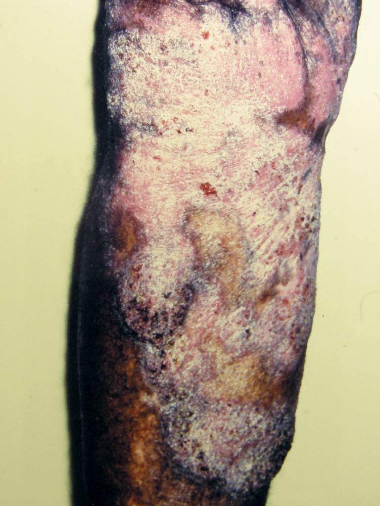
Initial symptoms of blastomycosis are flulike and typically resolve within days, though the patient may disregard their mild nature; therefore, the initial infection may go undiagnosed. In addition, asymptomatic infection occurs in about 50% of infected persons. Acute or chronic pneumonia can occur and, in elderly or immunocompromised patients, acute respiratory distress syndrome can result. The extrapulmonary disease can occur after the dissemination of Blastomyces dermatitidis to other organs, with the skin being the most common site involved. Clinically, cutaneous blastomycosis typically starts as papules that evolve into crusted, vegetative plaques often with central clearing or ulceration. Lymphangitis and lymphadenopathy may be present. Bone lesions are the next most common finding, occurring in 25% of extrapulmonary cases, and are typically lytic. Though osteomyelitis can involve any bone, the lower spine and pelvis are most commonly affected. Other findings of an extrapulmonary disease can include prostatitis, orchitis, or epididymitis. The central nervous system is involved in 5% to 10% of cases, including cases of meningitis and intracranial or epidural abscesses.
Evaluation
Direct visualization of Blastomyces dermatitides is essential for a definitive diagnosis. Sputum specimens stained with 10% potassium hydroxide or a fungal stain have an approximate 80% yield. Biopsy and histopathological examination of skin lesions reveal pseudoepitheliomatous hyperplasia with neutrophilic abscesses. Organisms can be difficult to identify and are often found within histiocytes or abscesses in the dermis. The yeasts are 8 to 15 micrometers in diameter with thick, double-contoured walls and display broad-based budding. Culture is the most sensitive method for detecting and diagnosing blastomycosis. [4]Growth is typically detected in 5 to 10 days but can take up to 30 days if few organisms are present in the specimen.
Chest radiologic imaging can be used to screen for pulmonary involvement, though findings are variable and lack specificity. Of all the systemic fungal infections, blastomycosis is most likely to resemble cancer on lung radiography. Lumbar puncture and cerebrospinal fluid analysis may be ensued to determine central nervous system involvement, though the diagnosis is also difficult and rarely definitive. Lastly, a chemiluminescent DNA probe is commercially available and can produce results within hours.[7] It will, however, produce a positive result with Paracoccidioides brasiliensis, though this is usually not a problem in the US as this organism is found almost exclusively in South and Central America.
Treatment / Management
Although spontaneous remission can occur, it is recommended that all patients with the mild or moderate disease be treated to avoid dissemination and recurrence. Itraconazole is the treatment of choice for all forms of the disease, except in severe, life-threatening cases. It is recommended that 600 mg be given orally daily for three days, followed by 200 mg to 400 mg per day for 6 to 12 months.[8] Itraconazole has relatively low toxicity and good efficacy, though it is important to remember that gastric acidity is required for its absorption. Other azoles, including ketoconazole and fluconazole, can be used but are not preferred due to lower efficacy and, in the case of ketoconazole, a worse side effect profile.[9] Voriconazole has good cerebrospinal fluid penetration and thus plays a role in CNS disease. Azoles are contraindicated in pregnancy. Amphotericin B is used in severe and life-threatening diseases at a high dose of 0.7 to 1 mg/kg/day to a total dose of 1.5 to 2 grams. Liposomal amphotericin B at a dose of 3 to 5 mg/kg per day can alternatively be used for severe infection and is preferred for CNS blastomycosis and treatment in pregnant women.[8][10] Amphotericin is associated with several notable adverse effects, particularly renal impairment, which is less likely with the lipid formulation. Other adverse effects include hypokalemia, anemia, fever, chills, nausea, and thrombophlebitis at the injection site.
Differential Diagnosis
The differential diagnosis of blastomycosis include the following:
- Pneumonia
- Tuberculosis
- Non-infectious pulmonary disease
- Cancer
Prognosis
The prognosis of patients who are immunocompetent is generally good as they have a decreased chance of suffering from complications of the infection. Successful treatment of this condition is achieved in approximately 80% to 95% of cases.
On the other hand, immunocompromised patients with blastomycosis have poor prognosis.
Complications
Complications include:
- Relapse/disease recurrence
- Abscesses
- Skin sores
- Side effects of treatment (amphotericin)
Deterrence and Patient Education
Immunocompromised patients should be educated on the avoidance of areas and activities that are associated with an increased risk of this infection.
Enhancing Healthcare Team Outcomes
The diagnosis and management of blastomycosis are complex and best done with an interprofessional team that includes the primary care provider, nurse practitioner, dermatologist, infectious disease expert, and a pulmonologist. The key is deciding on the treatment of patients with a mild disease because spontaneous remissions can occur. However, it is recommended that all patients with the mild or moderate disease be treated to avoid dissemination and recurrence. Itraconazole is the treatment of choice for all forms of the disease, except in severe, life-threatening cases. It is recommended that 600 mg be given orally daily for three days, followed by 200 to 400 mg per day for 6 to 12 months.[8]
The outlook for healthy patients with blastomycosis is good, but the recovery may be prolonged. In immunocompromised individuals, the outcomes are guarded. [11](Level V)