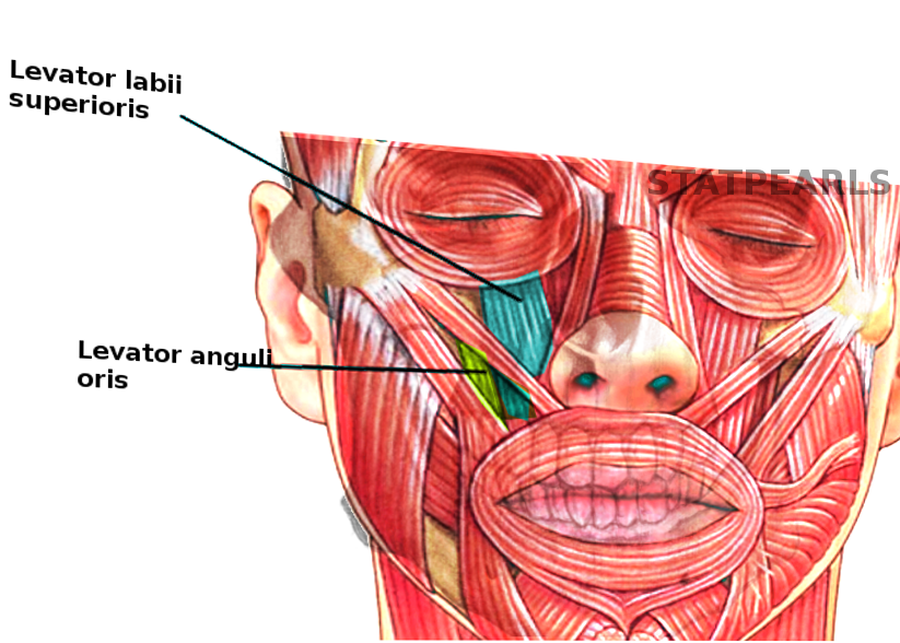
Anatomy, Head and Neck: Eye Levator Labii Superioris Muscle


Introduction

The levator labii superioris muscle, also known as the quadratus labii, contributes to facial expression and movement of the mouth and upper lip. It courses alongside the lateral aspect of the nose, and its primary function is elevation of the upper lip (see Image. Eye Levator Labii Superioris Muscle). It is also involved in movements such as expressions of disgust, sadness and disdain, nasal flaring, retching (vomiting), and even to show oral content. Its origin is on the lateral aspect of the nose, and it extends laterally towards the zygomatic bone. The levator labii superioris receives its blood supply from terminal branches of the facial artery and the infraorbital branch of the maxillary artery. The zygomatic branch of the facial nerve (cranial nerve VII) innervates the levator labii superioris. See Image. Levator Labii Superioris.

Structure and Function

The levator labii superioris is a thin, quadrilateral muscle that serves to elevate the upper lip, particularly during smiling, but it also provides eversion. It originates on the infraorbital rim of the maxilla and inserts into the orbicularis oris muscle.[1] Superior to the nasolabial fold, there is subcutaneous fat between the muscle and the dermis, but inferior to the fold, the muscle is more tightly adherent to the skin. The average levator labii superioris muscle is approximately 25 mm in length and 3.5 mm in thickness.[1]

Medial to the levator labii superioris is the levator labii superioris alaeque nasi muscle (sometimes called the angular head of the levator labii superioris muscle). The levator labii superioris alaeque nasi can be further subdivided into a furrow portion (nasolabial), an alar portion, and a lip portion. Some of the fibers lie superficial to the levator labii superiors, and some lie deep, with different authors reporting differing proportions of each.[2][3] The origin of the levator labii superioris alaeque nasi is the frontal process of the maxilla, near the medial palpebral ligament, and the muscle inserts into the facial soft tissue lateral to the nostril and upper lip, as well as interdigitating with fibers of the transverse portion of the nasalis muscle.[2] The main action of all three portions is to elevate the upper end of the nasolabial furrow, the lateral end of the nasal ala, and the middle portion of the upper lip.[3]

Lateral to the levator labii superioris is the levator anguli oris muscle, which raises the corner of the mouth. This belly arises from the maxilla, just lateral to the origin of the levator labii superioris, and it inserts on the modiolus of the orbicularis oculi. Its fibers run deep to the levator labii superioris, and for that reason, this muscle is innervated from its superficial surface. Also associated with the lateral aspect of the levator labii superioris muscle is the zygomaticus minor muscle, which elevates and lateralizes the upper lip while smiling. The zygomaticus minor lies superficial to the levator labii superioris muscle.[4]

Embryology

The muscles of the face begin to develop between the third and eighth weeks of embryonic development.[5][6] The muscles originate as a thickening of the mesoderm layer of the second branchial arch. The infraorbital lamina and occipital platysma are the first laminae to develop. Both infraorbital laminae give rise to the levator labii superioris, among many other facial mimetic muscles. Improper development may lead to congenital facial weakness, which most often presents with segmental dysfunction corresponding to the affected muscles, in contrast to traumatic or inflammatory facial paralysis, which is most often hemifacial.

Blood Supply and Lymphatics

The levator labii superioris receives its vascular supply from two arteries: the facial artery and the infraorbital artery.[1] Directly supplying the muscle is the angular artery, a terminal branch of the facial artery that arises from the superior labial artery; the facial artery is itself a branch of the external carotid artery. The infraorbital artery is a branch of the internal maxillary artery, a terminal branch of the external carotid artery. The infraorbital artery runs through the infraorbital foramen, along with the infraorbital nerve, a branch of the maxillary division of the trigeminal nerve (cranial nerve V2).[1] Venous outflow occurs through tributaries of the facial vein that correspond to the arterial inflow, and lymphatic drainage passes through nodes in the nasolabial region.[7]

Nerves

The zygomatic branch of the facial nerve innervates the levator labii superioris and the other midfacial muscles between the orbicularis oculi and the orbicularis oris, along with contributions from the buccal branch of the facial nerve.[1][7] The levator labii superioris muscle and the levator labii superioris alaeque nasi are innervated from their deep surfaces, but the levator anguli oris, just lateral to it, is innervated from its superficial surface.

Physiologic Variants

A 2018 study performed in Taiwan found that 31 out of 32 adult cadavers had zygomaticus minor muscle fibers inserting into the upper lip, originating from the zygomatic region and the orbicularis oculi muscle.[4] In 14 of the cadaver specimens (43.8%), some of the zygomaticus minor fibers blended with the lower border of the orbicularis oculi muscle. The muscle also attaches to the palpebral ligament medially, the face of the maxilla, the belly of the levator labii superioris alaeque nasi, and the depressor supercilii muscle as well.

Surgical Considerations

Excessive gingival display, or "gummy smile," is often caused by hyperactivity of the muscles of the upper lip, including but not limited to the levator labii superioris muscle. Correction of a gummy smile can be performed in multiple ways, depending on the severity, the case, and the amount of jaw protrusion. If the cause is hyperactivity of the upper lip muscles, myotomy and lip repositioning followed by orthodontic treatment can be an effective surgical intervention.[8][9]

Trauma or surgery for cutaneous malignancy can result in soft tissue defects of the nose; a pedicled levator labii superioris alaeque nasi flap has been described for reconstruction of these defects.[10]

Because of the critical role that the levator labii superioris muscle plays in producing a smile, increasing efforts have been made by facial reanimation surgeons to replace this muscle in addition to the zygomaticus major when attempting to rehabilitate a patient's smile. Described methods of reanimating the smile in patients with longstanding flaccid facial paralysis include the use of the sternohyoid and omohyoid muscles, the serratus anterior muscle, and the split gracilis muscle.[11][12]

Clinical Significance

There are alternative ways to treat a “gummy smile” or excessive gingival display. Injecting botulinum toxin is an effective treatment for patients with excessive gingival show caused by overactive upper lip elevators.[13] This option is a newer technique and is much less expensive and traumatizing to patients than performing surgery. The disadvantage of botulinum toxin treatment is that it requires repeat injections, as the drug effect lasts only 3 to 4 months, and it is only useful if a hyperactive upper lip is the cause of the excessive gingival display.[14] Additionally, infiltration of hyaluronic acid has been shown to be safe, effective, and long-lasting.[15]

Other Issues

Weakness of the levator labii superioris muscle is most often unilateral, but can also present bilaterally, and severity ranges from mild paresis to full paralysis. Most commonly, it results from damage to the main trunk of the facial nerve or the zygomatic/buccal branch of the facial nerve due to trauma or inflammation, such as Bell's palsy, Ramsay Hunt syndrome, or Lyme disease. Other causes of facial paralysis include multiple sclerosis, poliomyelitis, Guillain-Barré syndrome, and SARS-CoV-2 infection, among many others. Iatrogenic damage to the facial nerve can also lead to weakness of the levator labii superioris muscle.[16] Some patients are born with congenital facial paralysis, in which one or more mimetic muscles are atrophic or absent, such as in Möbius syndrome.[17] It is crucial to perform a full neurological examination to determine the cause of the weakness.

As an interesting side note, levator labii superioris muscle transposition has been described as a treatment for chronic oral maxillary sinus fistulas in horses.[18]


(Click Image to Enlarge)
<p>Levator Labii Superioris and Levator Anguli Oris

Levator Labii Superioris and Levator Anguli Oris. This illustration highlights 2 muscles that move the lips to dilate the mouth and make facial expressions.


Contributed by S Bhimji, MD


(Click Image to Enlarge)
<p>Eye Levator Labii Superioris Muscle</p>

Eye Levator Labii Superioris Muscle


StatPearls Publishing Illustration

Details

Author

Jeffrey Bloom

Editor:

Appaji Rayi

Updated:

9/26/2022 5:57:10 PM

References


[1]

Ferreira LM, Minami E, Pereira MD, Chohfi LM, Andrews Jde M. [Anatomical study of the levator labii superioris muscle]. Revista da Associacao Medica Brasileira (1992). 1997 Jul-Sep:43(3):185-8     [PubMed PMID: 9497543]


[2]

Hur MS, Hu KS, Park JT, Youn KH, Kim HJ. New anatomical insight of the levator labii superioris alaeque nasi and the transverse part of the nasalis. Surgical and radiologic anatomy : SRA. 2010 Oct:32(8):753-6. doi: 10.1007/s00276-010-0679-4. Epub 2010 May 30     [PubMed PMID: 20512646]


[3]

Daniel RK, Glasz T, Molnar G, Palhazi P, Saban Y, Journel B. The lower nasal base: an anatomical study. Aesthetic surgery journal. 2013 Feb:33(2):222-32. doi: 10.1177/1090820X12472695. Epub 2013 Jan 18     [PubMed PMID: 23335645]


[4]

Hur MS, Youn KH, Kim HJ. New Insight Regarding the Zygomaticus Minor as Related to Cosmetic Facial Injections. Clinical anatomy (New York, N.Y.). 2018 Oct:31(7):974-980. doi: 10.1002/ca.23272. Epub 2018 Oct 26     [PubMed PMID: 30194870]


[5]

Miles B, Srinivasan VN. Embryology, Pharyngeal Pouch. StatPearls. 2024 Jan:():     [PubMed PMID: 32491656]


[6]

Rinkoff S, Adlard RE. Embryology, Craniofacial Growth, And Development. StatPearls. 2024 Jan:():     [PubMed PMID: 34283522]


[7]

Westbrook KE, Nessel TA, Hohman MH, Varacallo M. Anatomy, Head and Neck: Facial Muscles. StatPearls. 2024 Jan:():     [PubMed PMID: 29630261]


[8]

Polo M. Myotomy of the levator labii superioris muscle and lip repositioning: a combined approach for the correction of gummy smile. Plastic and reconstructive surgery. 2011 May:127(5):2121-2122. doi: 10.1097/PRS.0b013e31820e930a. Epub     [PubMed PMID: 21532443]


[9]

Ishida LH, Ishida LC, Ishida J, Grynglas J, Alonso N, Ferreira MC. Myotomy of the levator labii superioris muscle and lip repositioning: a combined approach for the correction of gummy smile. Plastic and reconstructive surgery. 2010 Sep:126(3):1014-1019. doi: 10.1097/PRS.0b013e3181e3b6d4. Epub     [PubMed PMID: 20811233]


[10]

Moore K 2nd, Thompson R, Lian T. The pedicled levator labii superioris alaeque nasi flap: A durable single-stage option for reconstruction of full-thickness nasal defects. American journal of otolaryngology. 2019 Mar-Apr:40(2):279-281. doi: 10.1016/j.amjoto.2018.09.015. Epub 2018 Oct 2     [PubMed PMID: 30473167]


[11]

Vincent AG, Bevans SE, Robitschek JM, Groom KL, Herr MW, Hohman MH. Sterno-omohyoid Free Flap for Dual-Vector Dynamic Facial Reanimation. The Annals of otology, rhinology, and laryngology. 2020 Feb:129(2):195-200. doi: 10.1177/0003489419875473. Epub 2019 Oct 3     [PubMed PMID: 31578078]


[12]

Sakuma H, Tanaka I, Yazawa M, Shimizu Y. Multivector functioning muscle transfer using superficial subslips of the serratus anterior muscle for longstanding facial paralysis. Journal of plastic, reconstructive & aesthetic surgery : JPRAS. 2019 Jun:72(6):964-972. doi: 10.1016/j.bjps.2018.12.029. Epub 2018 Dec 23     [PubMed PMID: 30691992]


[13]

Hwang WS, Hur MS, Hu KS, Song WC, Koh KS, Baik HS, Kim ST, Kim HJ, Lee KJ. Surface anatomy of the lip elevator muscles for the treatment of gummy smile using botulinum toxin. The Angle orthodontist. 2009 Jan:79(1):70-7. doi: 10.2319/091407-437.1. Epub     [PubMed PMID: 19123705]


[14]

Suber JS, Dinh TP, Prince MD, Smith PD. OnabotulinumtoxinA for the treatment of a "gummy smile". Aesthetic surgery journal. 2014 Mar:34(3):432-7. doi: 10.1177/1090820X14527603. Epub     [PubMed PMID: 24676413]


[15]

Diaspro A, Cavallini M, Piersini P, Sito G. Gummy Smile Treatment: Proposal for a Novel Corrective Technique and a Review of the Literature. Aesthetic surgery journal. 2018 Nov 12:38(12):1330-1338. doi: 10.1093/asj/sjy174. Epub     [PubMed PMID: 30010767]


[16]

Hohman MH, Bhama PK, Hadlock TA. Epidemiology of iatrogenic facial nerve injury: a decade of experience. The Laryngoscope. 2014 Jan:124(1):260-5. doi: 10.1002/lary.24117. Epub 2013 Apr 18     [PubMed PMID: 23606475]


[17]

Gasteratos K, Azzawi SA, Vlachopoulos N, Lese I, Spyropoulou GA, Grobbelaar AO. Workhorse Free Functional Muscle Transfer Techniques for Smile Reanimation in Children with Congenital Facial Palsy: Case Report and Systematic Review of the Literature. Journal of plastic, reconstructive & aesthetic surgery : JPRAS. 2021 Jul:74(7):1423-1435. doi: 10.1016/j.bjps.2021.01.007. Epub 2021 Jan 31     [PubMed PMID: 33637466]

Level 3 (low-level) evidence

[18]

Brink P. Levator labii superioris muscle transposition to treat oromaxillary sinus fistula in three horses. Veterinary surgery : VS. 2006 Oct:35(7):596-600     [PubMed PMID: 17026543]