Anatomy, Thorax, Phrenic Nerves


Introduction

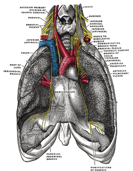
The phrenic nerve originates from the anterior rami of C3 through C5 and traverses the neck, heart, and lungs to reach the diaphragm. From its origin, the phrenic nerve descends vertically caudad and adjacent to the internal jugular vein. In the neck and upper thorax, the left phrenic nerve tracts proximal to the subclavian artery. The right phrenic nerve runs superficial to the anterior scalene muscle and the second part of the right subclavian artery. In the thorax, the right and left phrenic nerve will continue to descend anteriorly to the root of the lung and between the mediastinal surface of the parietal pleura and fibrous pericardium. The right phrenic nerve passes lateral to the right atrium and right ventricle and will continue to descend through the vena cava hiatus in the diaphragmatic opening at the level of T8. The left phrenic nerve descends anterior to the pericardial sac of the left ventricle and terminates at the central tendon of the diaphragm.[1][2][3]

Structure and Function

The phrenic nerve originates from the anterior rami of the C3 through C5 nerve roots and consists of motor, sensory, and sympathetic nerve fibers. It provides complete motor innervation to the diaphragm and sensation to the central tendon aspect of the diaphragm. The left phrenic nerve innervates the left diaphragmatic dome, and the right phrenic nerve innervates the right diaphragmatic dome, with the majority of nerve branching occurring on the inferior aspect of the diaphragm. The motor innervation activation will cause the diaphragm to contract with inspiration, resulting in a flattened diaphragm and increased intrapleural space. During exhalation, the diaphragm relaxes and returns to the dual dome shape. The phrenic nerve also provides touch and pain sensory innervation to the mediastinal pleura and the pericardium in addition to the intercostal nerves.

Embryology

The phrenic nerve is a peripheral nerve that originates from the neural crest cells and is derived from the neural plate. Neurulation, or the development of the neural plate, begins after the third week of fertilization. At weeks 5 through 6, the septum transversum, forming the thoracic diaphragm, descends from the cervical vertebrae to the thoracolumbar vertebrae. The phrenic nerve descends along with the septum transversum, carrying innervation from the ventral rami from C3 through C5.

Blood Supply and Lymphatics

The phrenic nerve is accompanied by the pericardiophrenic artery and superior phrenic vein throughout its course. The nerve, artery, and vein originate at the neck root and descend, parallel, along the lateral aspects of the pericardial sac, ultimately terminating at the superior aspect of the diaphragm. The pericardiophrenic artery is a branch of the internal thoracic artery. The superior phrenic vein drains into the azygos vein on the right and left.

Muscles

The phrenic nerves provide motor innervation to the diaphragm and work in conjunction with secondary respiratory muscles (trapezius, pectoralis major, pectoralis minor, sternocleidomastoid, and intercostals) to allow respiration.

The accessory phrenic nerve, if present, may provide motor innervation to the subclavius muscle. The subclavius muscle originates at the costochondral junction of the first rib and inserts at the subclavian groove of the clavicle. The subclavius muscle stabilizes the clavicle.

Physiologic Variants

In a small number of people, there may be an accessory branch of the phrenic nerve. An accessory phrenic nerve will follow the true phrenic nerve down its course to the diaphragm but often terminates at the pericardium. This variation will be located laterally and posteriorly to the main phrenic nerve and anteriorly to the subclavian vein. This variant is mostly C5 contribution and will branch off proximally at the root of the neck to provide motor innervation to the subclavius muscle.

Surgical Considerations

The phrenic nerve originates at the C3 through C5 nerve roots, which exits the spinal canal at the neck root and descends caudally, parallel to the pericardial sac to provide the motor innervation to the diaphragm. The phrenic nerve must be identified in cervical and thoracic surgical dissection to preserve the neuromuscular pathways. Superior to the clavicle, the phrenic nerve can be located in the posterior triangle of the neck, superficial to the anterior scalene. In the mediastinum, the phrenic nerves can be identified with lateral retraction of the lungs to reveal the pericardial sac where the phrenic nerves pass anteriorly to the root of the lung. The phrenic nerves are located on the lateral aspects with accompanying pericardiophrenic arteries and superior phrenic veins.

Lesions to either the left or right phrenic nerve will cause relative elevation of the ipsilateral dome of the diaphragm, ultimately paralyzing it from contraction and depression during inspiration. The right phrenic nerve is at risk of being severed with vena cava clamping as the phrenic nerve enters the caval diaphragmatic opening with the inferior vena cava at T8.[4][5][6]

Clinical Significance

The phrenic nerve supplies sensory innervation to the diaphragm. Pain arising from the diaphragm is often referred to the tip of the shoulder, also known as the Kehr sign. For example, a patient with a subphrenic abscess or a ruptured spleen may complain of pain in the left shoulder. The hiccup reflex is due to irritation of the phrenic nerve. It results from sudden spasms of the diaphragm which pull air against the closed fold of the larynx. The phrenic nerve must be identified during thoracic and open-heart surgery. It may be injured during the taking down of the internal mammary artery, which is used for coronary artery bypass. The phrenic nerve often is injured in infants undergoing congenital heart procedures. Once the phrenic nerve is injured, the diaphragm will become paralyzed. On a chest X-ray, the diaphragm will appear elevated. Ultrasound or fluoroscopy can be used to make the diagnosis of a paralyzed diaphragm. If only one side of the diaphragm is paralyzed, most patients can overcome the deficit and lead normal lives. If both sides are paralyzed, phrenic nerve stimulation, intercostal nerve transfer, or a permanent tracheostomy with ventilation dependence is required. Diaphragmatic plication is sometimes done in symptomatic patients when only one diaphragm is paralyzed. Patients who have spinal cord trauma may be able to breathe despite being paralyzed because the phrenic nerve has a higher origin at C3 through C5.[7][8]


(Click Image to Enlarge)
<p>The Anterior Divisions, The phrenic nerve and its relations with the vagus nerve, Pericardium, Superior vena cava, Ascendi

The Anterior Divisions, The phrenic nerve and its relations with the vagus nerve, Pericardium, Superior vena cava, Ascending aorta, Diaphragm


Henry Vandyke Carter, Public Domain, via Wikimedia Commons

Details

Editor:

John V. Ashurst

Updated:

7/24/2023 9:14:36 PM

References


[1]

Bains KNS, Kashyap S, Lappin SL. Anatomy, Thorax, Diaphragm. StatPearls. 2023 Jan:():     [PubMed PMID: 30137842]


[2]

Mahabadi N, Goizueta AA, Bordoni B. Anatomy, Thorax, Lung Pleura And Mediastinum. StatPearls. 2023 Jan:():     [PubMed PMID: 30085590]


[3]

Chaudhry R, Bordoni B. Anatomy, Thorax, Lungs. StatPearls. 2023 Jan:():     [PubMed PMID: 29262068]


[4]

Graves MJ, Henry BM, Hsieh WC, Sanna B, PĘkala PA, Iwanaga J, Loukas M, Tomaszewski KA. Origin and prevalence of the accessory phrenic nerve: A meta-analysis and clinical appraisal. Clinical anatomy (New York, N.Y.). 2017 Nov:30(8):1077-1082. doi: 10.1002/ca.22956. Epub 2017 Aug 16     [PubMed PMID: 28726261]

Level 1 (high-level) evidence

[5]

Wang J, Li J, Liu G, Deslauriers J. Nerves of the mediastinum. Thoracic surgery clinics. 2011 May:21(2):239-49, ix. doi: 10.1016/j.thorsurg.2011.01.006. Epub     [PubMed PMID: 21477774]


[6]

Khong P, Lazzaro A, Mobbs R. Phrenic nerve stimulation: the Australian experience. Journal of clinical neuroscience : official journal of the Neurosurgical Society of Australasia. 2010 Feb:17(2):205-8. doi: 10.1016/j.jocn.2009.06.012. Epub 2010 Jan 6     [PubMed PMID: 20056422]


[7]

Loukas M, Kinsella CR Jr, Louis RG Jr, Gandhi S, Curry B. Surgical anatomy of the accessory phrenic nerve. The Annals of thoracic surgery. 2006 Nov:82(5):1870-5     [PubMed PMID: 17062263]


[8]

Morgan JA, Morales DL, John R, Ginsburg ME, Kherani AR, Vigilance DW, Cheema FH, Smith CR Jr, Oz MC, Argenziano M. Endoscopic, robotically assisted implantation of phrenic pacemakers. The Journal of thoracic and cardiovascular surgery. 2003 Aug:126(2):582-3     [PubMed PMID: 12928662]