Introduction
The ability to perceive accurate and specific afferent sensory information from the external world is vitally important to human function, and there is a range of peripheral nerve fibers that perform this role. These specialized peripheral nerve fibers are classified into four subclasses according to their fiber diameter, conduction velocity, and extent of myelination. C-type fibers are unmyelinated fibers that form one of these groups, and they are involved in the afferent transfer of temperature, burning pain, and itch from the periphery to synapse, principally in lamina I and II of the dorsal spinal horn.
Structure and Function
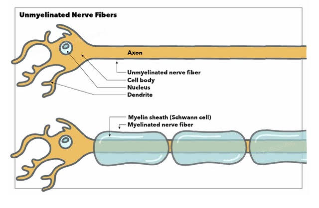
Peripheral nervous system axons, which relay action potentials elicited by various stimuli, can be myelinated to a differing degree by specialized Schwann cells that allow them to conduct the electrical signal ten times faster than unmyelinated fibers of the same diameter.[1] Despite the signal transduction advantages of myelinated axons, a large and important group of unmyelinated nerve fibers exist throughout the peripheral nervous system. This group of fibers is termed C-fibers and is further divided into functional subgroups based on the type of sensory information they relay.
Although they completely lack the myelin envelope, Schwann cells surround groups of unmyelinated C-fibers, creating an ultrastructure in which groups of C-fibers are bound together. These so-called Remak bundles vary in size and distribution between different anatomical sites, with L5 dorsal root ganglion bundles containing many more C-fibers than bundles in distal nerves, for example.[2]
Junction adhesion molecule 2 (JAM2) expression promotes local myelination inhibition, giving insight into how unmyelinated axons form in the nervous system. These contribute to the galectin-4 (gal-4), a galectin specifically sorted to axon membrane segments in a sulfatide-dependent process, forming the long, unmyelinated, discontinued segments along the axons, including the somatodendritic membrane.[3]
Unmyelinated fibers are widely distributed and are found in both hairy and glabrous skin. Mechanoafferent C tactile fibers are found in hairy skin and associated with hair follicles. These are also present in the glabrous skin of the glans penis and glans clitoris. C-fibers have greater innervation density compared to Type A-delta fibers. Torebjörk and Hallin introduced the concept of the nociceptive field of microneurography in 1970. This process produced a "marking" technique to discriminate single unit impulses by unmyelinated nerve fibers, including putative itch afferents and sympathetic efferents, based on a post-activation transmission delay of approximately several seconds vs. preceding impulses.[4]
C fibers are small-diameter fibers acting as nociceptors from the periphery to the central nervous system (CNS). The diameter of their axons ranges from 0.2 micrometers to 1.2 micrometers and can reach up to 3 micrometers in some cases.[5][6]
Conduction Properties of C-fibers
Unmyelinated C tactile fibers are slowly conducting. Their optimal response is through slow stroking stimulation with a velocity of 1 to 10cm/s (approximately 1 m/s). This signal produces the less well-localized, burning "second" pain after the Ad-fiber-related (approximately 15 m/s) pricking "first" pain.[6]
In unmyelinated fibers, conduction velocity is proportional to the square root of axonal diameter, which is proportional to the square root of channel density. This square root of channel density approximates the longitudinal number of sodium ion channels across the axon. On the other hand, conduction velocity approximates axonal diameter (not square root) in myelinated fiber sheathes. However, myelin in very thin fibers does not increase the conduction velocity.[7][5]
This conduction velocity equation is based on the new conductive model discussed in the study by Akaishi and colleagues (2017).[5] The differences in intracellular ionic concentration gradients around several ion (voltage-gated sodium, NaV) channels cause membrane charges across an axon. This ionic migration and conduction propagation, by increasing the Coulomb force between electrolytes from the inflowing sodium ions through voltage-gated sodium channels, generate the action potential at each channel. This increasing Coulomb force is inversely proportional to the sodium channel density on the axon membrane. Continuous transmission of this action potential thus causes nerve conduction in unmyelinated axons.
Categories of Unmyelinated Nerve Fibers
Most nociceptive afferents are unmyelinated C fibers. Four subclasses of C fiber unmyelinated nociceptive sensory afferents are based on responsiveness to mechanical and temperature stimuli. The majority include polymodal afferents. Others are mechanosensitive afferents and heat-sensitive afferents. Lastly, silent afferents do not respond to either mechanical or heat stimuli but become sensitized by inflammatory skin processes.[8]
Unmyelinated fibers transmit cutaneous and visceral peripheral signals to the CNS, processing important sensory and autonomic information; this is significant for good skin integrity, preventing pressure ulcers and accidental injuries. For example, the fibular nerve has 73% afferent (sensory) and 27% efferent (sympathetic) unmyelinated fibers.[9]
Multiple studies review the classification of C mechanoreceptors based on receptive and reflex properties of axonal conduction latency, slowing to repetitive electrical stimulation. One study classified C-mechanoreceptors into C tactile afferents (CT) and C-mechanosensitive nociceptors (CM). CT has less latency slowing than CM due to ionic channel (NaV) differences. Another study categorized C-mechanoreceptors into three main classes of C fibers. First, efferent sympathetic fibers (Symp). Next is afferent mechano-responsive fibers (CM) responsive to both mechanical and heat pain stimuli threshold and for temporal and spatial resolution of thermal pain. These fibers exhibit axonal conduction latency slowing during repetitive electrical stimulation of the skin. The third class, the afferent mechano-insensitive fibers (CMi), is not responsive to mechanical and heat but responsible for neurogenic inflammation (e.g., hyperalgesia) with axon reflex flare.[10][11]
Sensation
Unmyelinated afferent C fibers are the oldest peripheral component of the somatosensory system, responding to noxious stimuli. They are nociceptors, allowing feedback to CNS, and their spatial location across the body surface is crucial for motor defense. Their inputs are processed with a gross, functionally distinct somatotopic organization of nociceptive projections in the CNS, including in the somatosensory and multimodal cortical areas. Unmyelinated fibers from the dorsal root ganglion also mediate itch.[12][13]
Activation of mTORC1 signaling (active and phosphorylated) mediates protein translation in a small population of C fiber sensory fibers found in the skin and dorsal root, especially in response to pain. This activation takes place by deleting its negative regulator, Tsc2, resulting in an increase in the cell body and axon diameter of C fibers. Also, Tsc2 deletion resulted in a decrease in peptidergic nociceptors with an increase of nonpeptidergic nociceptors (IB4-positive neurons) and caused Cre expression (Cre recombinase expression) predominantly in C-nociceptors. This change reflects reduced noxious heat sensitivity and cold hypersensitivity.[14]
Touch and Pain
Unmyelinated mechanoafferent C tactile fibers in hairy skin mediate gentle touch by responding to slow, gentle, moving stroking (1 to 10 cm/s), light force, and temperatures approximately 32 degrees C, but not for tickle sensation. It reflects a comforting, protective interpersonal touch defined in the "social touch hypothesis." This hypothesis correlates low threshold (low mechanical indentation forces less than 5 mN) mechanosensitive C tactile fibers specifically to the positive affective, subjective pleasantness of human touch. The transmission of mechanical stimuli is from multiple interneurons, synapses, and connections within the dorsal horn expressing vesicular glutamate transporter VGLUT3.
The signals are then sent to the somatosensory system and affect processing brain areas, including the contralateral posterior insular cortex or the medial prefrontal cortex. High ratings on pleasantness by C tactile signaling transmitted to the pregenual anterior cingulate cortex and low ratings on intensity (weak) by A beta (Aß) fibers encoded to the primary and secondary somatosensory cortex (S1 contralateral, S2 bilateral) are observed and known as the peripheral neural mechanisms in erotic touch sensation. These fibers also transmit signals to reward-processing brain areas (putamen and orbitofrontal cortex) and social stimuli processing (posterior superior temporal sulcus).[15][16][17]
Heat and Pain
Unmyelinated C fibers have thermal and pain sensations. They can be slowly adapting or quickly adapting thermonociceptors. The basis of this classification is on cortical activity during EEG "frequency tagging" upon sustained ultraslow (0.2 Hz) and long-lasting (75s) sinusoidal activation of these fibers with heat stimulation of the skin. This process is assumed to trigger periodic activity within higher-order neurons processing this thermonociceptive input. Slow-adapting thermonociceptors respond gradually after sustained heat stimulus onset and do not (or minimally) adapt when heat stimulus remains sustained over time.
For example, the maximum response of these thermonociceptors approaches 1 second (s) after heat onset adapting slowly to a stable level after 10s. They have a response latency shorter than the response latency of A-delta fibers. On the other hand, quickly adapting thermonociceptors respond immediately and adapt rapidly following onset and sustained heat stimulus over time. This modification is significant where slowly adapting fibers sensitize more than quickly adapting nerve fibers after mild burn injury. Another study showed that slow, passive heat targeted to deep skin after intermittent contact using a thermode creates a high temporal summation of unmyelinated fibers.[8][18]
Clinical Significance
Chronic Pain
A reduction of pain perception (analgesic effect) is recorded as reduced noxious-evoked brain activity in electroencephalography (EEG) in infants after stroking (at 3 cm/s), possibly due to inhibited nociceptive C fiber input. This emphasizes the impact of social touch in which parents stroke their babies instinctively at optimal velocity is crucial for bonding. The mechanoafferent tactile fibers cause spinal inhibition of nociceptive neurons, even for heat pain perception. Also, activation of A-delta nociceptors produces spinal and cortical inhibition to C nociception. C tactile fibers also have implications in mechanical and cold allodynia and pain gating; this could be an area of research for non-pharmacological interventions, even in early life.[17][18][10]
Fibromyalgia is a chronic pain syndrome affecting widespread musculoskeletal areas, presenting significant treatment challenges due to its overlap with musculoskeletal and psychological pain etiologies and resistance to conventional pharmacological therapies.[19] Studies have identified that small fiber pathology (i.e., C-fibers) have a significant role in over 50% of patients with fibromyalgia and, therefore, represents a significant unmet therapeutic potential for this condition.[20] The extent to which interventions to address small fiber pathology will benefit all patients with fibromyalgia, or whether this, in fact, represents a distinct subgroup of patients with this condition, remains to be seen and will need to be elucidated with future experimental evidence.
Nerve Injury
Myelination is a crucial positive factor in determining the regeneration and repair of injured nerve fibers. It enables the afferent axons to regenerate into the peripheral nerve stump along the Schwann cell tubes. On the other hand, unmyelinated axons have lesser regenerative potential. Following injury, half of the regenerated unmyelinated afferent axons have chronic electrically hyperexcitable, continuous discharge properties, abnormal mechanosensitivity, and thermosensitivity. These continuous discharges in afferent C fibers may have originated from the injured axons as their physiologic stimuli and not from cell bodies of dorsal root ganglia. The regeneration is inefficient in that only one-third of unmyelinated afferents regenerate to reach the skin compared to almost two-thirds in A-fibers. The rest of the unmyelinated fibers die by 80 days after the cross-union. This could provide significant insight into the crucial role of injured axons (especially injured muscular afferents) as peripheral components of neuropathic pain and peripheral nerve injury for possible therapeutic targets.[21]
Sexual Stimulation
C tactile stimulation at a frequency appropriate for C-fiber stimulation, plus appropriate social context, was demonstrated to increase somatosensory sexual feelings and possible erotic perception.[22] Thus C-fibers may play a crucial role in the nervous transmission of peripheral sexual stimuli to higher cortical sexual arousal areas.