Continuing Education Activity
Juvenile idiopathic arthritis (JIA), a heterogeneous group of chronic arthritis, is the most common chronic rheumatological condition in children. There are seven different JIA subtypes with distinct phenotypes, genetic predispositions, pathophysiology, laboratory findings, disease course, and prognosis. This activity reviews the evaluation and management of JIA and reviews the role of the healthcare team in evaluating and treating patients with this condition.
Objectives:
- Describe the classic presentation and typical physical exam findings associated with juvenile idiopathic arthritis.
- Explain the imaging findings associated with JIA and the most common tests for the evaluation of juvenile idiopathic arthritis.
- Outline the imaging findings associated with JIA and the most common tests for the evaluation of juvenile idiopathic arthritis.
- Review the importance of collaboration amongst the interdisciplinary team to improve outcomes for patients affected by juvenile idiopathic arthritis.
Introduction
Juvenile idiopathic arthritis (JIA) is a heterogeneous group of idiopathic inflammatory arthritis affecting children younger than 16 years of age and lasting six weeks or longer. The terminology of chronic arthritis in children has evolved from juvenile chronic arthritis (JCA) and juvenile rheumatoid arthritis (JRA) to JIA since 1995. According to the consensus conference of the International League of Associations for Rheumatology (ILAR) in 2001, there are seven JIA categories: a) oligoarthritis; b) rheumatoid factor (RF) positive polyarthritis; c) RF negative polyarthritis; d) systemic arthritis; e) psoriatic arthritis; f) enthesitis-related arthritis; g) undifferentiated arthritis.[1] These subtypes have distinct phenotypes, genetic predispositions, pathophysiology, laboratory findings, disease course, and prognosis. Although chronic arthritis is mandatory for all subtypes, the extraarticular and the systemic manifestations characterized every specific subtype. Recently, a new preliminary data-driven classification for JIA is proposed and being formally validated by the Pediatric Rheumatology International Trial Organization (PRINTO).[2]
Etiology
The cause and trigger of chronic arthritis in JIA remain unclear. Abnormal immune responses triggered by the interactions between environmental factors in a genetically susceptible individual are speculative. Some environmental factors such as antibiotic exposure and C-section deliveries are potential risks; however, breastfeeding and household siblings are possible protectives.[3] The roles of microorganisms such as Parvovirus B19, Epstein-Barr virus, enteric bacteria, Chlamydophila pneumoniae, and streptococcal infections are still inconclusive.[4]
Based on familial aggregation studies and the concordance rate of 25% to 40% in monozygotic twins, genetic factors play a significant role.[5] Specific HLA alleles and non-HLA genes may be vulnerable to particular JIA subtypes and uveitis.[6] HLA-A2, HLA-DRB1:11, and HLA-DRB1:08 are associated with oligoarticular JIA and RF negative polyarticular JIA. HLADRB1:01 and HLADRB1:04 are associated with RF-positive polyarthritis. HLADRB1:04 and DRB1:11 are associated with systemic JIA. HLA-B27:05 and HLA-B27:04 are the most common HLA-B27 subtypes associated with ERA.[7] HLADRB1:01 and DQA1:01 are related to psoriatic JIA. HLADRB1:11 and HLADRB1:13 are associated with uveitis. The higher number of the HLA-DR risk alleles predisposes to the earlier development of JIA.[8]
Epidemiology
JIA is the most common rheumatic disease reported in children of the Western world. The incidence and prevalence are varied among 1.6 to 23 new cases for 100000 children, and 3.8 to 400 cases per 100000 children depending upon study designs, disease categories, and geographical areas.[9] In a US and Canada study, the incidence of JIA is 0.041 to 0.061 per 1000 children.[10] The Utah Population Database provides the prevalence of 1.2 per 1000 in white populations.[6] The relative risk of JIA in siblings varies from 15 to 30, similar to the relative risk of type 1 diabetes.[6]
The frequencies of different subtypes are 50% to 60% for oligoarthritis, 11% to 28% for RF negative polyarthritis, 2% to 7% for RF positive polyarthritis, 10% to 20% for systemic arthritis, 2% to 15% for psoriatic arthritis, 1% to 7% for enthesitis-related arthritis.[11] Specific subtypes are more common in some geographical regions. RF-negative polyarthritis is more common in North America; oligoarthritis is more common in southern Europe. Systemic arthritis and enthesitis-related arthritis are more common in southeast Asia. Uveitis is highest in northern Europe and southern Europe, but it is lowest in Latin America, Africa, the Middle East, and Southeast Asia.[12] Most JIA subtypes occur predominantly in females except enthesitis-related arthritis mainly affects males, and systemic JIA affects males and females equally.[13]
Pathophysiology
The imbalance of regulatory T cells, Th1 (IFN-gamma secreting T cells), and Th17 (interleukin -17 secreting T cells) of adaptive immunity is the feature of most subtypes of JIA. IL-17 induces proinflammatory cytokines and matrix metalloproteinases, leading to joint damage in oligoarthritis, polyarthritis, and psoriatic arthritis.[14][15] In ERA, IL-23 is a crucial cytokine that leads to inflammation through IL-17 and tumor necrosis factor (TNF), and new bone formation through IL-22.[16] In contrast, the critical immunopathophysiology of systemic arthritis is persistent activation of innate immunity, including monocytes[17], macrophages, and neutrophils.[18] As a result, innate proinflammatory cytokines such as IL-1 beta, IL-6, and IL-18 contribute to symptoms and signs of systemic arthritis.[19]
Histopathology
The synovial membrane of JIA shows a high degree of infiltrating inflammatory cells, including T and B lymphocytes, plasma cells, macrophages, and dendritic cells, villous hypertrophy, and hyperplasia, hyperplasia of synoviocytes, endothelial hyperplasia, and activation, and increased vascularisation with hyperemia.[20][10] The findings are non-specific and similar to rheumatoid arthritis in adults.
History and Physical
The disease course of JIA is highly unpredictable: in some patients is observed a self-limiting disease while in others, there is an unremitting disease with a high risk of joint destruction.[21]
The JIA has the general pattern of inflammatory joint disease (synovitis, joint effusion, soft tissue swelling, osteopenia, bone edema, and erosions) with some additional elements related to developmental age, such as epiphyseal growth disturbances, premature physeal fusion, and limb length inequality.[22]
The thorough history taking, including the age of onset, the affected joints, the duration of arthritis, the associated symptoms or diseases, and physical and MSK examinations are essential for diagnosis and classification of JIA.
A diagnosis of JIA is considered in any children younger than 16 years with arthritis for at least six weeks and exclusion of other causes of chronic arthritis.
Evaluation
There is no specific test for diagnosis and predicting disease activity in JIA.
Laboratory
Initial laboratory tests should include CBC, ESR, CRP, ANA, RF, anti-cyclic citrullinated peptide antibodies (anti-CCP), and HLA-B27. The typical inflammatory markers are common, especially in oligoarthritis. A positive RF or anti-CCP provides little value for the diagnosis but may indicate a poorer disease course and outcome. Ferritin, fibrinogen, AST, triglyceride are recommended when there is a concern of macrophage activation syndrome (MAS). Other tests for excluding other diseases may be considerations depending on the differential diagnosis.
Imaging
Imaging serves to improve the certainty of a diagnosis of JIA, narrow the differential diagnosis, and evaluate joint damage. The heterogeneity of clinical manifestations usually requires a multimodal imaging approach.
Radiography remains initial imaging used for symptomatic joints; however, the radiographic changes are undetectable in an early stage of JIA. The indirect signs of arthritis in radiography are soft tissue swelling, increased density of soft tissue as well as dislocation of fat folds. Other features are periarticular osteoporosis, joint space narrowing, bone erosion and deformity, and joint subluxation or ankyloses in an advanced stage.
Ultrasound (US) is an imaging modality easily accessible and non-irradiating that plays a significant role in the imaging of JIA. Also, the US allows the comparison with the contralateral side and the dynamic evaluation of joints. The US is capable of assessing synovial thickening, joint effusion, tenosynovitis, enthesitis, and bone erosions. US evaluation of the synovial thickening and the synovitis is particularly important for the diagnosis. They appear as abnormally hypoechoic tissue associated with joint lines or surrounding tendons. In addition to detecting synovitis, the US accurately guides for intra-articular corticosteroid injections. Ultrasound allows evaluation without sedating the patient.
Magnetic resonance imaging (MRI) is the modality gold standard for the study of JIA. All joints affected by pathological inflammatory phenomena can be easily examined in all possible plans and with an excellent contrast resolution of bone and soft tissues. It is the most sensitive imaging technique detecting synovitis. The standard MR imaging protocol needs to include: T1 spin-echo (SE) sequence, fat-suppressed sequence (classic T2 fat-sat; short tau inversion recovery - STIR; DIXON fat-suppression sequence); T1 fat-suppressed sequence precontrast and postcontrast. MRI is the only modality able to objective bone marrow edema and the most sensitive to detect bone erosions.[22]
Clinical-Laboratory Classification
The clinical manifestations and the test results of RF and HLA-B27 will be used to categorized JIA subtypes based upon the ILAR classification
Oligoarthritis is defined as chronic arthritis affecting four joints or less during the first six months of disease. Persistent oligoarthritis is defined as the affected joints being four or less after the first six months, and extended oligoarthritis is defined as more than four affected joints after the first six months.
RF negative polyarthritis is defined as arthritis affecting five joints or more during the first six months of disease with a negative IgM RF.
RF positive polyarthritis is arthritis affecting five joints or more during the first six months of disease with a positive IgM RF on at least two tests three months apart.Systemic arthritis is arthritis with or preceded by a fever of at least a 2-week duration and accompanied by at least 1 of the following: evanescent erythematous rash, generalized lymph node enlargement, hepatomegaly, and/or splenomegaly, or serositis (pericarditis and/or pleuritis and/or peritonitis).Psoriatic arthritis is defined as chronic arthritis with psoriasis or chronic arthritis with at least 2 of the following: dactylitis, nail pitting, onycholysis, or psoriasis in a first-degree relative.Enthesitis related arthritis (ERA) is defined as arthritis with enthesitis, or arthritis or enthesitis with at least 2 of SI joint tenderness and/or inflammatory lumbosacral pain, a positive HLA-B27, the onset of arthritis in a male over six years of age, acute anterior uveitis, history of ankylosing spondylitis, ERA, sacroiliitis with inflammatory bowel disease, reactive arthritis or acute anterior uveitis in a first-degree relativeUndifferentiated arthritis is defined as chronic arthritis, which does not fulfill criteria in any subtype or fulfills two or more subtypes.
Treatment / Management
Treatment of JIA requires anti-inflammatory and immunomodulatory drugs and physical therapy, and eventually, surgery, nutritional support, and psychosocial support may be needed. The choice of pharmacological treatment depends on the disease subtypes, disease severity and damage, associated disease, and family acceptance. Nonsteroidal anti-inflammatory drugs (NSAIDs) are the mainstay of initial symptomatic treatment for all subtypes. The NSAID use in JIA has decreased over time with modern aggressive treatment, including methotrexate and biologics.
Physical therapy emphasizes range of motion with minimal stress on joints. Swimming is often a good option. Patients should participate in moderate fitness, flexibility, and strengthening exercises.
Differential Diagnosis
The differential diagnosis of chronic arthritis is broad, depending on the clinical presentation and the JIA subtypes. Since JIA is a diagnosis of exclusion, any patients with a positive review of the system should be considered for possible diseases.
The following differential diagnosis should be considered:
A) Oligoarthritis need to exclude post-streptococcal reactive arthritis, Lyme arthritis, acute rheumatic fever, reactive arthritis, toxic synovitis, septic arthritis, pyomyositis, steroid-induced osteonecrosis, sickle cell disease, hemophilia, scurvy, osteomyelitis, chronic nonbacterial osteomyelitis (CNO), sports injury, non-accidental injury, pigmented villonodular synovitis (PVNS), osteoid osteoma, bone tumors, neuroblastoma, leukemia, and lymphoma.
B) Polyarthritis need to exclude post-streptococcal reactive arthritis, Lyme arthritis, acute rheumatic fever, reactive arthritis, scurvy, chronic nonbacterial osteomyelitis (CNO) or chronic recurrent multifocal osteomyelitis, non-accidental injury, systemic lupus erythematosus (SLE), Mixed connective tissue disease (MCTD), Sjögren syndrome, scleroderma, sarcoidosis, Blau syndrome, arthritis associated with inflammatory bowel diseases, Farber disease, benign hypermobility joint syndrome and amplified musculoskeletal pain syndrome
C) Systemic arthritis need to exclude infections (mycoplasma, cat scratch disease, bacterial endocarditis, Lyme disease), acute rheumatic fever, syndrome of periodic fever, aphthous stomatitis, pharyngitis and cervical adenitis (PFAPA syndrome), autoinflammatory syndromes, systemic vasculitis (polyarteritis nodosa, Kawasaki disease), inflammatory bowel disease, malignancy (leukemia, lymphoma, neuroblastoma), Castleman disease
D) Enthesitis-related arthritis. Apophysitis (especially Osgood-Schlatter, Sever disease), inflammatory bowel diseases (IBD), chronic recurrent multifocal osteomyelitis (CRMO), amplified musculoskeletal pain syndrome.
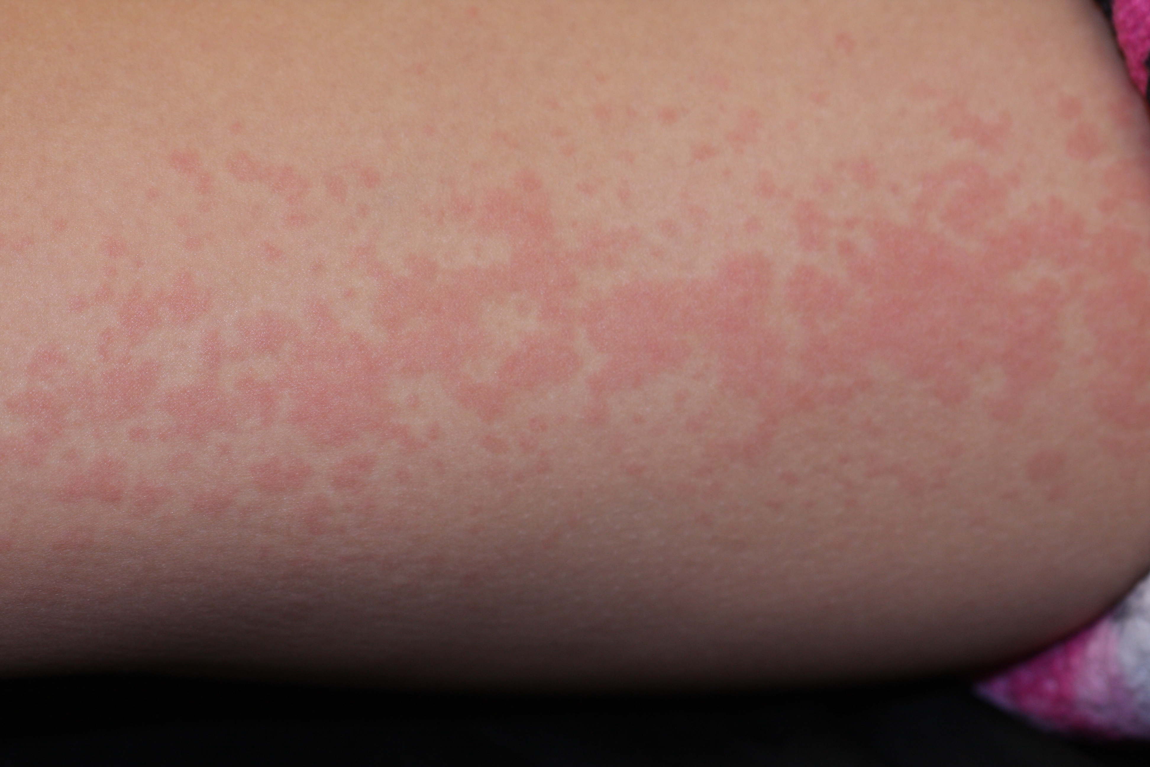
Arthralgias are common early in the course of sJIA, but arthritis is not always prominent. Any number of joints may be involved when arthritis becomes apparent. Disease in the wrists, knees, and ankles is most typical. Hands, hips, cervical spine, and temporomandibular joints are also sometimes affected, unlike the oligoarticular and polyarticular subtypes of JIA
Arthralgias are common early in the course of sJIA, but arthritis is not always prominent. Any number of joints may be involved when arthritis becomes apparent. Disease in the wrists, knees, and ankles is most typical, but the hands, hips, cervical spine, and temporomandibular joints are also sometimes affected. Unlike the oligoarticular and polyarticular subtypes of JIA
MSK complaints in children with joint pain are common in general practice and increase significantly after ten years of age in general practice.[23]
The most common presentation of musculoskeletal complaints is joint pain and soft tissue pain (65%), and the most common cause of all ages of children is trauma (45%).[24]
Toxicity and Adverse Effect Management
Nonsteroidal anti-inflammatory drugs (NSAIDs) inhibit cyclooxygenases, a necessary enzyme for prostaglandin synthesis. Physiologic roles of prostaglandins are initiating and maintenance of the protective mucosal barrier of the stomach and promoting intrarenal plasma flow and electrolyte balance. The potential gastrointestinal (GI) toxicities and nephrotoxicity should be addressed with the family. A survey of pediatric rheumatologists’ experience reveals significantly more abdominal pain, easy bruising, epistaxis, headaches, and fatigue for nonselective NSAIDs than for selective COX-2 NSAIDs. However, a study in safety profile, including GI toxicity between nonselective NSAIDs (COX1 and COX2 inhibitors) and selective COX-2 inhibitor (celecoxib) is no different. Since over 50% of children with JIA developed of GI symptoms related to NSAID, corticosteroid, and methotrexate use, reviewing the medical history and previous or concurrent use of NSAIDs, corticosteroid or methotrexate is necessary to assess and minimize the potential risk of gastrointestinal toxicity. The potential nephropathy, such as acute interstitial nephritis or acute papillary necrosis, was rarely reported. The prevalence of nephrotoxicity in children is 0.4%, which is five times less than the prevalence in adults. The benefit of routine monitoring (blood and urine) of asymptomatic children with JIA receiving only NSAIDs is unclear.
Prognosis
The prognosis of JIA has changed dramatically in recent years thanks to the availability of novel drugs, which can inhibit the biological mechanisms responsible for persistent inflammation selectively. Prompt and accurate diagnosis and treatment are essential to prevent permanent joint damage and preserve joint functionality. Some studies support the possibility of the existence of a “window of opportunity” in early disease, during which prompt treatment induces higher rates of remission and improves long-term outcomes.[21]
A recent study on 168 patients showed the remission off medication in 48.8% of cases, the remission on medication (or minimal disease activity) in 49.9% of cases, and only 1.3% of subjects were no-responders. No association was found between the state and duration of remission and age of patients, clinical features, disease course, or laboratory findings.[25]
Complications
The most common complications of JIA are leg-length discrepancy and joint contracture. An extremely fearsome complication is macrophage activation syndrome, due to the uncontrolled activation and proliferation of T lymphocytes and macrophages. The frequency of this syndrome in patients with JIA is unknown, but some studies report that it occurs in up to 10% of cases.
Other important complications are growth retardation, low bone mineral density for chronologic age, severe hip involvement with the need oh hip prosthesis, and amyloidosis.[25]
Deterrence and Patient Education
A chronic disabling pathology such as JIA requires proper child education and family support. Numerous experiences have been made on this topic. Among the most recent and interesting new studies, there is the use of comics for the education of children and artificial intelligence.[26][27]
Pearls and Other Issues
Any child with a history of persisting limp after a minor trauma should look for other diseases, including JIA and leukemia. The JIA has to be suspected in any children younger than 16 years with arthritis for at least six weeks and exclusion of other causes of chronic arthritis.
Enhancing Healthcare Team Outcomes
A recent study was realized by the collaboration between physicians and patient associations. A questionnaire was proposed to 622 parents in 23 European countries. The questions covered various domains of JIA care, including demographics, diagnosis, referrals to different health care professionals, access to pain and fatigue management and support groups, the information they received about the disease, and awareness of and participation in research.
The data furnished a view on JIA patients’ and their parents’ perspectives regarding the care they receive. It’s essential to increase the awareness of the existence of supportive care, such as pain and fatigue management, as well as support groups, or to start new initiatives where these are lacking.[28] [Level 4].
Pediatric pharmacists review medications and provide patient and family education about the importance of compliance and potential side effects, and report any concerns to the clinician staff. Pediatric and rehabilitation nurses provide direct care and facilitate communication between the interprofessional health team members. This type of collaboration among team members will lead to better outcomes for these patients. [Level 5]