Continuing Education Activity
Pars interarticularis defects also referred to as spondylolysis, are a common cause of axial back pain in adolescents, especially young athletes. The pars interarticularis (pars) lies between the superior and inferior articular process bilaterally at each zygapophyseal joint. A pars interarticularis defect is a unilateral or bilateral fracture involving the pars interarticularis of the posterior vertebral arch. This injury occurs almost exclusively in the lower lumbar region, most often at L5. This activity highlights the interprofessional management of this condition.
Objectives:
- Review typical presenting symptoms of pars interarticularis defects.
- Describe the evaluation of pars interarticularis defects.
- Outline the complications associated with untreated pars interarticularis defects.
- Describe the need for a well-integrated, interprofessional team approach to improve care for patients with pars interarticularis defects.
Introduction
Pars interarticularis defect (otherwise referred to as spondylolysis) represents a common cause of axial back pain in adolescents, especially in the case of young athletes. The pars interarticularis (pars) lies between the superior and inferior articular process bilaterally at each vertebral level. Anatomically, one can describe the pars as the region between two, one superior and one inferior, zygapophyseal joints. The definition of pars interarticularis defect is a unilateral or bilateral overuse or fatigue stress fracture involving the pars interarticularis of the posterior vertebral arch. This injury occurs almost exclusively in the lower lumbar region, most often at L5 [1]. Though history can be suggestive, especially in the case of young athletes involved in higher-risk sport (see below), diagnosis is made radiographically by the presence of fracture through the pars interarticularis. In cases of bilateral pars interarticularis defects, there is the potential for anterior or posterior spondylolisthesis (the slipping of one vertebral body relative to the adjacent segment). Spondylolisthesis can be graded based upon the percent degree of displacement of one vertebral body compared to the other. Grading of spondylolisthesis is included below in “staging.”
Two common clinical presentations of a pars defect include the imaging of an asymptomatic adolescent or adult in whom there is the incidental discovery of a pars defect. The second common presentation is an adolescent athlete involved in a sport requiring repetitive lumbar loading in extension and rotation, presenting with acute or insidious onset low back pain that is aggravated by continued lumbar loading. Although this history is typical, there is a broad differential diagnosis that might explain these symptoms. As such, the diagnosis of a pars interarticularis defect confirmation is only with radiographic support. Depending on the time of presentation and degree of injury, most cases of pars defects respond well to conservative treatment and relative rest from sport.
Etiology
The exact cause is still unclear. Currently, the most accepted theory is repetitive mechanical stress, specifically lumbar extension and rotation, which results in overuse or stress fracture to the pars interarticularis.[2] This theory garners support from the fact that, as noted below in epidemiology, the research observed zero cases of pars defects in 500 newborns and zero cases of pars defects in 143 non-ambulatory patients, suggesting this pathology develops as a result of repetitive axial loading over time.[1][3] Additionally, this theory is supported by the progression of unilateral pars defects into bilateral pars defects with age, again suggesting repetitive axial loading over time, both leading to the initial injury as well as disease progression.[4] As discussed, although generally thought to be the result of chronic repetitive stress to the pars region, these injuries can also occur due to a single acute overload injury.[5]
The pars interarticularis is most susceptible to chronic axial loading injury because it is a weak point in the vertebrae, and this region bears the highest stress load in extension/flexion.[6] The weakness of the pars region is multifactorial, with a hereditary and an acquired mechanical component. Mechanical factors include the physically narrow structure of the pars interarticularis as compared to other regions of the vertebrae. Furthermore, the pars in the lower lumbar vertebra characteristically have uneven trabeculation and cortication. The inherent mechanical flaws of the pars interarticularis in combination with the high-stress loads seen in the lower lumbar region render this region prone to stress fractures.
Additionally, there has been a strong association reported with spina bifida occulta.[1]
Epidemiology
- Overall incidence 4to 6% in adolescents and adults[1]
- Whites 2 to 3x more likely than African Americans[1]
- Males 2 to 3x more likely than females[1]
- Per Roche & Lowe’s study of 4200 cadavers, an overall incidence of 4.2%[7]
- 8% to 15% incidence in asymptomatic adolescent athletes[8]
- 47% incidence in adolescent athletes with concomitant low back pain[9]
- Per Morita et al. study, 185 adolescents less than 19 years old with known spondylolysis, the study found 180 were participating actively in sports[10]
- Per Standaert 2000, L5 level affected 85 to 95%[11]
- Per Standaert 2000, L4 level affected 5 to 15%[11]
- Per Fredrickson 1984, 500 newborn radiographs revealed 0 spondylolysis[1]
- Per Rosenberg 1981, 143 non-ambulatory patients, mostly due to cerebral palsy, spondylolysis[3]
- Per Jackson 1976, 100 female gymnasts 11% incidence of spondylolysis[12]
- Radiographically visualized spondylolysis has an association with spondylolisthesis approximately 25% of the time[13]
- Associated with spina bifida occulta
Pathophysiology
As previously noted, pars interarticularis defects or spondylolysis are generally thought to be the result of mechanical stress. Most commonly, fatigue or stress fractures result from repetitive loading and stress through the weak and high-stress load region of the pars. Extension and rotational loading place particular stress through this region. Though these injuries typically occur as a result of repetitive loading or movements, a single stressor or trauma can result in a pars defect. See “staging” below for details regarding the classification of pars defects. Though history and exam might be suggestive of the diagnosis, imaging ought to be used to confirm and assess severity. Fracture through the pars interarticularis is classically described on oblique radiographs as the “collar sign” on the “Scottie dog.” See the evaluation section below for details.
Histopathology
Sagi et al. demonstrated, via histomorphic analysis, the pars interarticularis begins to ossify at 12 to 13 weeks gestation by endochondral ossification. In the lower lumbar vertebra, the ossification center originates in the pars region resulting in uneven distribution of trabeculation and cortication in this region. As a result of this uneven distribution of isthmic ossification, this region is potentially more susceptible to fatigue fracture. In contrast, in the upper lumbar vertebra, the ossification center arises at the end of the pedicle, which results in more uniform trabeculation throughout the pars.[14]
History and Physical
As previously mentioned, there are two common clinical presentations associated with pars defects. First, imaging of an asymptomatic child or adolescent in whom there is an incidental finding of pars defect. In this case, the physical exam likely will not reveal any findings attributable to the pars defect.[9] Incidental discovery of pars defects may additionally be visible on CT or MRI imaging of the abdomen.
The second common presentation is an adolescent athlete involved in a sport requiring repetitive lumbar loading in extension and rotation, presenting with acute or insidious onset low back pain that is aggravated by continued lumbar loading. The incidence of pars defect in this pediatric athlete population presenting with low back pain is as high as approximately 50%. In light of that, this particular population merits a high index of suspicion followed by an appropriate diagnostic workup.[5]
In the symptomatic patient, classic history will include a child or adolescent athlete playing a sport that requires repetitive lumbar extension and rotation. The onset of pain may be either acute or insidious over several weeks. Patients will report their low back pain increases with strenuous activity or hyperextension and improves with relative rest. Pain typically remains located in the low back with occasional radiation to the buttock and/or proximal lower extremities, while neurologic symptoms such as numbness/tingling in the lower extremities are uncommon.[15]
Physical exam ought to include single-leg hyperextension, otherwise known as the Stork test. The patient should be instructed to stand on one leg while simultaneously hyperextending the low back. A positive single-leg hyperextension test is elicited by the reproduction of pain during this maneuver, typically worse when standing on the leg ipsilateral to the side of the pars defect. This maneuver is the only potentially pathognomonic physical exam finding.[5]
Potential additional physical exam findings include the hyperlordotic lumbar curve and tight hamstrings.[16] If clinically indicated, a complete neurologic exam should is necessary, though isolated pars defects typically present with no neurological findings.
Evaluation
Given the relatively nonspecific physical exam findings and the known high incidence in particular patient populations, if pars defect is suspected, additional workup ought to include appropriate imaging studies. There is no universally accepted imaging standard for the diagnosis of pars defect. Given the high-risk population includes children and adolescents, close attention to radiation exposure is warranted. With that in mind, initial workup generally begins with AP/lateral/oblique plain radiographs of the lumbar spine. AP/lateral radiograph is sufficient to detect most pars defects, though estimates are that 20% require oblique views to appreciate the lesion.[17][11] Dynamic plain radiographs may additionally be indicated when there is a concern for instability in the case of spondylolisthesis.
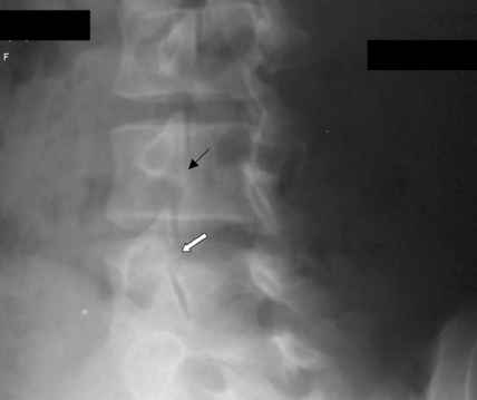
On oblique plain radiographs, the common descriptive term for a fracture to the pars interarticularis is as the “collar” on the “Scottie dog.” (figure 1).[18]
Plain radiographs can miss some lesions, especially acute injury. Other useful imaging modalities include CT, MRI, and bone scan. A bone scan is the most sensitive modality, and best detects early pars defects. This test should be utilized when there is high clinical suspicion for pars lesion, and initial imaging studies are equivocal. After plain radiographs, the imaging modality of choice is either MRI without contrast or CT scan without contrast. CT scan is the best imaging tool for determining fracture size and extent and is the most appropriate modality for follow-up assessment of healing. CT has the downside of additional radiation exposure, which is particularly concerning in the pediatric & adolescent population. Similar to a bone scan, MRI can be useful for early detection of acute lesions by the presence of bone marrow edema on T2 weighted sequences. Additionally, MRI has the benefit of no radiation exposure. MRI is somewhat limited, though, in its ability to adequately depict the cortical integrity of incomplete fractures.[19]
Treatment / Management
There is insufficient evidence to support the natural course or treated pars defect as the preferred management. Management generally reflects the following treatment algorithm.
- Nonoperative[20][21][20]
- Observation without activity limitations
- Indication
- Asymptomatic patients with spondylolysis or low-grade spondylolisthesis
- May participate in contact sports
- Physical therapy plus activity restriction
- Indications
- Symptomatic spondylolysis (type II)
- Symptomatic low-grade spondylolisthesis
- Technique
- Physical therapy program for 6 months and include
- Hamstring stretching
- Pelvic tilts
- Core strengthening
- TLSO bracing for 6 to 12 weeks
- Indications
- Acute pars stress reaction
- Spondylolysis (type II) that has failed to improve with physical therapy
- Low-grade spondylolisthesis that has failed to improve with physical therapy
- Outcomes
- Brace immobilization is superior to activity restriction alone for acute stress reaction
- Operative[22]:
- Indications
- Pars defect that has failed nonoperative management
- Multiple pars defects
- Low-grade spondylolisthesis (Myerding grade I & II) that fails conservative treatment, is progressive, has neurologic deficits, or likely to progress
- High-grade spondylolisthesis (Myerding grade III, IV, V)
- Potential approaches to surgery
- Direct pars interarticularis repair
- Posterolateral fusion
- Bucks fusion of facet joints
Additionally, on the discovery of the condition, vitamin D should be checked, and repleted levels are low.
Differential Diagnosis
- Lumbosacral myofascial pain
- Sacroiliac joint mediated pain
- Facet mediated pain
- Discogenic mediated pain
- Pars stress reaction (pre-spondylolysis)
- Spondylolisthesis
Staging
Pars defects (spondylolysis) subdivide into five categories according to the Wiltse-Newman Classification[22]:
- Dysplastic: congenital abnormalities/attenuated pars (approximately 20%)
- Isthmic: lesions in the pars resulting from a stress fracture or acute fractures (approximately 50%)
- Type II-A: pars fatigue fracture
- Type II-B: pars elongation due to a healed fracture
- Type II-C: pars acute fracture
- Degenerative: degeneration of the intervertebral discs that results in segmental instability and alterations of the articular processes
- Traumatic: an acute fracture that results in fractures to various regions of the neural arch
- Pathological: bone disease such as tumors and infections that result in lesions to the pars
Classification of spondylolisthesis is according to the Meyerding Classification.[22] This is a measure of the percent of antero or retro slippage/transitional displacement of one vertebral body on the vertebral body below observed on lateral radiographs.
- Grade I less than 25%
- Grade II 25 to 50%
- Grade III 50 to 75%
- Grade IV 75 to 100%
- Grade V greater than 100% (spondyloptosis)
Prognosis
The general prognosis for pars interarticularis defects is quite good. The overwhelming majority of cases are asymptomatic,[1] estimated at 80%. The majority of these asymptomatic cases will not lead to a progressive lesion. In the case of symptomatic isthmic pars defect in adolescent patients, roughly 75% to 95% will improve with appropriate conservative management.[10][23] Estimates are that 9% to 15% of symptomatic pars defects will eventually require surgical intervention.[24] Roche and Rowe’s study demonstrated between 20 and 80 years there is no evidence of increased incidence of pars defects, suggesting the highest risk is through adolescents, after which time the bones have fully ossified and risk plateaus.[7] Radiographically visualized spondylolysis is associated with spondylolisthesis approximately 25% of the time.[13]
Seitsalo et al. found that 23% of their 272 children/adolescents demonstrated slip progression greater than 10% at approximately 16 years follow up.[25] Another study, Danielson et al., found 3% of their 311 patients demonstrated slip progression greater than 20% at approximately four years follow up.[26] Patients are likely most vulnerable to slip progression during growth spurts.[27]
Complications
- Neurologic deficits
- Pseudoarthrosis
- Progression of slippage
- Hardware failure
- Chronic pain
Postoperative and Rehabilitation Care
As previously discussed, in cases where physical therapy is warranted, a structured PT program should be carried out for roughly six months. This program should focus on hamstring stretching to offload additional extension forces, core strengthening, and pelvic tilts. Additionally, the PT program should begin with a flexion-based program with slow progression into extension exercises once these are no longer aggravating.
Consultations
Consultation for possible spinal surgery is the recommendation for the cases described above in the “Treatment / Management” section under “II. Operative.”
Deterrence and Patient Education
High-risk sports:
- Gymnasts
- Football linemen
- Weightlifters
- Wrestlers
- Dancers
- Divers
- Volleyball
- Soccer
Pearls and Other Issues
Pars defects are surprisingly common in the pediatric/adolescent population who participate in sports and present with low back pain (as high as 50%). With that in mind, approach these patients with an appropriate index of suspicion and work up with imaging. If initial radiographs are negative (AP/lateral/oblique), follow up with MRI or CT. Early identification correlates with a significantly improved outcome.
Enhancing Healthcare Team Outcomes
Pars interarticularis defects can pose both diagnostic and treatment challenges for healthcare providers. An interprofessional team may improve detection and outcomes. Diagnosis can be challenging because the typical patient is a child or adolescent who the clinician would prefer to spare from radiation exposure. Yet, confirmation of suspected pars defects relies on imaging support. Additionally, imaging, in conjunction with symptoms, should guide management. No universal algorithm for workup exists for this pathology; therefore, workup must be approached in a case by case fashion. Primary care providers initiate an evaluation. Referal to orthopedists and sport medicine physicians is appropriate. Orthopedic nurses are involved with education, monitoring patients, and arrange to follow up. Physical therapists and nurses should report back to the team. Challenges arise when treating the pediatric population; providers would especially like to spare these patients from unnecessary radiation exposure and know that the majority of pars defects can be managed conservatively, so many patients undergo management without a full workup. Generally, when pars defect is high on the differential, regardless of age, appropriate plain radiographs should be obtained. If initial imaging is equivocal, again, regardless of age, imaging should progress to MRI or CT depending on the clinical history and provider preference. Patients and families must be educated on the importance of appropriate workup (including imaging) given outcomes are improved with early detection. [Level 5]