Continuing Education Activity
Eczema, or atopic dermatitis, is a common chronic skin condition that can lead to recurrent infections and poor quality of life if left untreated. Recognized as the "itch that rashes" due to the rash that results from scratching or rubbing, the hallmark of eczema is dry, itchy skin prone to infections. This activity explores the pathogenesis of eczema, acknowledging the intertwined roles of genetic and environmental factors. Learners will gain valuable insights into recognizing eczema across different age brackets, effective evaluation techniques, proactive management of flare-ups, and preventative measures against recurring infections associated with untreated eczema. The course discussion also highlights the role of interprofessional collaboration in improving outcomes for patients with this condition.
Objectives:
Identify the pathophysiology of eczema.
Evaluate the adverse effects of poorly controlled eczema.
Implement appropriate treatment options for eczema.
Communicate the importance of improving care coordination among the interprofessional team to improve outcomes for patients with eczema.
Introduction
Eczema, or atopic dermatitis, is the most common form of dermatitis.[1] Many factors, including genetic and environmental factors, are thought to play a part in the pathogenesis of eczema. It is most commonly seen in children but can be seen in adults as well. People with eczema tend to have dry, itchy skin prone to infection. The condition is commonly known as the "itch that rashes" because dry, itchy skin leads to a rash due to scratching or rubbing the skin.
Etiology
The exact etiology of eczema is not entirely understood, but it is believed to be a combination of genetic and environmental factors.[2]
Genetic Factors
There is a strong genetic component to eczema, with a family history of eczema, asthma, or allergies commonly found in affected individuals. Several genes associated with eczema have been identified, including those involved in the skin barrier function and the immune system.
Filaggrin Gene
One of the most well-known genes associated with eczema is the filaggrin gene (FLG). This gene provides instructions for making a protein called filaggrin, which is important in maintaining the skin barrier function. Mutations in this gene have been linked to eczema and other skin conditions and are thought to increase susceptibility to environmental irritants and allergens.[3][4]
Other Skin Barrier Genes
In addition to the filaggrin gene, other genes involved in the skin barrier function have been implicated in the development of eczema. These include genes involved in lipid synthesis and transport, such as the ceramide synthase gene and the ABCA12 gene.[5]
Immune-related Genes
Several genes involved in the immune response have also been associated with eczema, including genes that regulate T-cells, cytokines, and immunoglobulins. These genes include interleukin (IL)-4, IL-13, IL-31, signal transducer and activator of transduction (STAT)3, and Fc fragment of immunoglobulin (Ig)E receptor Ig (FCER1G).
Overall, the genetic factors involved in eczema are complex and likely involve multiple genes and genetic pathways. Although genetic testing is not routinely used to diagnose eczema, understanding the disease's genetic basis can help identify individuals at increased risk and guide treatment approaches.
Environmental Factors
Environmental factors also play a role in the development of eczema. Patients with eczema have a defect in their skin barrier function, leading to increased water loss and susceptibility to environmental irritants and allergens. Common triggers for eczema flares include exposure to irritants such as detergents, soaps, solvents, and allergens such as dust mites, pet dander, and certain foods. Other factors that can exacerbate eczema symptoms include stress, changes in temperature and humidity, and infections.
Immune System Activation
In addition to genetic and environmental factors, the immune system is also thought to play a role in the development of eczema. Patients with eczema have an overactive immune response to environmental triggers, leading to inflammation and skin damage.
Epidemiology
The lifetime prevalence of eczema is about 15% to 30% in children and 2% to 10% in adults. About 60% of cases will develop the disease within the first year of life. The prevalence of eczema is more common in rural areas than in urban ones. This incidence emphasizes the link between lifestyle and environmental factors in the mechanisms of AD.
Eczema is part of the triad known as "the atopic march." This relates to the association between patients with atopic dermatitis, asthma, and allergic rhinitis. About 50% of patients with severe eczema will develop asthma, and 75% will develop allergic rhinitis.[6]
Pathophysiology
Research shows there is a genetic component to eczema. One common mutation has been observed in FLG, a vital gene for skin cell maturity. This gene is responsible for creating the tough, flat corneocytes that form the outermost protective layer of skin. In a patient with normal skin cells, the corneocytes are tightly packed in an organized manner. A patient with an FLG mutation will have a dysfunctional skin barrier due to the haphazard organization of the skin cells.[7] This dysfunction causes a "leaky" skin barrier, allowing water loss and decreased protection from harmful substances.
People with eczema also have reduced numbers of β-defensins in the skin. β-defensins are host defense peptides vital for fighting off certain bacteria, viruses, and fungi. A decrease in these peptides leads to increased colonization and infection, especially with Staphylococcus aureus (S. aureus).[8]
Histopathology
The histopathology seen in eczema is nonspecific. In the acute-phase lesions, characterized by intensely pruritic, erythematous papules, histopathology reveals mild epidermal hyperplasia, infiltrations of lymphocytes and macrophages along the venous plexus in the dermis, and intercellular edema of the epidermis (spongiosis).
Lesions biopsied in chronic eczema, characterized by lichenification and fibrotic papules, may reveal increased hyperplasia and hyperkeratosis of the skin. There is also persistent dermal inflammatory cell infiltration with lymphocytes and macrophages. The chronic phase lacks the edema or spongiosis that is present in acute-phase lesions.[9]
History and Physical
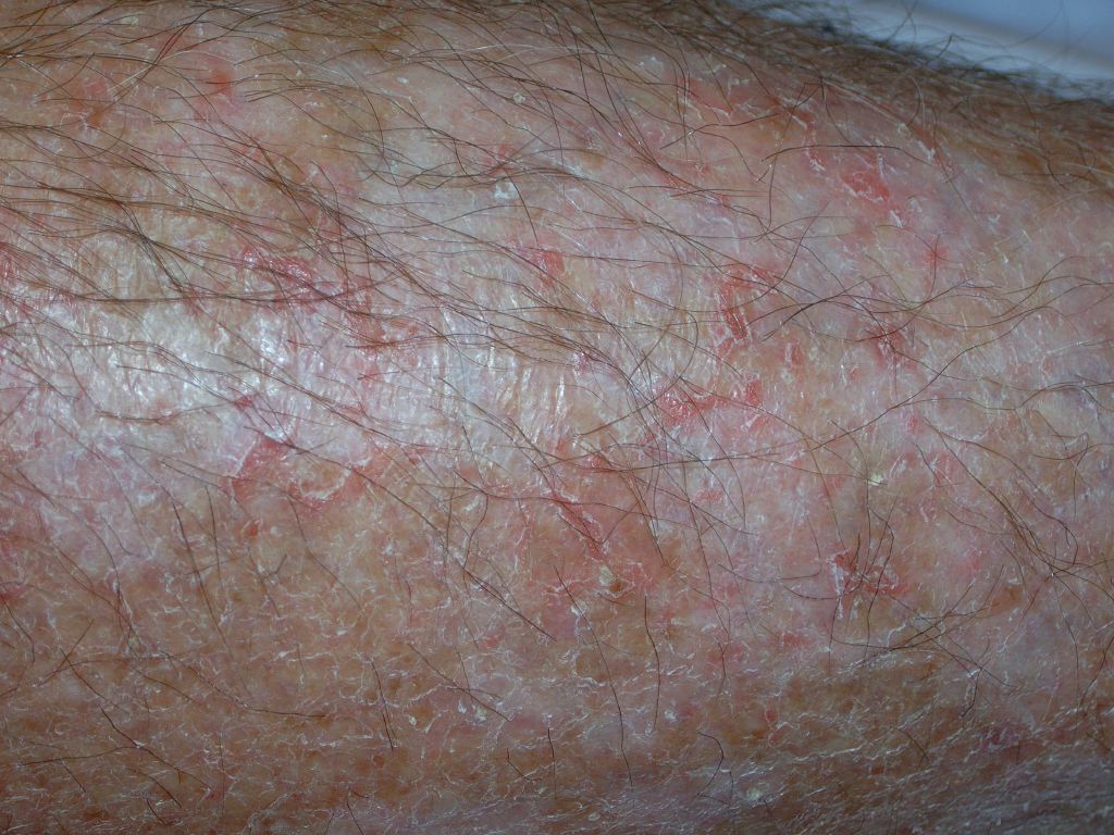
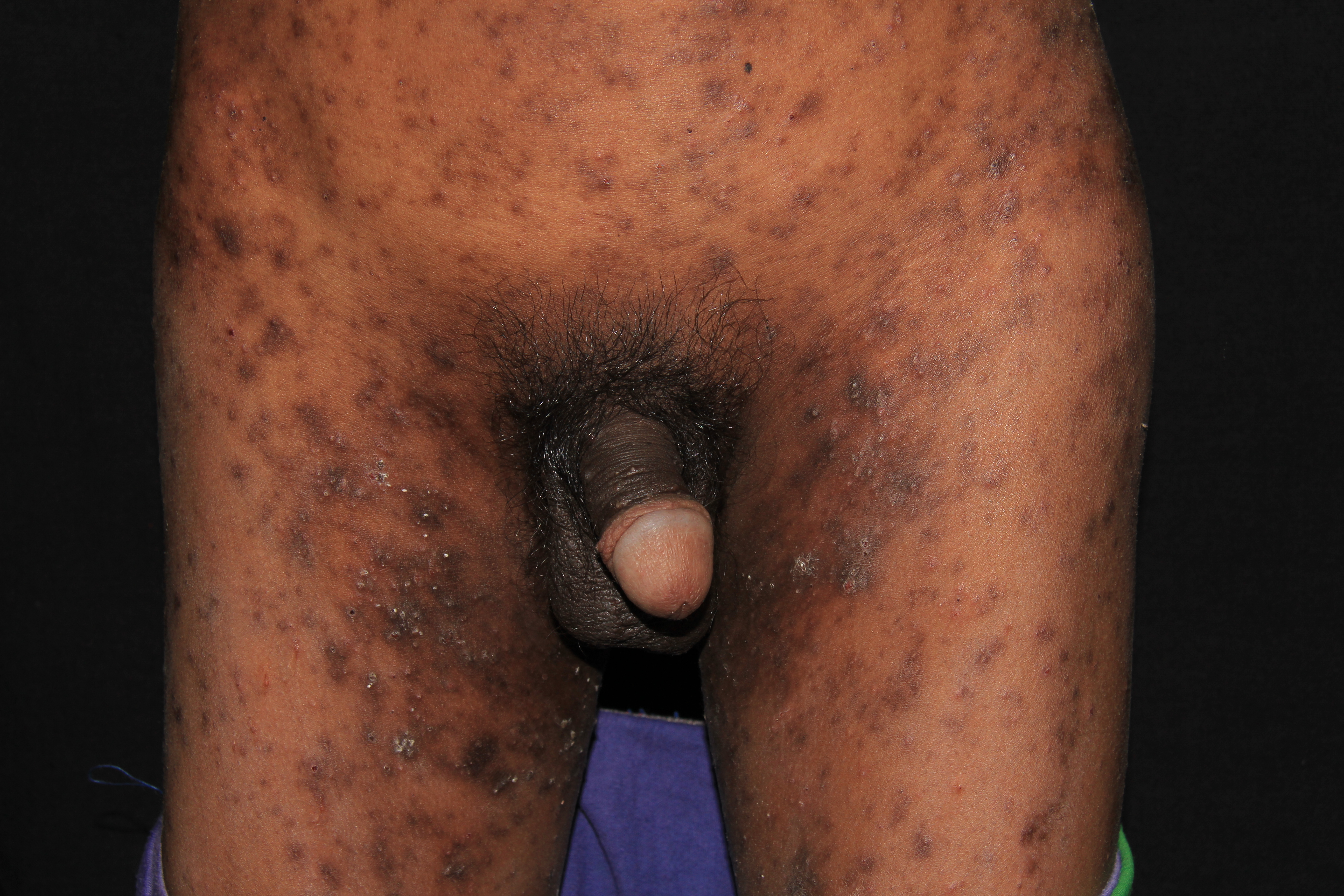
Acutely, the eczematous rash is intensely pruritic with erythematous papules and excoriations. With continued itching and rubbing, the skin starts to thicken; on physical exam, there may be lichenification (thickening of the skin with exaggeration of the typical skin markings due to scratching or rubbing).
The distribution of the rash varies depending on patient age. Infants tend to have widely distributed, dry, scaly, and erythematous patches with small excoriations. They also tend to have lesions on their face, especially the cheeks. With age, the rash becomes more localized; areas affected include the extensor surfaces such as the wrists, elbows, ankles, and knees. School-aged children tend to follow the pattern seen in adults: the involvement of the flexural surfaces usually affects the anticubital and popliteal fosse.
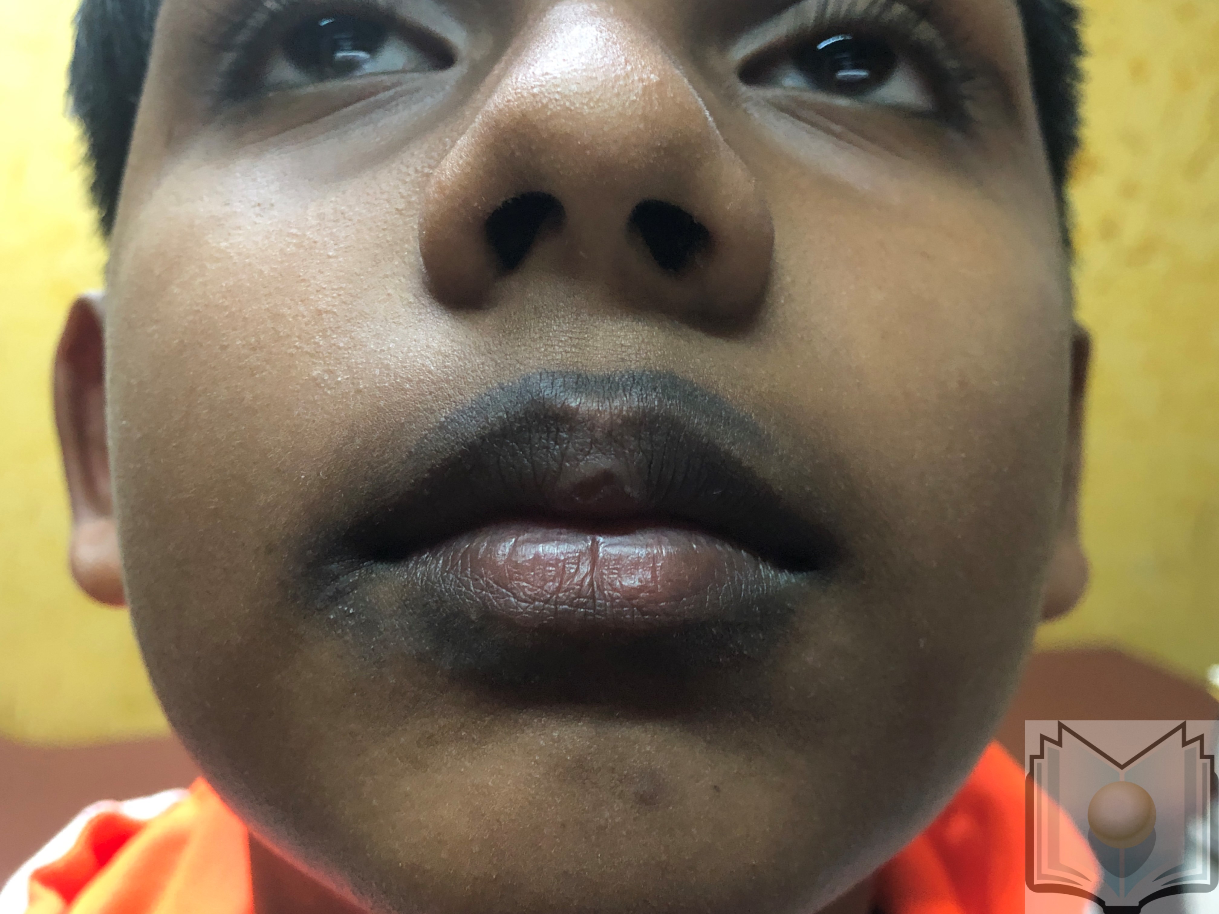
In addition to a rash, other physical exam findings may indicate that a person has eczema. Examination of the face may reveal Dennie-Morgan lines. [10] These are crease-like wrinkles just below the lower eyelid. This exam finding can be seen in up to 25% of patients with eczema. People with eczema may have coexisting pityriasis alba and hypopigmented patches or fine scaling plaques, most commonly seen on the face. On examination of the hands, there may be an increased number and depth of skin lines known as hyperlinear palms. People who have eczema and allergic rhinitis may have a transverse crease formed across their nose. This line is called the "allergic salute" and is caused by habitually rubbing the nose in an upward direction.[11]
Evaluation
Diagnosis is typically clinical based on the appearance of the rash and the history given by the patient. Routine labwork is usually not indicated. If the diagnosis is unclear, allergy testing and patch testing may be performed.
Treatment / Management
The main management and treatment protocols for eczema include hydration and topical anti-inflammatory medications for flare-ups. The priority in treatment is focusing on a daily skin moisturizing regimen with a fragrance-free ointment with limited preservatives.[12] An ointment is preferred over a cream due to the high proportion of oil to water in lotions. Patients and caregivers should also identify and address any triggers. They should be instructed to avoid any environmental allergens, harsh soaps, detergents, fragrances as well as rough or nonbreathable fabrics.
Skin flare-ups can be treated with topical anti-inflammatory medications, such as topical steroids or steroid-free products like pimecrolimus, tacrolimus, or Eucirsa. In children, itching tends to be worse at nighttime. Oral antihistamines can be used intermittently at bedtime for disturbed sleep due to itch; however, antihistamines are no longer recommended for daytime use for itching in eczema. Patients with poorly controlled eczema have a higher risk of cutaneous infections. Patients and caregivers may be instructed to take diluted bleach baths or intranasal mupirocin to decrease the number of cutaneous infections.[13]
Differential Diagnosis
The differential diagnosis for eczema includes many eczematous dermatitides, including:[14]
- Contact dermatitis
- Cutaneous fungal infections
- Seborrheic dermatitis
- Drug eruptions
- Scabies
- Psoriasis
- Ectodermal dysplasia
- Hyper-IgE syndrome
- Netherton syndrome
- Wiskott-Aldrich syndrome
Toxicity and Adverse Effect Management
It is crucial that patients and caregivers understand that the use of topical steroids should only be used for active lesions. Topical steroids can also be used prophylactically by applying them to the affected skin a few times a week to prevent flare-ups.
Topical steroids are not recommended for daily use, as long-term use can cause atrophy (thinning of the skin), stretch marks (striae), acne, telangiectasia (widened blood vessels), and rebound dermatitis or rosacea. The strength and formulation of the topical steroid need to be carefully determined based on the location of the body affected. Less potent steroids should be used on the face and intertriginous regions.
Prognosis
Most children will outgrow eczema, and their symptoms will be resolved by adulthood. However, children with the already persistent disease, later onset, or more severe disease have increased persistence.[15]
Complications
Due to the dysfunctional skin barrier seen in eczema, patients are at increased risk for infection from bacterial, viral, and fungal pathogens.[16] About 10% of healthy individuals are colonized with S. aureus compared with over 90% of patients with eczema.[17] The density of S. aureus colonization correlates with the severity of dermatitis.[18] Infection with S. aureus may cause furuncles, impetigo, or cellulitis. Patients with repeated bacterial infections may require treatment with diluted bleach baths and intranasal mupirocin to reduce the number of bacteria on their skin.
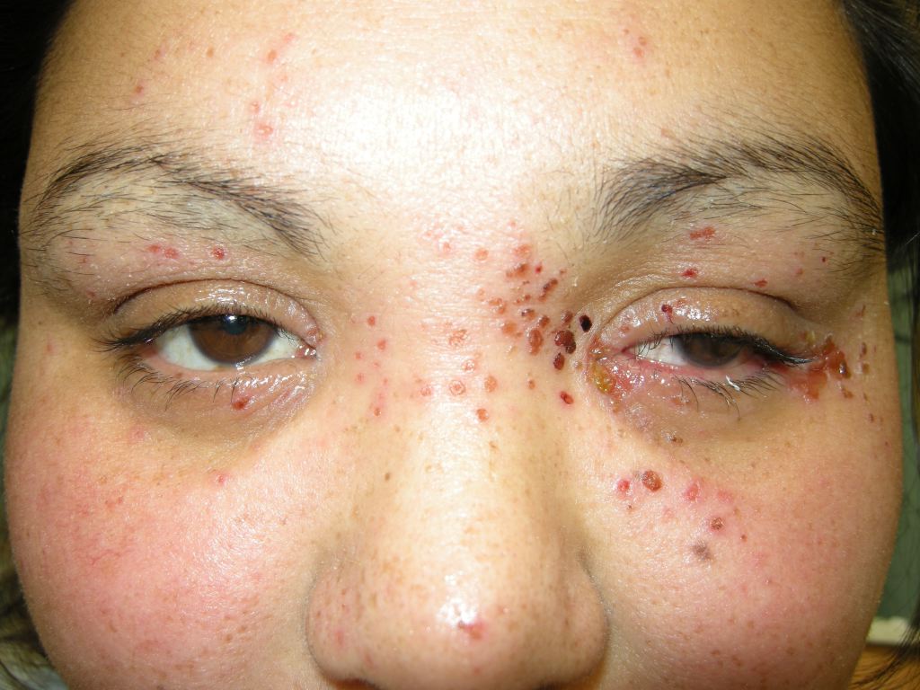
Patients with eczema are also more susceptible to viral infections. Eczema herpeticum is a life-threatening infection caused by herpes simplex virus-1. Patients present with widespread blisters, fever, and fatigue. The blisters appear in clusters and can cover a large body area. Eczema herpeticum is a medical emergency with complications including keratoconjunctivitis, meningitis, encephalitis, or secondary bacterial sepsis.[19]
Another life-threatening viral infection seen in patients with eczema is eczema cosackium.[16] Eczema coxsackie is a variant of hand, foot, and mouth disease classically associated with the enterovirus coxsackievirus A16. Instead of the typical presentation of blisters and erosions on the hands, feet, and hard palate, patients with eczema have widespread blisters and abrasions that tend to appear in regions previously affected by eczema. The rash can appear similar to eczema herpeticum. However, patients will lack fevers, decreased appetite, or fatigue. Parents may report a brief history of diarrhea or fever a week before the rash appears. Rare complications include aseptic meningitis.[20]
Deterrence and Patient Education
For patients with eczema, it is essential to determine what triggers the condition. Reduction or elimination of these triggers is an important step in treatment. Patients may see an improvement in their skin and reduced flare-ups if they avoid allergens (commonly dust mites, egg, peanuts, milk, fish, soy, rice, and wheat) and irritants (particularly chemicals, heat, soaps, humidity, acrylic, and wool).
Enhancing Healthcare Team Outcomes
Eczema is a common condition seen in pediatric and family medicine offices. Typically, patients with mild-to-moderate eczema can be treated in the office by their primary care provider with standard therapy. Patients with moderate-to-severe cases may require referral to dermatology for systemic treatments. If patients do not respond to typical treatment regimens, they may benefit from a consult with an allergist for a patch or skin scratch testing. An interprofessional team of a specialty-trained dermatology nurse and specialty-trained dermatology clinician will provide the best patient care.