Continuing Education Activity
Rosacea is a common chronic inflammatory disease that presents with recurrent flushing, erythema, telangiectasia, papules, or pustules on nose, chin, cheeks, and forehead. Although usually limited to the skin, an association of rosacea with systemic comorbidities has been reported. Prompt diagnosis and treatment are required to prevent permanent scarring, persistent erythema, and ocular sequelae. This activity reviews the evaluation and treatment of rosacea and highlights the role of the interprofessional team in evaluating and treating patients with this condition.
Objectives:
- Describe the pathophysiology of rosacea.
- Summarize the typical presentation of a patient with rosacea.
- Outline some management options available for rosacea.
- Review the importance of improving care coordination amongst interprofessional team members to improve outcomes for patients affected by rosacea.
Introduction
Rosacea is a common chronic inflammatory disease that presents with recurrent flushing, erythema, telangiectasia, papules, or pustules on nose, chin, cheeks, and forehead. There are four clinical subtypes of rosacea based on the predominant signs and symptoms: erythematotelangiectatic, papulopustular, phymatous, and ocular. The subtypes are not mutually exclusive. Patients can present with features of multiple subtypes, and the predominant features and areas of involvement can change over time. Fifty to seventy-five percent of patients with rosacea have eye involvement with symptoms including dryness, redness, tearing, tingling/burning sensation, foreign-body sensation, light sensitivity, and blurred vision. In addition to the skin and eye symptoms, rosacea can cause anxiety, embarrassment, and depression and can have a significant impact on the quality of life. Although usually limited to the skin, an association of rosacea with systemic comorbidities such as neurologic diseases, inflammatory bowel disease, and cardiovascular diseases has been reported.[1][2][3][4]
Etiology
The exact etiology of rosacea is not fully understood. Genetics, immune reaction, microorganisms, environmental factors, and neurovascular dysregulation are among the known etiological factors for the development of rosacea. In addition, besides the known effect of ultraviolet (UV) exposure as a trigger for rosacea, it may also play a role in the etiology of the disease.[5] A genetic predisposition is supported by a higher incidence of disease in patients with a family history of rosacea. Furthermore, specific human leucocyte antigen (HLA) loci have been identified in patients with rosacea.[6]
Among microorganisms, Demodex mites appear to play a role in rosacea as they are seen in higher numbers on rosacea-affected skin, though it is not clear if this is a cause or consequence of rosacea.[4] Helicobacter pylori is another organism with reported association with rosacea.[7]
Epidemiology
As the diagnosis of rosacea is mainly based on clinical judgment, a large number of patients, especially those with mild disease, may remain undiagnosed. It is estimated that the worldwide incidence of rosacea is higher than 5% of the population. It favors adults between 30 and 50 years of age, affects females more than males, and is more commonly diagnosed in individuals with fair skin (phototypes I and II), affecting more than 10% of Whites.[4][8]
Pathophysiology
Neurovascular dysregulation, activation of the immune system, and infestation with Demodex mites are among the pathophysiological mechanisms postulated for rosacea.
Dilation of lymphatic and blood vessels with exposure to extreme temperatures, spices, and alcohol has been observed in rosacea. Elevated expression of nonspecific cation channels such as transient receptor protentional vanilloid 1 (TRPV-1) and ankyrin 1 on sensory neurons and keratinocytes and release of vasoactive peptides following exposure to triggers is the proposed mechanism for the erythema and flushing.[2]
Activation of the adaptive and innate immune system response by overexpression of Th1/Th17 and toll-like receptor 2 (TLR-2), respectively, are other known pathomechanisms for rosacea. TLR-2 activation results in increased activity of mast cells via an increase in LL-37 production. In addition, expression of matrix metalloproteinases and vascular endothelial growth factor is increased in rosacea.[2][4] In rosacea, microbes may trigger activation of the immune response. This hypothesis is supported by an increased number of organisms, such as Demodex folliculorum on the skin and helicobacter pylori infection in the gut of patients with rosacea.[2][6][8]
Histopathology
Rosacea is a clinical diagnosis, and biopsy is not typically necessary. Histologic findings of rosacea can vary across different subtypes. Solar elastosis, telangiectasia, edema, and perivascular lymphohistiocytic infiltration can be seen in the erythematotelangiectatic subtype. Papulopustular rosacea presents with neutrophilic infiltration in hair follicles. Hyperplasia of sebaceous glands, fibrosis, and dilation of hair follicles are observed in phymatous rosacea. Granuloma formation is seen in the granulomatous rosacea subtype.[9]
History and Physical
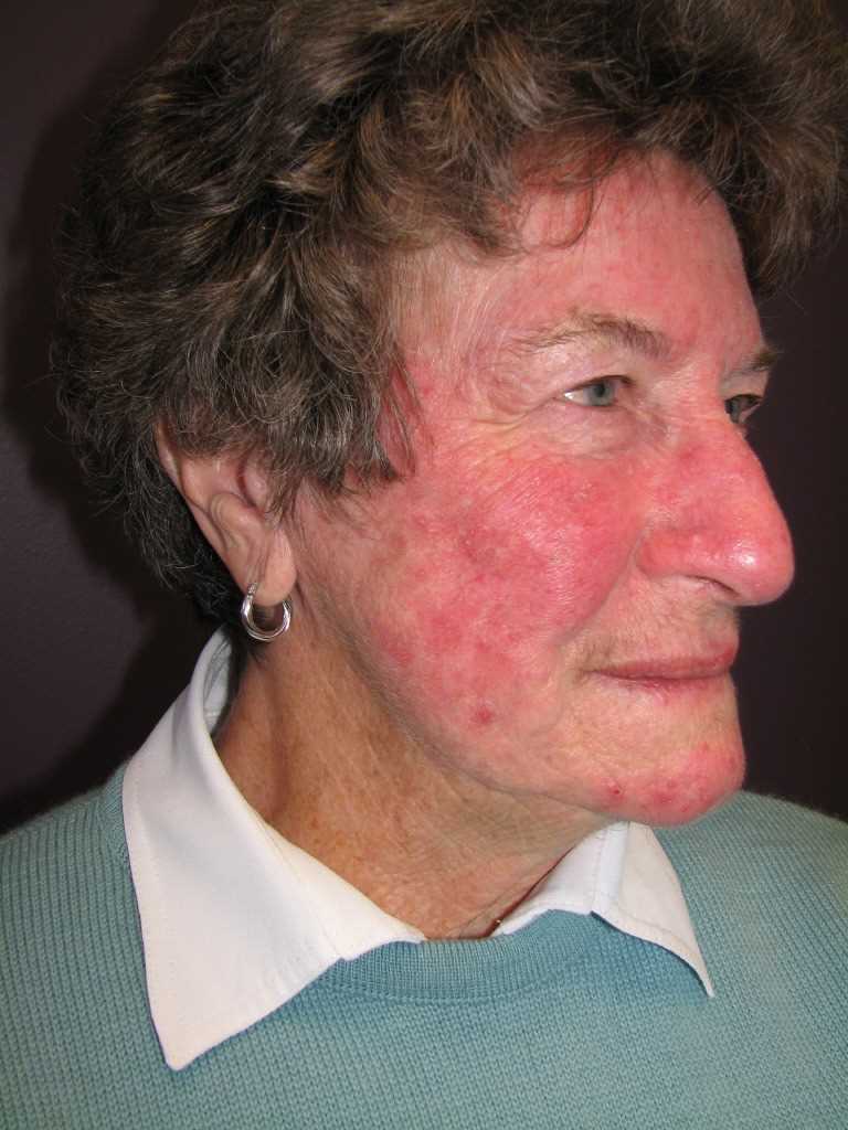
Rosacea typically presents during adulthood, though it can rarely affect children. Rosacea presents with 4 different clinical subtypes during the disease. The erythematotelengiectatic subtype, which presents with persistent erythema with intermittent flushing of nose and cheeks, is usually the first clinical manifestation of rosacea. In the papulopustular form, patients present with eruptions of papules and pustules on the affected area on the face. This subtype is sometimes called “adult acne” due to the similarity in the appearance of lesions. Notably, rosacea lacks comedones, helping to differentiate it from true acne. The phymatous subtype manifests with fibrosis and hypertrophy of sebaceous glands. It is typically seen on the nose of male patients (rhinophyma), but can also affect the cheeks, chin, and glabella. Although phyma is typically a late manifestation of rosacea, de novo cases with no prior skin changes have been reported. Ocular rosacea presents with tearing, dry eye, gritty sensation, pruritus, hordeola, and blepharitis.
Based on the latest diagnostic guidelines by the National Rosacea Society Expert Committee, one of the following clinical presentations is considered diagnostic for rosacea:
Fixed centrofacial erythema in a characteristic pattern that may periodically intensify
Phymatous changes
Two of the following major criteria below are also considered diagnostic:
Flushing
Papules and pustules
Telangiectasia
Ocular manifestations including lid margin telangiectasia, interpalpebral conjunctival injection, spade-shaped infiltrate in the cornea, and scleritis and sclerokeratitis.[3]
Evaluation
Rosacea is a clinical diagnosis. Patients should be asked about potential triggers. Ophthalmic evaluation is necessary for patients with ocular symptoms.
Treatment / Management
The first step in the treatment of rosacea is to advise the patient to identify and then avoid triggers such as UV light, spices, weather changes, and alcoholic beverages. Universal skin care recommendations for all patients with rosacea include pH-balanced skin cleansers (as opposed to soaps), broad-spectrum sunscreen with SPF 30 or higher and regular use of moisturizers. Rosacea often causes the skin to become sensitive and irritable, and products that cause irritation should be avoided. Cosmetics containing green pigment are best for masking persistent erythema.[10] The choice of therapy is guided by the signs and symptoms present for the individual patient. The majority of the therapies aim to reduce inflammation. Though they provide anti-inflammatory properties, topical steroids should be avoided in rosacea as they are associated with rebound flaring or induction of rosacea-like perioral dermatitis.[11] The persistent erythema and telangiectasias are not completely secondary to inflammation and often require treatment targeting the skin vasculature, such as brimonidine, oxymetazoline, or vascular laser. The phymatous changes of rosacea result in irreversible changes to the skin that require surgical intervention when indicated.
Topical Treatment
Erythema
- Brimonidine tartrate (alpha-2 agonist) 0.33% gel (Daily application on the face)
- Oxymetazoline hydrochloride (alpha-1 agonist) 1% cream (Daily application on face)
- Inflammatory papules and pustules
- Ivermectin 1% cream (daily application)
- Azelaic acid 15% gel, foam or 20% cream (daily 1 to 2 times application)
- Metronidazole 0.75% and 1% gel or cream (daily 1 to 2 times application)
Ocular Involvement
Artificial tears
- Fusidic acid gel (daily 1 to 2 times application on eyelids) limited data available for efficacy
- Metronidazole 0.75% gel (daily 1 to 2 times application on eyelids) limited data available for efficacy
- Cyclosporine 0.05% eyedrops, (one drop every 12 hours) limited data available for efficacy
Systemic Treatment
Flushing
- Propranolol (20 to 40 mg 2 to 3 times/day), carvedilol (6.25mg 2 to 3 times/day)
- Clonidine (50 mcg twice daily)
Inflammatory papules and pustules
- Subantimicrobial-dose doxycycline, modified-release (40 mg daily, 30 mg immediate-release and 10 mg delayed-release beads, for 8 to 12 weeks)
- Minocycline (50 to 100 mg twice daily for 8 to 12 weeks)
- Tetracycline (250 to 500 mg twice daily for 8 to 12 weeks)
- Azithromycin (250-500 mg 3 times weekly for 4 to 8 weeks)
- Isotretinoin (0.25 to 0.3 mg/kg/day for 12 to 16 weeks)
Phyma (inflamed)
- Doxycycline (100 mg 1 to 2 times daily for 8 to 12 weeks)
- Tetracycline (250 to 500 mg twice daily for 8 to 12 weeks)
- Isotretinoin (0.25-0.3 mg/kg/day for 3 to 4 months)
Ocular Involvement
- Subantimicrobial-dose doxycycline, modified-release (40 to 100 mg daily)
Procedures/Interventions
- Erythema/telangiectasia
- Intense pulsed light therapy
- NdYAG laser
- PDL pulsed dye laser 585 to 595 nm
Phyma (non-inflamed)
- CO2 laser 10,600 nm
- Surgical resection
- Electrosurgery
Referral to an ophthalmologist is recommended if the patient shows any ocular involvement, especially severe symptoms or visual disturbance. Topical treatment is recommended in pregnant women. Azithromycin, erythromycin, and clarithromycin are considered to be safe in pregnant women with mild-severe inflammatory rosacea. Systemic therapies are often used for flares that don’t respond to topical therapy alone. Continuation of topical treatment is recommended to maintain remission after controlling the flareup.[3][4][6][12][13]
Differential Diagnosis
Acne: While the two diseases share papules and pustules, the presence of comedones is unique to acne and helps distinguish the two.
Seborrheic dermatitis presents with erythema and greasy scaling on the scalp and face. It tends to be distributed more in the nasolabial folds and hair-bearing areas of the face. Since the two conditions are common, many patients will present with both seborrheic dermatitis and rosacea. Treatment of one may unmask the other.
Keratosis pilaris rubra typically affects adolescent patients with tiny follicular papules of the lateral cheeks and neck on erythematous patches.
Flushing: Flushing in rosacea is usually limited to the face. Flushing that involves other areas warrants further attention.
Acute cutaneous lupus erythematosus: The malar rash of lupus can appear similar to rosacea, but it usually spares the nasolabial folds and lacks papulopustules.
Drug-induced acneiform eruption: Onset is usually abrupt and temporally related to medication intake. Lesions are typically monomorphic (all papules or pustules in the same stage) and involve the trunk.
Prognosis
Rosacea is not a life-threatening disease, and the overall prognosis of rosacea is good. However, it can lead to depression and anxiety. If left untreated, patients can develop permanent scarring and persistent erythema. In addition, ocular sequelae could be a complication of untreated, ocular rosacea. Recent studies elaborated on the possible correlation of rosacea with neurologic, cardiovascular, endocrine, and gastrointestinal comorbidities. Consideration of these comorbidities in patients with rosacea may be warranted, though currently no evidence-based recommendations for screening have been established.[1][14]
Deterrence and Patient Education
Patients should be advised to make a diary of triggering factors and to avoid them. Strict UV protection with daily use of sunscreen, preferably mineral products, gentle skin care, using soap-free and non-comedogenic cleansers, and avoiding irritant cosmetic and skincare products recommendations providers should discuss with patients.
Enhancing Healthcare Team Outcomes
Patients with rosacea are usually seen by primary care physicians initially, where first-line therapy can be initiated. Since early recognition and treatment may help prevent disfigurement, any patient with an uncertain diagnosis or lack of response to therapy should be referred to a dermatologist. Screening for ocular symptoms should be performed in all rosacea patients, and referral to an ophthalmologist should be considered with any ocular symptoms.