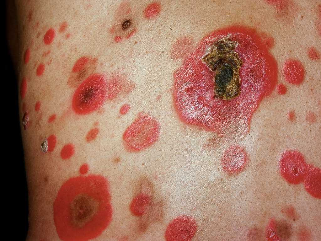
Mycosis Fungoides

Earn CME/CE in your profession:


Continuing Education Activity

The early identification, diagnosis and management of Mycosis fungoides is imperative in a dermatology out patient setting. This activity reviews the etiolopathogenesis, epidemiology, clinical and histological features, diagnosis and treatment of Mycosis fungoides.

Objectives:

  • Identify the various clinical stages of mycosis fungoides.
  • Describe the histopathology of mycosis fungoides.
  • Outline the treatment of mycosis fungoides.
  • Explain the importance of collaboration and communication among the interprofessional team to ensure mycosis fungoides is appropriately treated thereby improving outcomes.

Introduction

Primary cutaneous lymphomas are the second most common extranodal non-Hodgkin Lymphomas. They may be of either T cell, B cell, or NK cell origin. Cutaneous T Cell lymphomas (CTCL) comprise a group of heterogeneous lymphomas which clinically differ from systemic lymphomas, even though they might show similar histology. [1][2][3]

Mycosis fungoides is the most common type of CTCL. It is a cutaneous lymphoma that originates in the peripheral epidermotropic T-cells, specifically the memory T-cells (CD45RO+), which express the T-cell receptor (TCR) and CD4+ immunophenotype.[4]

Etiology

The cause of mycosis fungoides is unclear. The following are various hypotheses proposed:[5]

  • Genetic and epigenetic abnormalities, most commonly deletions and translocations involving several different chromosomes or chromosomal segments.[6][7]
  • Environmental and occupational exposure to solvents and chemicals has been implicated in the etiology of the disease. [8]
  • An infectious etiology that is of human T-lymphotropic virus Type 1 has been suggested, but conclusive evidence has still not been found. [9]
  • Cytokines such as IL-2 and IL-4 may play a role, as they show higher levels in patients with mycosis fungoides and Sezary syndrome.[10]

Epidemiology

Mycosis fungoides has an incidence of around 6 cases per million per year in Europe and the United States. This accounts for 4% of all non-Hodgkin lymphoma cases. It is more common in adults over 50 years of age, with a male:female ratio between 1.6 and 2. The disease is more common amongst Blacks than Caucasians or Asians.[11][12]

Pathophysiology

Among the cutaneous lymphomas, nearly two-thirds are of the T cell type. The most common immunophenotype is CD4 positive. In mycosis fungoides, there is a clonal expansion of CD4 cells that often lack the normal T cell antigens like CD7, CD5 or CD 2. These cells are attracted towards the skin by keratinocytes. As the cells accumulate in the dermis, they cluster around the langerhan cells- which is known as Pautrier microabscesses. Some of the malignant cells travel to the local lymph nodes and then pass into the bloodstream where they circulate with other CLA-positive T cells.[13][14]

Histopathology

Histopathology of mycosis fungoides varies within stages of the disease. Superficial lymphoid infiltrate, epidermotropism with absent or rare spongiosis spongiosis, and lymphoid atypia are the predominant features.[4][15][16]

  1. Patch stage: In the early patch stage. it may be difficult to diagnose as infiltrates may be very scant. In more established patches, a lymphocytic infiltrate may be seen around the basal cell layer. The cells are usually small, well-differentiated lymphocytes with rounded or cerebriform nuclei. The epidermis shows hyperkeratosis and acanthosis. Basal cell degeneration with melanin pigment incontinence may be seen. A dense, mixed perivascular infiltrate may be seen, along with fibrosis of dermal papillae.
  2. Plaque stage: In the plaque stage, the epidermis shows acanthosis with a psoriasiform hyperplasia and absent or rare spongiosis. The upper dermis may show a dense, band-like lymphocyte infiltrate with cerebriform nuclei and prominent epidermotropism. A third of cases may show Pautrier's abscesses.
  3. Tumor stage: A loss of epidermotropism may be noticed in the tumor stage. The infiltrate is a dense dermal infiltrate with large cerebriform lymphocytes, with a diminished number of T lymphocytes and dendritic cells. 

Immunohistochemistry

Mycosis fungoides tumor cells are characterized by epidermotropic peripheral T lymphocytes whose phenotype is CD2+, CD3+, CD4+, and CD5+. In a minority of patients with mycosis fungoides, T lymphocytes may be CD4- and CD8+, CD4- and CD8-, or CD4+ and CD8+.[15]The loss of CD7 expression can be observed even in the early phases of the disease. However, isolated negativity for CD7 is not a sufficient criterion for diagnosis as it can be shown in inflammatory dermatoses. The loss of CD26 expression seems to be specific to mycosis fungoides neoplastic cells.[17] 

The immunohistochemical pattern of Sezary syndrome is CD3+, CD4+, CD7-, and CD8- cells, which is identical to mycosis fungoides. Immunostaining for MUM-1 (multiple myeloma oncogene) might be used to differentiate these as it is positive in Sezary syndrome and negative in mycosis fungoides.[17]

History and Physical

The clinical presentation of mycosis fungoides varies with the stage of the disease.

  1. Patch stage: The earliest lesion seen in mycosis fungoides is an erythematous, or brownish scaly patch, which may show slight atrophy. Single or multiple lesions of variable diameters and locations develop, often in covered areas and particularly in the gluteal region and on the proximal thighs. The term sometimes given to them of "plaque parapsoriasis" is a misnomer because the lesions are actually patches. "Small-plaque" parapsoriasis and "large-plaque" parapsoriasis are set apart based on the clinical and histopathological correlation.[16]Small plaque parapsoriasis lesions are 2 to 6 cm in diameter, located on the trunk, and do not present atrophy or telangiectasia. Small-plaque parapsoriasis shows minimum potential to become infiltrated or tumoral.Large-plaque parapsoriasis presents with lesions that are larger than 6 cm in diameter, with signs of atrophy and poikiloderma. They are seen on the trunk, gluteal region, proximal extremities and mammary region. The histopathological picture may resemble mycosis fungoides, in fact, large-plaque parapsoriasis does progress to frank mycosis fungoides (i.e infiltrated and/or tumoral lesions) in around 7.5% to 14% of cases.[16]
  2. Plaque stage: In the plaque stage, lesions now appear larger with evident infiltration, along with the appearance of new lesions. The lesions are annular or horseshoe-shaped with an infiltrated base, raised, well-defined edges and asymmetrical distribution. They may also affect the face and the scalp. [18]
  3. Tumor stage: In the tumor stage, erythematous-purplish papules or nodules of larger diameter are seen. [19]

The frequency of lymph nodal or visceral dissemination increases as the lesions progress from the patch stage to the plaque stage and finally the tumor stage.

Less Common or Rare Clinical Variants

  1. Bullous/vesicular lesions[20] 
  2. Purpuric lesions[21]
  3. Poikilodermatous[22] 
  4. Syringotropic[23]
  5. Hypopigmented lesions (mainly in children)[24]

Clinical Variants (as described in the WHO-EORTC Classification for Cutaneous Lymphomas)[25]

  1. Follicular or folliculotropic mycosis fungoides
  2. Pagetoid reticulosis or Woringer- Kolopp type
  3. Granulomatous slack skin

Sezary syndrome:

Sezary syndrome constitutes 3% of all cutaneous lymphomas and is characterized by a triad of manifestations: erythroderma with pruritus, lymphadenopathy, and atypical circulating lymphocytes (referred to as Sezary or Lutzner cells). The syndrome is understood as a leukemic phase of T-cell cutaneous lymphomas and bone marrow compromise is rarely found in advanced forms of the disease only. Associated clinical manifestations include lagophthalmos, alopecia, palmoplantar hyperkeratosis and onycodystrophy. Sezary Syndrome must be differentiated from erythroderma in mycosis fungoides progression. In the WHO-EORTC classification, MF and SS are listed as seperate diseases.[26][27]

Evaluation

Recommended evaluation of the patient with mycosis fungoides/Sezary syndrome:[3][28]

Complete Physical Examination

  • Type and extent of skin lesions, with an estimation of body surface area, must be noted. Look for ulceration in the lesions.
  • For lesions in the tumor stage, evaluate the number, size, and sites of the lesions.
  • Look for lymphadenopathy and organomegaly.

Skin biopsy

  • The biopsy site must be chosen carefully. Usually, the most indurated area is selected.
  • Immunophenotyping for the following markers is performed: CD2, CD3, CD4, CD5, CD7, CD8, and a B-cell marker like CD20. 
  • Evaluation for clonality of TCR gene rearrangement

Blood tests

  • CBC, liver function tests, LDH, etc.
  • TCR gene rearrangement.
  • Abnormal lymphocyte evaluation may be performed by Sezary cell count with the determination of the absolute number of Sezary cells and flow cytometry.

Radiologic tests

  • Chest X-rays, ultrasounds of peripheral nodal groups, chest CTs, fluorodeoxyglucose (FDG)-PET scans, and MRIs may be performed to evaluate for any potential lymphadenopathy or visceral involvement, depending upon the stage of the disease.

Lymph Node Biopsy

  • In clinically suspicious lymph nodes, such as firm, irregular, or fixed nodes, or large nodes of diameter greater than or equal to 1.5 cm in diameter, an excisional biopsy may be performed. It is performed from the largest draining lymph node or as guided by an FDG-PET, the node with the highest standardized uptake value (SUV).
  • Cervical lymph nodes are the preferred sites, followed by axillary and then inguinal. The biopsy specimen is analyzed for histopathological examination, flow cytometry, and T-cell receptor gene rearrangement.

Treatment / Management

Early stage:

  • For patients with early-stage mycosis fungoides (IIA or below), the various treatment options are topical corticosteroids, topical nitrogen mustards (mechlorethamine, HN2), topical bexarotene, imiquimod, psoralen-ultraviolet A (PUVA) therapy or ultraviolet B (UVB) therapy.  Local radiation therapy may be used for localized lesions (such as in pagetoid reticulosis). [28]
  • For patients with highly symptomatic, generalized thickened plaques in whom a prompt response is needed, the recommendation is total skin electron beam therapy (TSEBT) or skin-directed therapies in combination with systemic therapies rather than HN2 or UVB alone. [29]
  • Systemic therapies, such as retinoids or interferons, histone deacetylase (HDAC) inhibitors, or low-dose methotrexate are used if skin-directed therapies fail, if skin symptoms are extensive/severe, or if patients have a worse prognostic profile such as folliculotropic mycosis fungoides, large cell transformation, or early blood involvement. [30][31][32]

Advanced Stage

  • Advanced stage (IIB to IV) mycosis fungoides is a heterogeneous group that encompasses those patients that present with extracutaneous disease or advanced skin lesions (e.g., tumors). It is often a chronic or persistent disease with a relapsing course. The main goals of therapy are long-term disease control, prompt symptom relief, and management of life-threatening (aggressive) disease.[28]
  • For most patients with cutaneous tumors involving a limited percent body surface area (typically less than 10%), treatment with localized radiation to the tumors plus skin-directed therapy as needed for concurrent patch/plaque disease is recommended.[28]
  • For patients with generalized tumors, total skin electron beam therapy (TSEBT) or systemic therapies are acceptable treatment options. TSEBT can be followed by other skin-directed therapies or systemic therapies to prolong response duration. [33]
  • For more extensive disease, combinations of skin-directed therapies and systemic therapies are used. The various systemic options available include methotrexate, bexarotene, targeted immunotherapy (such as alemtuzumab), polychemotherapy, etc.[34]
  • Patients with disease that is difficult to control should be considered for allogeneic hematopoietic cell transplantation with curative intent.[35]

Differential Diagnosis

It is easy to confuse mycosis fungoides for common skin disorders such as eczema, psoriasis, parapsoriasis, photodermatitis, or drug reactions. The important differentials to keep in mind include:[36]

  1. Atopic dermatitis, contact dermatitis, drug eruptions, or erythrodermic psoriasis, all of which may cause pruritis and erythroderma resembling mycosis fungoides
  2. Psoriasis
  3. Sezary syndrome which may resemble erythrodermic mycosis fungoides
  4. Other lymphomas such as subcutaneous panniculitis-like T cell lymphoma, primary cutaneous anaplastic large cell lymphoma, cutaneous gamma/delta T cell lymphoma, or cutaneous B cell lymphoma.

Staging

The standard staging system for mycosis fungoides was the TNMB system, which was the strongest prognostic indicator for mycosis fungoides. Olsen et al. (2007) published the staging norms of mycosis fungoides and SS as a result of the ISCL-EORTC discussions, and modified the existing TNMB staging system in view of advances in cellular and molecular biology in diagnostic methods, as given below.[37]

Skin

  • T1: Limited patches, papules, and/or plaques covering less than 10% of the skin surface. May further stratify into T1a (patch only) vs. T1b (plaque +/- patch).
  • T2: Patches, papules or plaques covering 10% or more of the skin surface. May further stratify into T2a (patch only) vs. T2b (plaque +/- patch).
  • T3: One or more tumors (1 cm or more in diameter)
  • T4: Confluence of erythema covering 80% or more of body surface area

Lymph Nodes

N0: No clinically abnormal peripheral lymph nodes; biopsy not required

N1: Clinically abnormal peripheral lymph nodes; histopathology Dutch grade 1 or NCI LN0-2

  • N1a: Clone negative (A T-cell clone is defined by PCR or Southern blot analysis of the T-cell receptor gene)
  • N1b: Clone positive

N2: Clinically abnormal peripheral lymph nodes; histopathology Dutch grade 2 or NCI LN3

  • N2a: Clone negative
  • N2b: Clone positive

N3: Clinically abnormal peripheral lymph nodes; histopathology Dutch grades 3 to 4 or NCI LN4; clone positive or negative

Nx: Clinically abnormal peripheral lymph nodes; no histologic confirmation

Visceral

M0: No visceral organ involvement

M1: Visceral involvement (must have pathology confirmation and organ involved should be specified)

Blood

B0: Absence of significant blood involvement: 5% or less of peripheral blood lymphocytes are atypical (Sezary) cells

  • B0a: Clone negative
  • B0b: Clone positive

B1: Low blood tumor burden: more than 5% of peripheral blood lymphocytes are atypical (Sezary) cells but does not meet the criteria of B2

  • B1a: Clone negative
  • B1b: Clone positive

B2: High blood tumor burden: 1000/microL or more Sezary cells with positive clone

Prognosis

The prognosis of mycosis fungoides and Sezary syndrome is variable. The TNMB staging is the strongest prognostic factor. The following factors may be associated with a poor prognosis:[5]

  1. Presence of extracutaneous disease (stage IV),
  2. Older age (greater than 60),
  3. Presence of large cell transformation (LCT), and
  4. Increased LDH
  5. Folliculotropic mycosis fungoides
  6. Tumor distribution at diagnosis

Complications

  • Marked pruritus
  • Lymphadenopathy
  • Onychodystrophy
  • Panniculitis
  • Skin ulcers
  • Scaring alopecia

Enhancing Healthcare Team Outcomes

The management of mycosis fungoides is best done with an interprofessional team of healthcare workers that includes an oncologist, internist, dermatologist, nurses, pharmacists, and social workers. The disorder has no cure, and the aim is to improve the quality of life. The patient should be encouraged to use ample moisturizer to prevent skin dryness and pruritus. The patient should be told to avoid the sun and wear long-sleeved garments when going out. A cool humidified environment is highly recommended. The patient should also be educated about the other treatment options like irradiation, Photodynamic therapy and UV light treatment enhanced with psoralen. Finally, the patient should be seen by a dietitian as many people develop moderate nausea from the treatment and lose significant weight. A high-calorie diet with regular exercise is recommended. [38][35](Level V)

Outcomes

Mycosis fungoides is an incurable disorder unless the patient has very early stage disease. The mortality and morbidity increase as the tumor advances. While early-stage patients have a 95% survival over ten years, those who advanced cancer only have a 3-4 year survival. Those with the advanced extra-cutaneous disease only have a survival of fewer than 18 months. Poor prognostic factors include male gender, advanced age, and elevated LDH.[39][33] (Level V)


(Click Image to Enlarge)
<p>Mycosis Fungoides</p>

Mycosis Fungoides


DermNet New Zealand

Details

Author

Tanvi Vaidya

Editor:

Talel Badri

Updated:

7/31/2023 8:36:25 PM

References


[1]

Scarisbrick JJ, Quaglino P, Prince HM, Papadavid E, Hodak E, Bagot M, Servitje O, Berti E, Ortiz-Romero P, Stadler R, Patsatsi A, Knobler R, Guenova E, Child F, Whittaker S, Nikolaou V, Tomasini C, Amitay I, Prag Naveh H, Ram-Wolff C, Battistella M, Alberti-Violetti S, Stranzenbach R, Gargallo V, Muniesa C, Koletsa T, Jonak C, Porkert S, Mitteldorf C, Estrach T, Combalia A, Marschalko M, Csomor J, Szepesi A, Cozzio A, Dummer R, Pimpinelli N, Grandi V, Beylot-Barry M, Pham-Ledard A, Wobser M, Geissinger E, Wehkamp U, Weichenthal M, Cowan R, Parry E, Harris J, Wachsmuth R, Turner D, Bates A, Healy E, Trautinger F, Latzka J, Yoo J, Vydianath B, Amel-Kashipaz R, Marinos L, Oikonomidi A, Stratigos A, Vignon-Pennamen MD, Battistella M, Climent F, Gonzalez-Barca E, Georgiou E, Senetta R, Zinzani P, Vakeva L, Ranki A, Busschots AM, Hauben E, Bervoets A, Woei-A-Jin FJSH, Matin R, Collins G, Weatherhead S, Frew J, Bayne M, Dunnill G, McKay P, Arumainathan A, Azurdia R, Benstead K, Twigger R, Rieger K, Brown R, Sanches JA, Miyashiro D, Akilov O, McCann S, Sahi H, Damasco FM, Querfeld C, Folkes A, Bur C, Klemke CD, Enz P, Pujol R, Quint K, Geskin L, Hong E, Evison F, Vermeer M, Cerroni L, Kempf W, Kim Y, Willemze R. The PROCLIPI international registry of early-stage mycosis fungoides identifies substantial diagnostic delay in most patients. The British journal of dermatology. 2019 Aug:181(2):350-357. doi: 10.1111/bjd.17258. Epub 2018 Nov 25     [PubMed PMID: 30267549]


[2]

Su C, Tang R, Bai HX, Girardi M, Karakousis G, Zhang PJ, Xiao R, Zhang G. Disease site as a prognostic factor for mycosis fungoides: an analysis of 2428 cases from the US National Cancer Database. British journal of haematology. 2019 May:185(3):592-595. doi: 10.1111/bjh.15570. Epub 2018 Sep 14     [PubMed PMID: 30216417]

Level 3 (low-level) evidence

[3]

Prince HM, Querfeld C. Integrating novel systemic therapies for the treatment of mycosis fungoides and Sézary syndrome. Best practice & research. Clinical haematology. 2018 Sep:31(3):322-335. doi: 10.1016/j.beha.2018.07.007. Epub 2018 Jul 18     [PubMed PMID: 30213403]


[4]

Hossain C, Jennings T, Duffy R, Knoblauch K, Gochoco A, Chervoneva I, Shi W, Alpdogan SO, Porcu P, Pro B, Sahu J. The histological prevalence and clinical implications of folliculotropism and syringotropism in mycosis fungoides. Chinese clinical oncology. 2019 Feb:8(1):6. doi: 10.21037/cco.2018.10.02. Epub     [PubMed PMID: 30818957]


[5]

Lim HLJ, Tan EST, Tee SI, Ho ZY, Boey JJJ, Tan WP, Tang MBY, Shen L, Chan YH, Tan SH. Epidemiology and prognostic factors for mycosis fungoides and Sézary syndrome in a multi-ethnic Asian cohort: a 12-year review. Journal of the European Academy of Dermatology and Venereology : JEADV. 2019 Aug:33(8):1513-1521. doi: 10.1111/jdv.15526. Epub 2019 Mar 27     [PubMed PMID: 30801779]


[6]

Bergallo M, Daprà V, Fava P, Ponti R, Calvi C, Montanari P, Novelli M, Quaglino P, Galliano I, Fierro MT. DNA from Human Polyomaviruses, MWPyV, HPyV6, HPyV7, HPyV9 and HPyV12 in Cutaneous T-cell Lymphomas. Anticancer research. 2018 Jul:38(7):4111-4114. doi: 10.21873/anticanres.12701. Epub     [PubMed PMID: 29970537]


[7]

Väisänen E, Fu Y, Koskenmies S, Fyhrquist N, Wang Y, Keinonen A, Mäkisalo H, Väkevä L, Pitkänen S, Ranki A, Hedman K, Söderlund-Venermo M. Cutavirus DNA in Malignant and Nonmalignant Skin of Cutaneous T-Cell Lymphoma and Organ Transplant Patients but Not of Healthy Adults. Clinical infectious diseases : an official publication of the Infectious Diseases Society of America. 2019 May 17:68(11):1904-1910. doi: 10.1093/cid/ciy806. Epub     [PubMed PMID: 30239652]


[8]

Slodownik D, Moshe S, Sprecher E, Goldberg I. Occupational mycosis fungoides - a case series. International journal of dermatology. 2017 Jul:56(7):733-737. doi: 10.1111/ijd.13589. Epub 2017 Mar 3     [PubMed PMID: 28255994]

Level 2 (mid-level) evidence

[9]

Blaizot R, Ouattara E, Fauconneau A, Beylot-Barry M, Pham-Ledard A. Infectious events and associated risk factors in mycosis fungoides/Sézary syndrome: a retrospective cohort study. The British journal of dermatology. 2018 Dec:179(6):1322-1328. doi: 10.1111/bjd.17073. Epub 2018 Oct 12     [PubMed PMID: 30098016]

Level 2 (mid-level) evidence

[10]

Fujii K. New Therapies and Immunological Findings in Cutaneous T-Cell Lymphoma. Frontiers in oncology. 2018:8():198. doi: 10.3389/fonc.2018.00198. Epub 2018 Jun 4     [PubMed PMID: 29915722]


[11]

Amorim GM, Niemeyer-Corbellini JP, Quintella DC, Cuzzi T, Ramos-E-Silva M. Clinical and epidemiological profile of patients with early stage mycosis fungoides. Anais brasileiros de dermatologia. 2018 Jul-Aug:93(4):546-552. doi: 10.1590/abd1806-4841.20187106. Epub     [PubMed PMID: 30066762]

Level 2 (mid-level) evidence

[12]

Amorim GM, Niemeyer-Corbellini JP, Quintella DC, Cuzzi T, Ramos-E-Silva M. Hypopigmented mycosis fungoides: a 20-case retrospective series. International journal of dermatology. 2018 Mar:57(3):306-312. doi: 10.1111/ijd.13855. Epub 2018 Jan 10     [PubMed PMID: 29318586]

Level 2 (mid-level) evidence

[13]

Eder J, Rogojanu R, Jerney W, Erhart F, Dohnal A, Kitzwögerer M, Steiner G, Moser J, Trautinger F. Mast Cells Are Abundant in Primary Cutaneous T-Cell Lymphomas: Results from a Computer-Aided Quantitative Immunohistological Study. PloS one. 2016:11(11):e0163661. doi: 10.1371/journal.pone.0163661. Epub 2016 Nov 28     [PubMed PMID: 27893746]


[14]

Tardío JC, Arias D, Khedaoui R. Indeterminate Cell Histiocytosis and Mycosis Fungoides: A Hitherto Unreported Association. The American Journal of dermatopathology. 2019 Jun:41(6):461-463. doi: 10.1097/DAD.0000000000001154. Epub     [PubMed PMID: 30024412]


[15]

Yamashita T, Abbade LP, Marques ME, Marques SA. Mycosis fungoides and Sézary syndrome: clinical, histopathological and immunohistochemical review and update. Anais brasileiros de dermatologia. 2012 Nov-Dec:87(6):817-28; quiz 829-30     [PubMed PMID: 23197199]


[16]

Pimpinelli N, Olsen EA, Santucci M, Vonderheid E, Haeffner AC, Stevens S, Burg G, Cerroni L, Dreno B, Glusac E, Guitart J, Heald PW, Kempf W, Knobler R, Lessin S, Sander C, Smoller BS, Telang G, Whittaker S, Iwatsuki K, Obitz E, Takigawa M, Turner ML, Wood GS, International Society for Cutaneous Lymphoma. Defining early mycosis fungoides. Journal of the American Academy of Dermatology. 2005 Dec:53(6):1053-63     [PubMed PMID: 16310068]


[17]

Robson A. Immunocytochemistry and the diagnosis of cutaneous lymphoma. Histopathology. 2010 Jan:56(1):71-90. doi: 10.1111/j.1365-2559.2009.03457.x. Epub     [PubMed PMID: 20055906]


[18]

Burg G, Dummer R, Nestle FO, Doebbeling U, Haeffner A. Cutaneous lymphomas consist of a spectrum of nosologically different entities including mycosis fungoides and small plaque parapsoriasis. Archives of dermatology. 1996 May:132(5):567-72     [PubMed PMID: 8624155]


[19]

Keehn CA, Belongie IP, Shistik G, Fenske NA, Glass LF. The diagnosis, staging, and treatment options for mycosis fungoides. Cancer control : journal of the Moffitt Cancer Center. 2007 Apr:14(2):102-11     [PubMed PMID: 17387295]


[20]

Bowman PH, Hogan DJ, Sanusi ID. Mycosis fungoides bullosa: report of a case and review of the literature. Journal of the American Academy of Dermatology. 2001 Dec:45(6):934-9     [PubMed PMID: 11712043]

Level 3 (low-level) evidence

[21]

Georgala S, Katoulis AC, Symeonidou S, Georgala C, Vayopoulos G. Persistent pigmented purpuric eruption associated with mycosis fungoides: a case report and review of the literature. Journal of the European Academy of Dermatology and Venereology : JEADV. 2001 Jan:15(1):62-4     [PubMed PMID: 11451328]

Level 3 (low-level) evidence

[22]

Lindae ML, Abel EA, Hoppe RT, Wood GS. Poikilodermatous mycosis fungoides and atrophic large-plaque parapsoriasis exhibit similar abnormalities of T-cell antigen expression. Archives of dermatology. 1988 Mar:124(3):366-72     [PubMed PMID: 3257858]


[23]

Zelger B, Sepp N, Weyrer K, Grünewald K, Zelger B. Syringotropic cutaneous T-cell lymphoma: a variant of mycosis fungoides? The British journal of dermatology. 1994 Jun:130(6):765-9     [PubMed PMID: 8011503]


[24]

El-Shabrawi-Caelen L, Cerroni L, Medeiros LJ, McCalmont TH. Hypopigmented mycosis fungoides: frequent expression of a CD8+ T-cell phenotype. The American journal of surgical pathology. 2002 Apr:26(4):450-7     [PubMed PMID: 11914622]


[25]

Willemze R, Jaffe ES, Burg G, Cerroni L, Berti E, Swerdlow SH, Ralfkiaer E, Chimenti S, Diaz-Perez JL, Duncan LM, Grange F, Harris NL, Kempf W, Kerl H, Kurrer M, Knobler R, Pimpinelli N, Sander C, Santucci M, Sterry W, Vermeer MH, Wechsler J, Whittaker S, Meijer CJ. WHO-EORTC classification for cutaneous lymphomas. Blood. 2005 May 15:105(10):3768-85     [PubMed PMID: 15692063]


[26]

Lopez AT, Bates S, Geskin L. Current Status of HDAC Inhibitors in Cutaneous T-cell Lymphoma. American journal of clinical dermatology. 2018 Dec:19(6):805-819. doi: 10.1007/s40257-018-0380-7. Epub     [PubMed PMID: 30173294]


[27]

PDQ Adult Treatment Editorial Board. Mycosis Fungoides (Including Sézary Syndrome) Treatment (PDQ®): Health Professional Version. PDQ Cancer Information Summaries. 2002:():     [PubMed PMID: 26389288]


[28]

Wain T, Venning VL, Consuegra G, Fernandez-Peñas P, Wells J. Management of cutaneous T-cell lymphomas: Established and emergent therapies. The Australasian journal of dermatology. 2019 Aug:60(3):200-208. doi: 10.1111/ajd.13011. Epub 2019 Feb 26     [PubMed PMID: 30809800]


[29]

Dairi M, Dadban A, Arnault JP, Lok C, Chaby G. Localized mycosis fungoides treated with laser-assisted photodynamic therapy: a case series. Clinical and experimental dermatology. 2019 Dec:44(8):930-932. doi: 10.1111/ced.13936. Epub 2019 Mar 1     [PubMed PMID: 30825216]

Level 2 (mid-level) evidence

[30]

Cho A, Jantschitsch C, Knobler R. Extracorporeal Photopheresis-An Overview. Frontiers in medicine. 2018:5():236. doi: 10.3389/fmed.2018.00236. Epub 2018 Aug 27     [PubMed PMID: 30211164]

Level 3 (low-level) evidence

[31]

Brazzelli V, Bernacca C, Segal A, Barruscotti S, Bolcato V, Michelerio A, Tomasini CF. Photo-photochemotherapy in Juvenile-onset Mycosis Fungoides: A Retrospective Study on 9 Patients. Journal of pediatric hematology/oncology. 2019 Jan:41(1):34-37. doi: 10.1097/MPH.0000000000001277. Epub     [PubMed PMID: 30130275]

Level 2 (mid-level) evidence

[32]

Photiou L, van der Weyden C, McCormack C, Miles Prince H. Systemic Treatment Options for Advanced-Stage Mycosis Fungoides and Sézary Syndrome. Current oncology reports. 2018 Mar 23:20(4):32. doi: 10.1007/s11912-018-0678-x. Epub 2018 Mar 23     [PubMed PMID: 29572582]


[33]

Jang BS, Kim E, Kim IH, Kang HC, Ye SJ. Clinical outcomes and prognostic factors in patients with mycosis fungoides who underwent radiation therapy in a single institution. Radiation oncology journal. 2018 Jun:36(2):153-162. doi: 10.3857/roj.2017.00542. Epub 2018 Jun 29     [PubMed PMID: 29983036]

Level 2 (mid-level) evidence

[34]

Alpdogan O, Kartan S, Johnson W, Sokol K, Porcu P. Systemic therapy of cutaneous T-cell lymphoma (CTCL). Chinese clinical oncology. 2019 Feb:8(1):10. doi: 10.21037/cco.2019.01.02. Epub     [PubMed PMID: 30818958]


[35]

Berg S, Villasenor-Park J, Haun P, Kim EJ. Multidisciplinary Management of Mycosis Fungoides/Sézary Syndrome. Current hematologic malignancy reports. 2017 Jun:12(3):234-243. doi: 10.1007/s11899-017-0387-9. Epub     [PubMed PMID: 28540671]


[36]

Olisova OY, Grekova EV, Varshavsky VA, Gorenkova LG, Alekseeva EA, Zaletaev DV, Sydikov AA. [Current possibilities of the differential diagnosis of plaque parapsoriasis and the early stages of mycosis fungoides]. Arkhiv patologii. 2019:81(1):9-17. doi: 10.17116/patol2019810119. Epub     [PubMed PMID: 30830099]


[37]

Olsen E, Vonderheid E, Pimpinelli N, Willemze R, Kim Y, Knobler R, Zackheim H, Duvic M, Estrach T, Lamberg S, Wood G, Dummer R, Ranki A, Burg G, Heald P, Pittelkow M, Bernengo MG, Sterry W, Laroche L, Trautinger F, Whittaker S, ISCL/EORTC. Revisions to the staging and classification of mycosis fungoides and Sezary syndrome: a proposal of the International Society for Cutaneous Lymphomas (ISCL) and the cutaneous lymphoma task force of the European Organization of Research and Treatment of Cancer (EORTC). Blood. 2007 Sep 15:110(6):1713-22     [PubMed PMID: 17540844]


[38]

O'Brien JS, Manning T, Perera M, Prince HM, Lawrentschuk N. Blueprint unknown: a case for multidisciplinary management of advanced penile mycosis fungoides. The Canadian journal of urology. 2017 Dec:24(6):9139-9144     [PubMed PMID: 29260643]

Level 3 (low-level) evidence

[39]

Lebowitz E, Geller S, Flores E, Pulitzer M, Horwitz S, Moskowitz A, Kheterpal M, Myskowski PL. Survival, disease progression and prognostic factors in elderly patients with mycosis fungoides and Sézary syndrome: a retrospective analysis of 174 patients. Journal of the European Academy of Dermatology and Venereology : JEADV. 2019 Jan:33(1):108-114. doi: 10.1111/jdv.15236. Epub 2018 Sep 25     [PubMed PMID: 30176169]

Level 2 (mid-level) evidence