Large Bowel Obstruction

Earn CME/CE in your profession:


Continuing Education Activity

Large bowel obstruction usually presents as an emergency with sepsis, dehydration, and hemodynamically compromised state. The most common cause of large bowel obstruction is colorectal cancer. This activity explains the risk factors, evaluation, and management of large bowel obstruction and highlights the importance of an interprofessional team in enhancing care for affected patients.

Objectives:

  • Identify the pathophysiology of large bowel obstruction.
  • Outline the appropriate history and physical examination that should be done for patients with suspected large bowel obstruction.
  • Summarize the treatment options available for patients with large bowel obstruction.
  • Review interprofessional team strategies for improving care coordination and communication to advance care for patients with large bowel obstruction.

Introduction

Large bowel obstruction usually presents as an emergency. Depending on the etiology, the patient may have had various symptoms before the acute presentation. Patients can potentially be very ill, septic, dehydrated, and hemodynamically and cardiovascularly compromised, especially if they have a competent ileocecal valve, which prevents decompression of the large bowel into the small bowel and leads to a closed-loop obstruction.[1][2]

Etiology

The most common cause of large bowel obstruction is an underlying colorectal malignancy. Approximately 40% of colorectal cancers present as emergencies and large bowel obstruction is the most common presentation.

Benign causes of large bowel obstruction are strictures secondary to diverticular disease or inflammatory bowel diseases, as well as volvulus, most commonly of the sigmoid colon. Occasionally the sigmoid colon can get obstructed in a left inguinal hernia sac.

External compression occurs most likely in the pelvis, secondary to gynecological malignancies.[3][4]

Epidemiology

Colorectal malignancies start occurring more commonly in the fifth decade of life, although the incidence of colorectal cancer has recently stabilized and is even slightly regressive in most parts of the developed world. The risk is approximately a 6% lifetime risk. There now appears to be an increase in presentation of patients younger than 50 with colorectal malignancies, and since this group of patients does not currently fall into screening recommendations, they may present with aggressive and advanced disease in the emergency setting.[5][6]

Pathophysiology

Closed-loop obstruction is a surgical emergency. If inflow and outflow of the colon are both obstructed (classical examples are a sigmoid volvulus or obstructing sigmoid colon cancer with a competent ileocecal valve), the obstructed bowel will continue to distend, owing to a significant amount of gas-forming bacteria trapped inside. This will eventually lead to vascular compromise, initially affecting the venous outflow, leading to further congestion until the arterial inflow ceases. Ischaemia, necrosis and eventual perforation of the obstructed segment occur. The part of the colon most at risk of perforation is the caecum as it has the thinnest wall compared to other parts of the colon.

While the colon is obstructed and distending, mucosal ischemia can lead to bacterial translocation of the gut flora, increasing the risk that the patient will present with bacteremia and sepsis.

History and Physical

History and presenting symptoms depend on the etiology, as well as the level of obstruction.

Obstruction due to underlying colorectal malignancy tends to occur on the left side (descending, sigmoid and rectum), as these are the more common locations for colorectal cancer. Also, the diameter of the bowel is smaller, and the feces tend to be solid. Patients may have experienced a change in bowel habit leading up to the obstruction, usually an alternating pattern of diarrhea and constipation, as only liquefied feces can pass through the narrowed lumen. Questioning patients carefully about changes in their bowel patterns often reveals that symptoms have been present for weeks or months before the acute presentation. They may report bleeding per rectum in addition to the change in bowel habits. Patients presenting with obstruction will experience abdominal distension, abdominal discomfort and are unable to pass feces and ultimately flatus. Patients may or may not have a positive family history of colorectal malignancy.

Large bowel obstruction due to a sigmoid volvulus usually presents more acutely with abdominal distension rather than a change in bowel habit, and in patients who are less mobile and have a tendency for constipation, for example, nursing home residents and bedbound patients.

Pain is not as prominent a feature as it is in small bowel obstruction, due to the less frequent contractions in the large bowel compared to the small bowel.

Patients with an incompetent ileocecal valve will decompress their large bowel into the small bowel and eventually present with vomiting, but a patient with a competent ileocecal valve will have ongoing distension of the obstructed colon, and will eventually present with significant right iliac fossa pain and impending caecal perforation.

Examination of the abdomen reveals significant distension, a tympanitic percussion note and potentially guarding and rebound over areas of impending perforation.

If the perforation has occurred already, the patient will be grossly peritonitic.

The digital rectal examination reveals an empty rectum.

Evaluation

The patient should have the standard blood work (full blood count and renal function tests), to aid resuscitation and assess the kidney function for a potential computed tomography scan with contrast.[7][8][9]

A plain abdominal x-ray will show the dilated colon, and a cutoff in the area of obstruction, as well as a paucity of gas in the rectum, or a classical coffee bean sign in the case of a volvulus.

If a colonic malignancy is suspected, a computed tomogram (CT) of thorax, abdomen, and pelvis will stage the disease and allow for further management.

Oral bowel preparation for colonoscopic evaluation is contraindicated in patients with bowel obstruction. If a histological diagnosis is required, the patient can receive a per rectal enema and an attempt to reach the obstructing lesion with minimal air insufflation using a colonoscope.

Treatment / Management

Before any surgical intervention, the patient needs to be adequately resuscitated with intravenous fluids and electrolyte imbalances corrected. A nasogastric tube helps to decompress the stomach in those patients with an incompetent ileocecal valve. In a patient with sigmoid volvulus, decompression should be attempted with a rectal flatus tube by the bedside, or during a flexible endoscopy if the bedside decompression fails. Those patients who cannot be decompressed will need surgical intervention.[1][10][11]

Patients with malignant obstruction will require surgery for definitive management. The procedure depends on the stage of the disease. In patients with metastatic disease, a defunctioning stoma is a preferred option, as this will ensure the timely start of chemotherapy. Patients without metastatic disease should undergo a definitive resectional and restorative procedure, followed by adjuvant therapy as necessary. In those patients planned for curative resection, a colonic stenting procedure or a defunctioning stoma in the emergency setting can allow elective resection of cancer and is a good option to offer the patient a laparoscopic resection once the colon is decompressed and the patient has resumed normal oral intake. A decompressive procedure, however, is only an option in patients without bowel ischemia and perforation, and should not be attempted in patients who are peritonitis or septic.

Patients with impending perforation, cecal ischemia or fecal peritonitis will require an emergency laparotomy and resection of the perforated colon as well as the obstructing lesion.

Differential Diagnosis

  • Megacolon
  • Diverticulitis
  • Small bowel obstruction
  • Abdominal hernia

Complications

  • Perforation
  • Bowel necrosis. ischemia, gangrene
  • Sepsis
  • Electrolyte alterations
  • Dehydration
  • Death

Pearls and Other Issues

In centers equipped for colonic stenting, a colonic stent can be inserted via a colonoscope under radiological guidance to decompress the bowel. This can be used in some patients to avoid an emergency operation and enable them to have a laparoscopic resection later. The patient should consent for stent complications such as migration and perforation, as well as the possibility that successful placement and decompression fails, in which case either a stoma or a resectional procedure are necessary.

Enhancing Healthcare Team Outcomes

Once bowel obstruction is diagnosed the care is usually interprofessional. Besides a general surgeon, a gastroenterologist, radiologist, oncologist, dietitian, nurse, and physical therapist are usually involved in the management. After treatment, surgical recovery can be long depending on the cause. Patients who have had an ileostomy or colostomy need to be followed by a stoma nurse. A dietitian should be consulted to educate patients with stomas on the type of foods they can eat. Since most patients are frail, physical therapy should be involved to help with functional recovery.  Patients with bowel obstruction as a result of cancer need follow up to ensure that they receive the right treatment.[12][13]

Outcomes

The morbidity and mortality after large bowel obstruction depend on the cause and presence of other comorbidities. The key is time to treat; in most cases, early treatment results in better outcomes. Mortality is significantly higher when there is bowel perforation or necrosis. Mortality rates of 15-30% have been reported in patients with perforation following large bowel obstruction. Recent use of self-expandable metallic stents for malignant bowel obstruction have yielded good short-term results, but long-term survival is not greatly altered. [14][6][Level 5]


(Click Image to Enlarge)
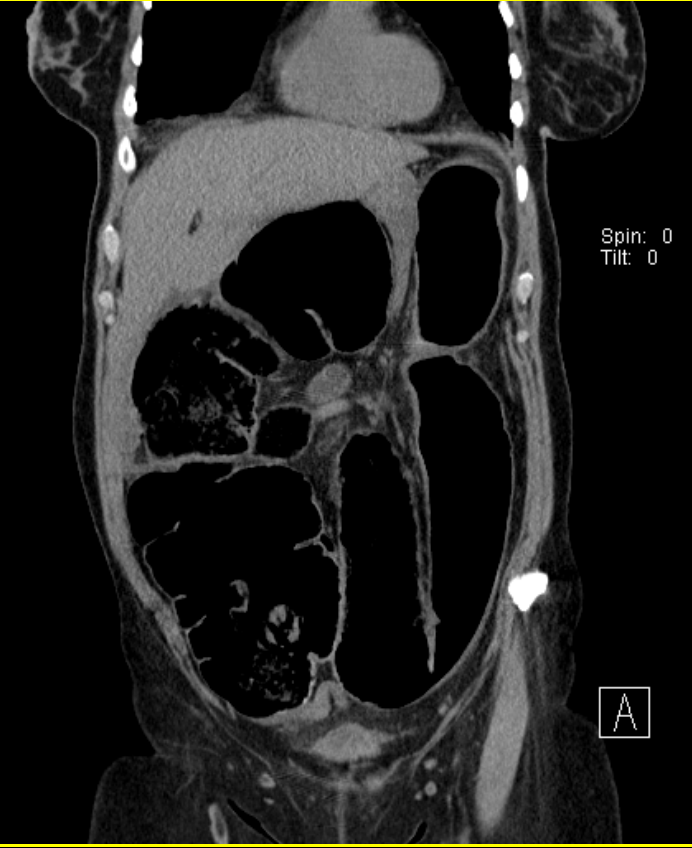
FIGURE 5: Coronal CT abdomen reveals cecal volvulus
FIGURE 5: Coronal CT abdomen reveals cecal volvulus. Usually a patient with a cecal volvulus will present with small and large bowel obstructions, with collapse of the distal large bowel, and with extensive dilation of the proximal small bowel.
Contributed by Wyckoff Height Medical Center 2017
Details

Updated:

8/8/2023 12:30:55 AM

References


[1]

Pisano M, Zorcolo L, Merli C, Cimbanassi S, Poiasina E, Ceresoli M, Agresta F, Allievi N, Bellanova G, Coccolini F, Coy C, Fugazzola P, Martinez CA, Montori G, Paolillo C, Penachim TJ, Pereira B, Reis T, Restivo A, Rezende-Neto J, Sartelli M, Valentino M, Abu-Zidan FM, Ashkenazi I, Bala M, Chiara O, De' Angelis N, Deidda S, De Simone B, Di Saverio S, Finotti E, Kenji I, Moore E, Wexner S, Biffl W, Coimbra R, Guttadauro A, Leppäniemi A, Maier R, Magnone S, Mefire AC, Peitzmann A, Sakakushev B, Sugrue M, Viale P, Weber D, Kashuk J, Fraga GP, Kluger I, Catena F, Ansaloni L. 2017 WSES guidelines on colon and rectal cancer emergencies: obstruction and perforation. World journal of emergency surgery : WJES. 2018:13():36. doi: 10.1186/s13017-018-0192-3. Epub 2018 Aug 13     [PubMed PMID: 30123315]


[2]

Toumi O, Hamida B, Njima M, Bouchrika A, Ammar H, Daldoul A, Zaied S, Ben Jabra S, Gupta R, Noomen F, Zouari K. Adenosquamous carcinoma of the right colon: A case report and review of the literature. International journal of surgery case reports. 2018:50():119-121. doi: 10.1016/j.ijscr.2018.07.001. Epub 2018 Jul 11     [PubMed PMID: 30103094]

Level 3 (low-level) evidence

[3]

Quinn K, Davis ME, Carter L, Shortell CK, Sommer C. Emergency General Surgery-A Misnomer? The American surgeon. 2018 Jul 1:84(7):1214-1216     [PubMed PMID: 30064591]


[4]

De Monti M, Cestaro G, Alkayyali S, Galafassi J, Fasolini F. Gallstone ileus: A possible cause of bowel obstruction in the elderly population. International journal of surgery case reports. 2018:43():18-20. doi: 10.1016/j.ijscr.2018.01.010. Epub 2018 Feb 4     [PubMed PMID: 29414501]

Level 3 (low-level) evidence

[5]

Shah D, Makharia GK, Ghoshal UC, Varma S, Ahuja V, Hutfless S. Burden of gastrointestinal and liver diseases in India, 1990-2016. Indian journal of gastroenterology : official journal of the Indian Society of Gastroenterology. 2018 Sep:37(5):439-445. doi: 10.1007/s12664-018-0892-3. Epub 2018 Oct 10     [PubMed PMID: 30306342]


[6]

Doshi R, Desai J, Shah Y, Decter D, Doshi S. Incidence, features, in-hospital outcomes and predictors of in-hospital mortality associated with toxic megacolon hospitalizations in the United States. Internal and emergency medicine. 2018 Sep:13(6):881-887. doi: 10.1007/s11739-018-1889-8. Epub 2018 Jun 12     [PubMed PMID: 29948833]


[7]

Cinar H, Berkesoglu M, Derebey M, Karadeniz E, Yildirim C, Karabulut K, Kesicioglu T, Erzurumlu K. Surgical management of anorectal foreign bodies. Nigerian journal of clinical practice. 2018 Jun:21(6):721-725. doi: 10.4103/njcp.njcp_172_17. Epub     [PubMed PMID: 29888718]


[8]

Orgul G, Soyer T, Yurdakok M, Beksac MS. Evaluation of pre- and postnatally diagnosed gastrointestinal tract obstructions. The journal of maternal-fetal & neonatal medicine : the official journal of the European Association of Perinatal Medicine, the Federation of Asia and Oceania Perinatal Societies, the International Society of Perinatal Obstetricians. 2019 Oct:32(19):3215-3220. doi: 10.1080/14767058.2018.1460350. Epub 2018 Apr 12     [PubMed PMID: 29606013]


[9]

Syrmis W, Richard R, Jenkins-Marsh S, Chia SC, Good P. Oral water soluble contrast for malignant bowel obstruction. The Cochrane database of systematic reviews. 2018 Mar 7:3(3):CD012014. doi: 10.1002/14651858.CD012014.pub2. Epub 2018 Mar 7     [PubMed PMID: 29513393]

Level 1 (high-level) evidence

[10]

Farkas N, Kaur V, Shanmuganandan A, Black J, Redon C, Frampton AE, West N. A systematic review of gallstone sigmoid ileus management. Annals of medicine and surgery (2012). 2018 Mar:27():32-39. doi: 10.1016/j.amsu.2018.01.004. Epub 2018 Jan 31     [PubMed PMID: 29511540]

Level 1 (high-level) evidence

[11]

Di Saverio S, Birindelli A, Segalini E, Novello M, Larocca A, Ferrara F, Binda GA, Bassi M. "To stent or not to stent?": immediate emergency surgery with laparoscopic radical colectomy with CME and primary anastomosis is feasible for obstructing left colon carcinoma. Surgical endoscopy. 2018 Apr:32(4):2151-2155. doi: 10.1007/s00464-017-5763-y. Epub 2017 Aug 8     [PubMed PMID: 28791424]


[12]

Utomo B, Alvarez C, Baldonedo RF. Management of Colorectal Cancer Patients Undergoing a Colonic Stenting: A Multidisciplinary Team Approach. Gastroenterology nursing : the official journal of the Society of Gastroenterology Nurses and Associates. 2017 Sep/Oct:40(5):342-349. doi: 10.1097/SGA.0000000000000255. Epub     [PubMed PMID: 28957966]


[13]

Costa G, Ruscelli P, Balducci G, Buccoliero F, Lorenzon L, Frezza B, Chirletti P, Stagnitti F, Miniello S, Stella F. Clinical strategies for the management of intestinal obstruction and pseudo-obstruction. A Delphi Consensus study of SICUT (Società Italiana di Chirurgia d'Urgenza e del Trauma). Annali italiani di chirurgia. 2016:87():105-17     [PubMed PMID: 27179226]

Level 3 (low-level) evidence

[14]

Matsuda A, Miyashita M, Matsumoto S, Sakurazawa N, Kawano Y, Yamahatsu K, Sekiguchi K, Yamada M, Hatori T, Yoshida H. Colonic stent-induced mechanical compression may suppress cancer cell proliferation in malignant large bowel obstruction. Surgical endoscopy. 2019 Apr:33(4):1290-1297. doi: 10.1007/s00464-018-6411-x. Epub 2018 Aug 31     [PubMed PMID: 30171397]