Continuing Education Activity
Fractures of the fifth metatarsal are common injuries that must be recognized and treated appropriately to avoid poor clinical outcomes for the patient. The clinician must recognize all injury patterns of the fifth metatarsal and initiate the appropriate treatment plan or referral process to avoid potential complications. Classification of these fractures is crucial to making management decisions. Metaphyseal arteries and diaphyseal nutrient arteries provide the blood supply to the fifth metatarsal base. A vascular watershed area exists in zone 2, contributing to the high nonunion rates seen with these fractures. This activity describes the evaluation and management of fractures of the fifth metatarsal, illustrates the zones of proximal base fractures and more distal fractures, and examines the interprofessional team's role in evaluating and treating patients with these fractures.
Objectives:
- Identify the etiology of fifth metatarsal fractures.
- Describe the zones of a proximal base of the fifth metatarsal fracture.
- Explain the treatment strategies for the different types of fifth metatarsal fractures.
- Review the importance of interprofessional collaboration in the management of fifth metatarsal fractures.
Introduction
Fractures of the fifth metatarsal are common injuries that must be recognized and treated appropriately to avoid poor clinical outcomes for the patient. Since orthopedic surgeon Sir Robert Jones first described these fractures in 1902, there has been an abundance of literature focused on the proximal aspect of the fifth metacarpal due to its tendency towards poor bone healing. Nevertheless, it is critical that the clinician recognizes all injury patterns of the fifth metatarsal and initiate the appropriate treatment plan or referral process to avoid potential complications.
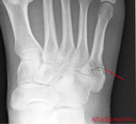
Classified by Lawrence and Bottle, the base, or proximal aspect, of the fifth metatarsal is broken up into three anatomical zones: zone 1, the tuberosity; zone 2, the metaphyseal-diaphyseal junction; and zone 3, the diaphyseal area within 1.5 cm of the tuberosity.[1] Fractures through zone 1 are called pseudo-Jones fractures, and fractures through zone 2 are referred to as Jones fractures. Additionally, a patient may sustain a shaft fracture greater than 1.5 cm distal to the tuberosity, a long spiral fracture extending into the distal metaphyseal area, the so-called dancer's fracture, or a stress fracture of the metatarsal.
Classification of these fractures is crucial to making management decisions. Metaphyseal arteries and diaphyseal nutrient arteries provide the blood supply to the fifth metatarsal base.[2] A vascular watershed area exists in zone 2, contributing to the high nonunion rates seen with these fractures.
Etiology
Zone 1 fractures are tuberosity avulsion fractures, also called pseudo-Jones fractures, and occur when the hindfoot gets forced into inversion during plantarflexion.[3] This acute injury pattern may occur after an athlete lands awkwardly after a jump. These fractures rarely involved the fifth tarsometatarsal joint and lay proximal to the fourth and/or fifth intermetatarsal joint
- Zone 2 injuries have the name Jones fractures. These acute injuries may occur with a significant adduction force to the foot with a lifted heel.[3] This type of injury pattern can occur with a sudden change of direction by an athlete. These fractures usually involve the fourth and/or fifth metatarsal articulation and have nonunion rates as high as 15 to 30%.
- Zone 3 injuries are chronic injuries of repetitive microtrauma, causing increasing pain with activity over months. There is an increased risk of nonunion with these fractures.
- The dancer's fracture, or long spiral fracture of the distal metatarsal, is typically caused by the dancer rolling over their foot while in the demi-pointe position or sustained while landing a jump.
Epidemiology
Fractures of the fifth metatarsal are the most prevalent metatarsal fractures. These fractures peak during the third decade of life for men and the seventh decade for women. There is a strong correlation between female gender and zone 1 fractures and dancer's fractures. Zone 1 injuries are typical of a twisting injury and are the most common fracture of the base of the fifth metatarsal.[4]
History and Physical
These patients typically present with pain about the lateral aspect of the forefoot that is worse with weight-bearing activity. This pain may occur in the setting of acute trauma or repetitive microtrauma over weeks to months. One should be suspicious of stress fracture with antecedent pain or pain of worsening quality or duration over time. The examiner must obtain a thorough past medical history and social history to make treatment decisions and optimize patients with surgical indications.
It is important to evaluate the skin for open injuries that may require more urgent debridement. Physical examination may reveal tenderness to palpation, swelling, and ecchymosis at the site of injury. Patients will also have pain with resisted foot eversion. It is critical to evaluate the patient for other injuries, including damage to the lateral ankle ligamentous structures and Lisfranc injury.
Evaluation
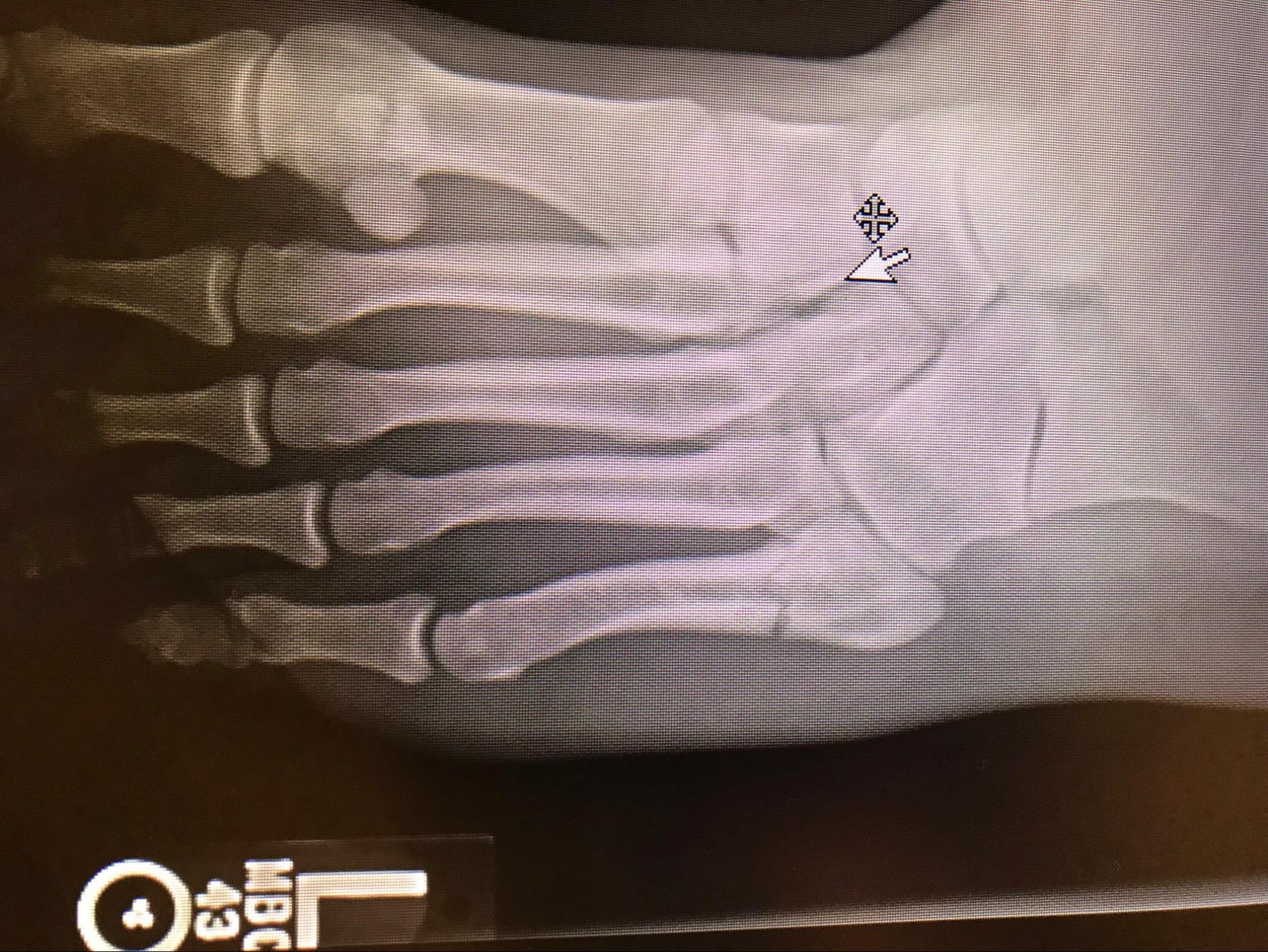
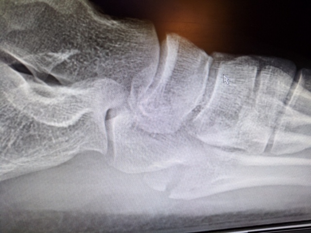
Radiographs are the initial imaging of choice used to evaluate for these injuries. AP, lateral, and oblique images of the foot are essential to making the diagnosis. In zone 1 injuries, the medial fracture line lies proximal to the fourth to the fifth intermetatarsal joint. In zone 2 injuries, the medial fracture line extends towards or even into the fourth and/or fifth intermetatarsal joint. In zone 3 injuries, the medial fracture line will typically exit distal to the fourth and/or fifth intermetatarsal joint, but some may be more proximal.[1] The usual fracture pattern seen in dancer fractures is an oblique spiral fracture beginning distal and lateral and extending proximal and medial.[5] Other distal diaphyseal fractures are generally seen on radiographs running in the transverse plane.
The radiographic appearance of fifth metatarsal base stress fractures classifies into three types based on the Torg classification system.[6] These classifications are as follows::
- Type I fractures:
- Early
- No intramedullary sclerosis
- Sharp fracture line with no widening
- Minimal cortical hypertrophy
- Minimal periosteal reaction
- Type II:
- Delayed
- Evidence of intramedullary sclerosis
- Widened fracture line with the involvement of both cortices
- Periosteal reaction present
- Type III:
- Nonunion
- Complete obliteration of the medullary canal by sclerotic bone
- Wide fracture line with new periosteal bone
Other imaging modalities such as CT and MRI may be considerations in the setting of delayed healing, nonunion, or high index of suspicion for a stress fracture with a normal radiograph, but these are not routine studies.
Treatment / Management
Treatment decisions have their basis on the anatomic zone of injury, the social and medical history of the injured patient, and evidence of radiographic signs of healing.
Nondisplaced zone 1 injuries can be treated conservatively with protected weight-bearing in a hard-soled shoe, walking boot, or walking cast. Progression to weight-bearing as tolerated can initiate as pain and discomfort subside over 3 to 6 weeks. Fractures involving 30% of the articular surface or with an articular step-off over 2 mm have treatment with open reduction, internal fixation, closed reduction, and percutaneous pinning or excision of the fragment.[7]
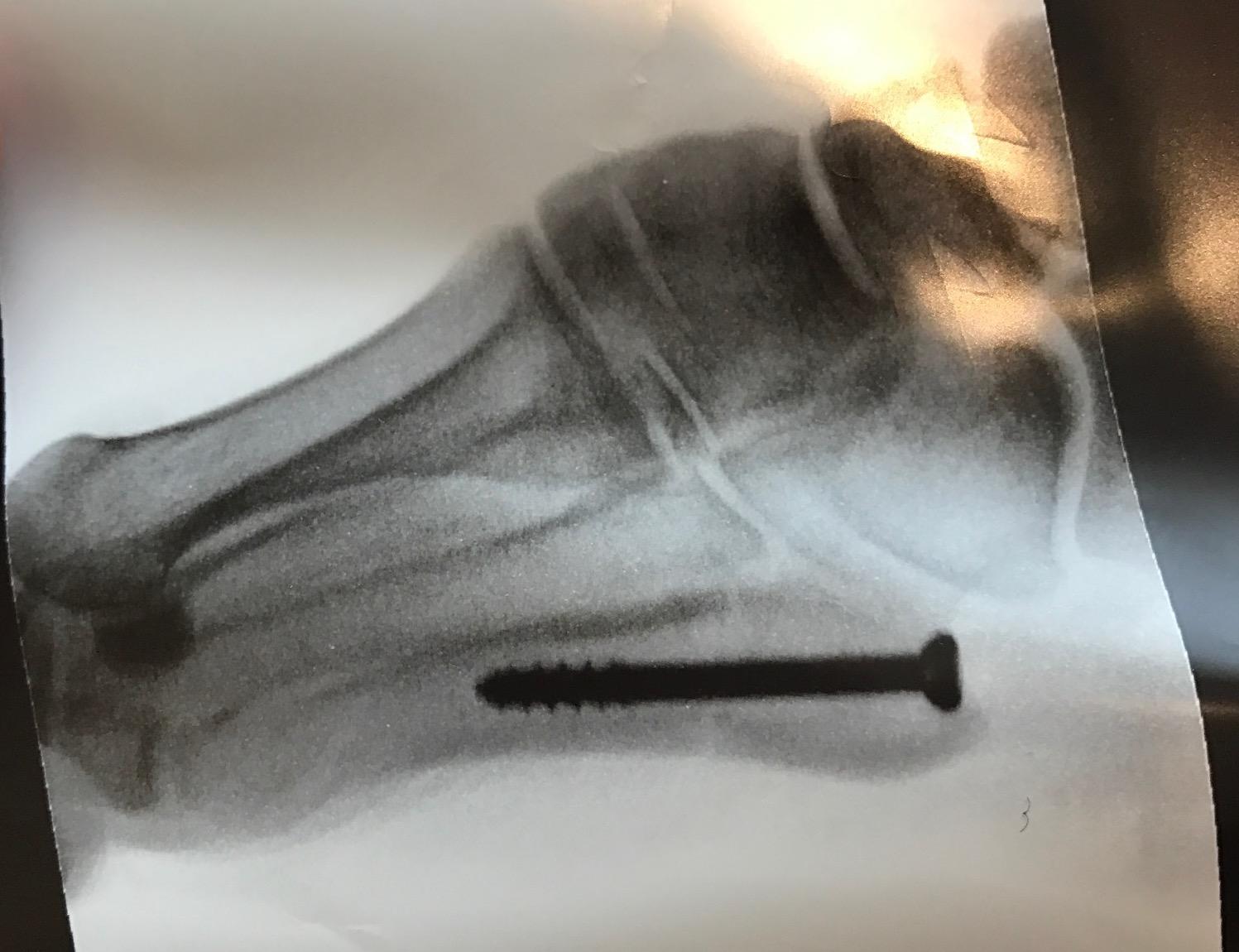
Nondisplaced zone 2 injuries, or Jones fractures, may also be treated conservatively with 6 to 8 weeks of non-weight bearing in a short leg cast. The physician may advance weight-bearing status as radiographic evidence of bone healing appears. Indications for surgical interventions include the high-performance athlete, the informed patient who elects to proceed with surgical treatment, or displaced fractures. There are many forms of surgical interventions, including intramedullary screw fixation, tension band constructs, and low-profile plates and screws. Surgical management of high-performance athletes minimizes the risk of nonunion and prevents prolonged restriction from physical activity.
Diaphyseal zone 3 stress fractures paint a more complicated picture for the patient and physician. A trial of conservative management with non-weight bearing in a short leg cast may be the initial therapy; however, immobilization for up to 20 weeks may be necessary before there is observable radiographic union, and even then, nonunion development is not uncommon. High-performance athletes or individuals with Torg Type II or III fractures may require surgical interventions. Surgical options include intramedullary screw fixation, bone grafting procedures, or a combination of the two.[7]
The bone grafting inlay technique requires removing a 0.7 by 2.0 cm rectangular section of bone at the fracture site and replacing it with an autogenous corticocancellous bone graft of the same dimensions taken from the anteromedial distal tibia. The medullary cavity must be curetted or drilled until all the sclerotic bone has been removed and the medullary canal reestablished prior to inserting the donor graft.
Nondisplaced dancer’s fractures and other fractures of the fifth metatarsal shaft and neck receive the same treatment as nondisplaced zone 1 injuries. Weight-bearing status can advance as tolerated by pain. Surgical interventions may be required if evidence of delayed union or nonunion exists. If there is more than 3 mm of displacement or angulation exceeds 10 degrees, the fracture should be reduced and splinted.[8] If the fracture remains malreduced or there is evidence of loss of reduction on follow-up radiographs, surgical interventions with percutaneous pinning or plate and screw fixation should be a consideration.
Patients treated with intramedullary screw fixation or bone graft inlay technique should remain non-weight bearing in a plaster splint or short leg cast for six weeks with a gradual return to sport or activity.[7]
Differential Diagnosis
Differential diagnosis of pain in this region includes:
- Bone tumor
- Bursitis
- Calluses
- Foreign body granuloma
- Ganglia
- Gout
- Hemangioma
- Metatarsalgia
- Morton neuroma
- Neuropathic osteoarthropathy
- Osteoarthritis
- Osteomyelitis
- Plantar fibromatosis
- Plantar plate disruption
- Rheumatoid arthritis
- Sesamoiditis
- Septic arthritis
- Stress fracture
- Subchondral insufficiency fracture
- Tendinosis
- Tenosynovitis
- Tendon rupture
- Tenosynovial giant cell tumor
- Turf toe
Prognosis
The majority of acute, nondisplaced fractures of the fifth metatarsal heal with conservative treatment by 6 to 8 weeks. Zones 2 and 3 injuries have a higher rate of nonunion due to the vascular watershed area mentioned earlier, with zone 2 injuries having a nonunion rate as high as 15 to 30%. Diaphyseal stress fractures may have a prolonged course of healing of up to 20 weeks.
Complications
There is an increased risk of nonunion with zones 1 and 2 injuries, as discussed earlier. There is also a risk of nonunion associated with using an intramedullary screw with a diameter under 4.5 mm or the use of a screw that is too long, causing fracture distraction or malreduction.[7] Fixation failure rates are higher in individuals who do not abide by non-weight bearing restrictions or who return to sport or activity before evidence of radiographic union.
Postoperative and Rehabilitation Care
Patients, especially high-level athletes, should be educated regarding the risk of nonunion in specific fracture patterns. The clinician should conduct a thorough discussion of both operative and nonoperative management.
Rehabilitation
The progression of rehabilitation is primarily dictated by the type and severity of the fracture, the resulting treatment approach selected by the orthopedic physician, and the established protocol provided by each referring orthopedic professional. Limited evidence is available regarding one specific rehabilitation protocol following a metatarsal fracture. As such, it is imperative for the rehabilitation team to coordinate with the referring orthopedic professionals on desired rehab timeline and components. In addition, the rehabilitation approach is also determined by the patient's initial status and overall goals, with a more aggressive approach instituted for athletes, including early limb loading and anti-gravity treadmills as means to avoid increased gaps in training.[9][10]
In general, rehabilitation consists of 3 phases: Phase 1 emphasizes ambulation and assistive device/orthotic training, range of motion (ROM) exercises, and pain/edema management. Phase 2 is a continuation of ROM exercises with the introduction and progression of resistance exercises, manual soft tissue mobilization and scar management, continued gait training and use of anti-gravity treadmill/assistive device/orthotic training, and exercises to promote and maintain the strength of lower extremities/upper extremities during rehab efforts. Phase 3 includes full ROM of affected joints in the foot and ankle, progression of resistance and strength training of both the affected joint and lower extremities, progression of gait training with an emphasis on normalizing gait pattern without assistive device use, implementation of stability and proprioceptive exercises, and progression of motions specific to the sport if rehabbing an athlete (including agility and coordination drills as appropriate).[10][11][9]
Deterrence and Patient Education
Patients must understand what they need to do depending on the type of injury sustained. In some cases, rest is the only treatment needed to promote the healing of a stress or traumatic fracture of a metatarsal bone. Patients must also avoid any offending or exacerbating activities. Casting, immobilization, or a rigid shoe may be required in certain instances. For both surgical and non-surgical cases, physical therapy, exercises, and rehabilitation may be scheduled to enable a return to normal activities. Patient compliance with therapy, exercise, and device use is crucial to success.[12]
Enhancing Healthcare Team Outcomes
An interprofessional team, including the orthopedic nurse, is most effective in appropriately managing fifth metatarsal fractures. Generally, the patient will present to their primary care physician or the emergency department complaining of typical signs and symptoms. Proper diagnosis directs the treatment and triage of these patients. Prompt referral to an orthopedic surgeon is paramount as appropriate treatment can lead to union rates as high as 97%.[13] [Level 4] Mismanagement of these fractures may lead to poor clinical outcomes and lifestyle alterations. An emergent referral to orthopedic surgery is necessary for an open fracture. Traditionally, intramedullary screw fixation has been an option when surgery is warranted, but newer techniques with planar plating have resulted in successful outcomes in elite athletes.[14]
With proper identification of fracture and interprofessional communication with an orthopedic referral, outcomes of this difficult-to-treat fracture can stand a better chance of full recovery, but this requires an interprofessional team approach, including clinicians, specialists, specialty-trained nurses, physical and occupational therapists, and pharmacists, all collaborating across disciplines to achieve optimal patient results. [Level 5]