Introduction
A joint is a point where two bones make contact. Joints can be classified either histologically or functionally. Histological classification is based on the dominant type of connective tissue, and functional classification is based on the amount of movement permitted. Histologically the three joints in the body are fibrous, cartilaginous, and synovial. Functionally the three types of joints are synarthrosis (immovable), amphiarthrosis (slightly moveable), and diarthrosis (freely moveable). The two classification schemes can be correlated: synarthroses are fibrous, amphiarthroses are cartilaginous, and diarthroses are synovial.[1][2]
Joints, comprising bones and connective tissue, are embryologically derived from mesenchyme. The bones either develop directly through intramembranous ossification or indirectly through endochondral ossification. Each specific joint has a unique vascular supply and innervation scheme; patterns do exist. Muscles provide stability to joints, and there is a direct correlation between muscle strength and joint stability, particularly with synovial joints.
Many pathophysiological conditions affect joints, and again, patterns exist by histological class. Because diseases that affect the joints are common across the lifespan, a thorough understanding of joint structure and function is of great clinical significance.
Structure and Function
The histological and functional classification schemes offer a broad understanding of joints. As aforementioned, histological classification is based on the dominant type of connective tissue, and functional classification is based on the amount of movement permitted.
Histologically the three joints in the body are fibrous, cartilaginous, and synovial. Functionally the three types of joints are synarthrosis (immovable), amphiarthrosis (slightly moveable), and diarthrosis (freely moveable). The two classification schemes can be correlated: synarthroses are fibrous, amphiarthroses are cartilaginous, and diarthroses are synovial.[1][2]
Within these categories, each specific joint type (suture, gomphosis, syndesmosis, synchondrosis, symphysis, hinge, saddle, planar, pivot, condyloid, ball, and socket) has a specific function in the body.
Fibrous Joints
A fibrous joint is a fixed joint where collagenous fibrous connective tissue connects two bones. Fibrous joints (synarthroses) are usually immovable and have no joint cavity. They are subdivided further into sutures, gomphoses, and syndesmoses.
Sutures are immobile joints found only in the cranium. The plate-like bones of the skull are slightly mobile at birth because of the connective tissue between them. The spaces between the bones are termed fontanelles. This initial joint flexibility allows the fetal head to pass through the birth canal at delivery and permits brain enlargement after birth. As the skull enlarges, the fontanelles reduce to a narrow layer of fibrous connective sutures that suture the bony plates together. This connective tissue is termed Sharpey fibers. Eventually, cranial sutures ossify. Finally, the two adjacent plates fuse to form one bone; this fusion is termed synostosis.
Gomphoses are the immobile joints found only between the teeth and their sockets in the mandible and maxillae. The periodontal ligament is the fibrous tissue that connects the tooth to the socket.
Syndesmoses are slightly movable joints (amphiarthroses). This type of fibrous joint maintains integrity between long bones and resists forces that attempt to separate the two bones. All syndesmoses are amphiarthroses, but each specific syndesmosis joint permits a different degree of movement. For example, the tibiofibular syndesmosis primarily provides strength and stability to the leg and ankle during weight-bearing; however, the antebrachial interosseous membrane of the radioulnar syndesmosis permits rotation of the radius bone during forearm movements. The interosseous membranes of the leg and forearm are also areas of muscle attachment.[3]
Cartilaginous Joints
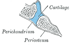
In cartilaginous joints, the bones attach by hyaline cartilage or fibrocartilage. Depending on the type of cartilage involved, the joints are further classified as primary or secondary cartilaginous joints.
A synchondrosis, or primary cartilaginous joint, only involves hyaline cartilage and can be temporary or permanent.
A temporary synchondrosis is an epiphyseal plate (growth plate). It functions to permit bone lengthening during development. The epiphyseal plate connects the diaphysis (shaft of the bone) with the epiphysis (end of the bone) in children. Over time, the cartilaginous plate expands and is replaced by bone, adding to the diaphysis. Eventually, when all the hyaline cartilage has ossified, the bone has completed its lengthening, and the diaphysis and epiphysis fuse in synostosis. Other temporary synchondroses join the ilium, ischium, and pubic bones of the hip. Over time, these also fuse into a single hip bone.
A permanent synchondrosis does not ossify with age; it retains its hyaline cartilage. Permanent synchondroses function to connect bones without movement as a synarthrosis joint. Examples are found in the thoracic cage, such as the first sternocostal joint: the first rib is joined to the manubrium by its costal cartilage. Other examples include the relationship between the anterior end of the other 11 ribs and the costal cartilage.[4]
A symphysis, or secondary cartilaginous joint, involves fibrocartilage. Fibrocartilage is thick and strong, so symphyses have a remarkable ability to resist pulling and bending forces. While the fibrocartilage firmly unites adjacent bones, the joint is still an amphiarthrosis joint and permits limited movement.
A symphysis can be narrow or wide. Narrow symphyses include the pubic symphysis and the manubriosternal joint. In females, the slight mobility of the pubic symphysis between the left and right pubic bones is critical for childbirth. A wider example of a symphysis is the intervertebral symphysis or intervertebral disc. The thick pad of fibrocartilage fills the gap between adjacent vertebrae and provides cushioning during high-impact activity.
Synovial Joints
Synovial joints (diarthroses) are freely mobile and are considered the main functional joints of the body. The synovial joint is characterized by the presence of a joint cavity. The primary purpose of the synovial joint is to prevent friction between the articulating bones of the joint cavity. The joint cavity is surrounded by the articular capsule, a fibrous connective tissue attached to each participating bone just beyond its articulating surface. The joint cavity contains synovial fluid secreted by the synovial membrane (synovium), which lines the articular capsule. Hyaline cartilage forms the articular cartilage, covering the entire articulating surface of each bone. The articular cartilage and the synovial membrane are continuous. Some synovial joints also have associated fibrocartilage between articulating bones. An example of this is the menisci of the knee.
While all synovial joints are diarthroses, the extent of movement varies among different subtypes and is often limited by the ligaments that connect the bones. Therefore, synovial joints are often further classified by the type of movements they permit. There are six such classifications: hinge (elbow), saddle (carpometacarpal joint), planar (acromioclavicular joint), pivot (atlantoaxial joint), condyloid (metacarpophalangeal joint), and ball and socket (hip joint).
Synovial Joint: Hinge
A hinge joint is an articulation between the convex end of one bone and the concave end of another. This type of joint is uniaxial because it only permits movement along one axis. In the body, this axis of movement is usually flexion and extension (bending or straightening). Examples include the elbow, knee, ankle, and interphalangeal joints.
Synovial: Condyloid
A condyloid joint, or an ellipsoid joint, is an articulation between the shallow depression of one bone and the rounded structure of one or more other bones. This type of joint is biaxial because it permits two axes of movement: flexion/extension and medial/lateral (abduction/adduction). An example is the metacarpophalangeal joints of the hand between the distal metacarpal and proximal phalanx, commonly known as the knuckle.
Synovial Joint: Saddle
A saddle joint is an articulation between two bones that are saddle-shaped - concave in one direction and convex in another. This type of joint is biaxial. One example is the first carpometacarpal joint between the trapezium (carpal) and the first metacarpal bone of the thumb. This arrangement permits the thumb to flex and extend (within the plane of the palm) as well as abduct and adduct (perpendicular to the palm). This dexterity gives humans the characteristic trait of “opposable” thumbs. The opposable thumb is critical for the complex motions involved in the use of the hand. Loss of the thumb by any mechanism severely limits the utility of the hand.
Synovial Joint: Planar
A planar joint, or gliding joint, is defined as an articulation between two bones that are both flat and of similar size. This type of joint is multiaxial because it permits many movements; however, surrounding ligaments usually restrict this joint to a small and tight motion. Examples include intercarpal joints, intertarsal joints, and the acromioclavicular joint.
Synovial Joint: Pivot
A pivot joint is an articulation within a ligamentous ring between the rounded end of one bone and another bone. This type of joint is uniaxial because, although the bone rotates within this ring, it does so around a single axis. An example is the atlantoaxial joint between C1 (atlas) and C2 (axis) of the vertebrae. This joint permits side-to-side head motion of the head. Another example is the proximal radioulnar joint. The radius sits in the annular radial ligament, which holds it in place as it articulates with the radial notch of the ulna, which permits pronation and supination.
Synovial Joint: Ball and Socket
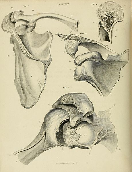
A ball and socket joint is an articulation between the rounded head of one bone (ball) and the concavity of another (socket). This type of joint is multiaxial: it permits flexion/extension, abduction/adduction, and rotation. The only two ball and socket joints of the body are the hips and the shoulder (glenohumeral). The shallow socket of the glenoid cavity permits a more extensive range of motion in the shoulder; the deeper socket of the acetabulum and the supporting ligaments of the hip constrain the movement of the femur.
Embryology
Joints, comprising bones and connective tissue, are embryologically derived from mesenchyme. The bones either develop directly through intramembranous ossification or indirectly through endochondral ossification. During direct development, the mesenchymal cells differentiate into bone-producing cells. During indirect development, the mesenchymal cells first differentiate into hyaline cartilage that is gradually displaced by bone. The connective tissue of the joint arises from the mesenchymal cells between the developing bones.
For the synovial joints of the limbs, the space between the developing long bones is termed the joint interzone. The interzone becomes apparent in the sixth week of embryonic development when a cellular condensation of mesoderm on either side, termed the paraxial blastema, chondrifies into hyaline cartilage models for the long bones. In the eighth week of embryonic development, mesenchymal cells at the margin of the interzone become the articular capsule; cell death in the center forms the joint cavity, which is filled with synovial fluid produced by mesenchymal cells. The articular cartilage is a remnant of the hyaline cartilage that, between gestational weeks 6 and 8, became the long bones via endochondral ossification.[5][6]
Blood Supply and Lymphatics
Every joint has a different blood supply; however, there are patterns based on the histological classification of joints.
Fibrous joints are usually supplied by perforating branches of the proximal vessels. For example, the blood supply of the tibiofibular joint is derived from branches of the anterior tibial artery and the peroneal (fibular) artery.
Cartilaginous joints only receive their vascular supply at the periphery because cartilage is an avascular tissue. Intervertebral discs, for example, are supplied at the margins by capillaries from the vertebral bodies.
Synovial joints receive vascular supply through a rich anastomosis of arteries extending from either side of the joint, termed the periarticular plexus. Some vessels penetrate the fibrous capsule to form a rich plexus deeper in the synovial membrane. This deeper plexus, the circulus vasculosus, forms a loop around the articular margins that supplies the articular capsule, synovial membrane, and terminal bone. The articular cartilage, which is avascular hyaline cartilage, is nourished by the synovial fluid.
Lymphatic vessels for every joint follow the lymph drainage of the surrounding tissue; some joints house lymph nodes, like the popliteal lymph nodes in the popliteal fossa of the knee.
Nerves
Every joint in the body has different innervation; however, the innervation of synovial joints is most extensively understood.
Sensory and autonomic fibers innervate synovial joints. The autonomic nerves are vasomotor in function, controlling the dilation or constriction of blood vessels through the actions of norepinephrine and epinephrine. These sympathetic nervous system fibers use alpha-1 adrenergic receptors to cause the arteriolar vascular smooth muscle to contract. The sensory nerves of the articular capsule and ligaments (articular nerves) provide proprioceptive feedback from Ruffini endings and Pacinian corpuscles. Proprioception of the joint permits reflex control of posture, locomotion, and movement. Free nerve endings convey pain sensation that is diffuse and poorly localized. The articular cartilage has no nerve supply.
Two general principles apply to synovial joint innervation: the Hilton law and the Gardner observation. The Hilton law states that the articular nerves supplying a joint are branches of the nerves that supply the muscles responsible for moving that joint. Therefore, irritation of articular nerves causes a reflex spasm of the muscles, which position the joint for the greatest comfort. These nerves also supply the overlying skin, providing a mechanism for referred pain from joint to skin. The Gardner observation indicates that the part of the articular capsule that is tightened by contraction of a group of muscles receives nerve supply from the same nerves that innervate the antagonist muscles. This relationship provides local reflex arcs that stabilize the joint.[7]
Muscles
Muscles are most critical in providing additional support for synovial joints. The muscles and their tendons which cross a joint resist the forces acting on that joint, behaving as a dynamic "ligament." Muscle strength is, therefore, essential to the stability of synovial joints, especially during high-stress activity, as well as for joints with weaker ligaments, for example, the glenohumeral joint.
Clinical Significance
Different pathologic conditions are associated with different joint types. Below is a review of the most common injuries that plague each histological class.
Fibrous Joints
Sutures, the immobile fibrous joints that bind the bony plates of the skull, can fuse too early in development, a condition termed craniosynostosis. The plates of the neonatal skull are not fused so as to permit space for the brain to grow in all planes; early fusion (synostosis) alters the shape of the head. For example, if the sagittal suture synostoses, the head will not develop width and will instead grow long and narrow (scaphocephaly). In addition to altered head shape, some children may experience symptoms secondary to high brain pressure due to more confined skull space. These include headaches, developmental delays, or problems with eyesight.[8]
A syndesmosis joint, the slightly mobile fibrous joint that connects long bones with an interosseous membrane, can be sprained. For example, in the leg, excessive external rotation can push the fibula away from the tibia, causing injury to the distal tibiofibular syndesmosis; this is termed a “high ankle sprain.” [9][3]
Cartilaginous Joints
Epiphyseal plates, an example of temporary synchondroses, are vulnerable to damage when there is an injury to the associated growing long bone. Such damage to the cartilage would stop bone lengthening and stunt bone growth.
Synovial Joints
Arthritis results in the destruction of the synovial joint. There are many types of arthritis, distinguished by different mechanisms of injury. The most common type of arthritis is osteoarthritis, which, by definition, is gradual damage to and subsequent thinning of the articular cartilage. This condition is considered a “wear and tear” injury and presents in older patients; it often correlates with prior injury to the joint and longstanding high-impact stress on the joint (due to sports or excessive body weight). Because the articular cartilage has no innervation, the degradation itself does not cause pain. Instead, as the articular cartilage becomes thinner, more pressure is placed on the bones. The joint responds by overproducing synovial fluid, which leads to swelling and inflammation that stretches the highly innervated articular capsule to cause pain and stiffness of the joint. The underlying bone also has a rich nerve supply that perceives pain.
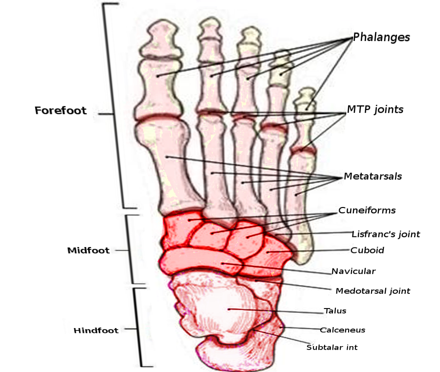
Gout is another form of arthritis caused by the deposition of uric acid crystals within a joint. Uric acid causes gout when there is an excessive amount in the body, either due to over-production or improper excretion by the kidneys. The most commonly affected joint is the metatarsophalangeal (MTP) joint of the big toe. This condition is termed podagra.[10][11][12] Patients often present with excruciating pain and swelling.[13] The consumption of alcohol may exacerbate podagra.[14][15][16]
Synovitis is inflammation of the synovial membrane that lines the articular capsule of synovial joints. The most common cause is the overuse of a synovial joint in an active, healthy person. Persistent synovitis in multiple joints can indicate rheumatoid arthritis, where the synovium is the target of the autoimmune attack. Patients with synovitis often present with pain out of proportion to examination; sometimes, the patient has pain without swelling or tenderness, or arthralgia.