Continuing Education Activity
Acromioclavicular (AC) and sternoclavicular (SC) joint injuries are most commonly seen in a contact sport setting (such as football, rugby, and ice hockey) or traumatic setting. The severity of these injuries can vary, the worst of which require surgical intervention. For those that do not require surgery, osteopathic manipulative treatment (such as muscle-energy) is a valuable option in the treatment of these injuries. This activity reviews the evaluation and treatment procedure of the AC and SC joints with muscle energy. It highlights the role of the healthcare team in treating patients with these injuries.
Objectives:
- Review the anatomy of the AC and SC joints.
- Describe the diagnostic steps to identify somatic dysfunctions of the AC and SC joints.
- Summarize the muscle energy procedure, along with its indications and contraindications, for the AC and SC joint.
- Outline the importance of communication amongst the interprofessional team to improve outcomes for patients receiving muscle energy treatment for the AC and SC joints.
Introduction
Acromioclavicular (AC) joint injuries are one of the more common injuries of the shoulder. A recent study by Nordin et al. found that AC Joint injuries have an incidence of 2 in every 10,000 people ages 18 to 75. Most of these injuries are in young men, with more severe AC joint injuries occurring in the elderly population.[1] AC joint injury severity is assessed radiographically. Images and patient characteristics provide indications for surgery. Damage to surrounding structures such as the AC ligament, coracoclavicular ligament, and muscle stabilizers of the shoulder are indicators of more severe damage to the AC Joint.[2]
The Tossy and Rockwood classifications are two of the most commonly used radiographic systems in the evaluation of AC joint injury severity. Tossy has three categories: AC sprain, AC subluxation, and AC dislocation.[3] Rockwood has six more specific categories: Damage to AC ligament (I), rupture of AC ligament and damage to coracoclavicular ligament (II), rupture of both AC and Coracoclavicular ligament (III), posterior dislocation of AC joint (IV), high-grade superior dislocation with rupture of dynamic stabilization mechanism (V), and inferior dislocations of AC joint (VI).[2] Non-surgical treatments are recommended for types I and II, and surgery is indicated for types IV and VI. There is controversy surrounding the surgical vs. non-surgical approach in types III and V.[4]
The sternoclavicular (SC) joint has its own variety of ligamentous injuries, similar to that of the AC joint. However, its anatomical position of dislocations is either anterior or posterior. Anterior dislocation can often be resolved with closed reduction versus posterior dislocation, which may need open reduction and is more severe due to the critical anatomy residing posterior to the clavicle.[5] Injury to the SC joint is much less common. One retrospective study found an incidence of just 0.9% of all shoulder girdle injuries seen at a level 1 trauma center over 19 years.[6]
This review aims to look at the non-surgical treatment of AC and SC joint injuries. Specifically, muscle energy techniques that are utilized in osteopathic manipulative treatment. Muscle energy is a technique typically used to treat hypertonic muscles and restricted joints. It applies to structures throughout the body, especially the AC and SC joints. The isometric variation of the muscle energy technique is most commonly used, and includes the following steps [7]:
- Localization of the restrictive barrier of the muscle/joint under evaluation
- The patient actively contracts muscle/joint in a specific direction for a specific amount of time (usually 3 to 5 seconds)
- Counterforce is being applied by the provider during this 3 to 5-second interval.
- The patient relaxes after this 3 to 5-second interval.
- The provider takes the muscle/joint being treated further into the restrictive barrier.
- Steps #2 through 5 are repeated (typically done 3 to 5 times total)
Anatomy and Physiology
The AC joint is just one of the complex structures that makes up the shoulder girdle. The acromion, which originates from the spine of the scapula, meets with the lateral aspect of the clavicle to form the AC joint. The clavicle and the acromion of the scapula have a flat articular surface, with an oval contour; the clavicular facet turns outwards and downwards, the acromial facet looks medially and upwards. Between the articular surfaces, there is a fibrocartilaginous disc with variable development; it is rarely complete. It is stabilized with the deltoid and trapezius muscles, as well as the AC ligament and coracoclavicular ligaments. The coracoclavicular ligament consists of the conoid and trapezoid ligaments, which provides vertical stability, whereas the AC ligament provides horizontal stability.[8]
The AC joint is a synovial joint (arthrodial joint) that only allows gliding motion. It assists in abduction and flexion of the shoulder. The AC joint receives blood supply from the suprascapular and thoracoacromial arteries, along with innervation from the suprascapular, axillary, and lateral thoracic nerves.[9] The acromioclavicular joint comes into play, simultaneously with the sternoclavicular joint and within the entire joint mechanism of the thoracic belt, to allow sliding movements through which the scapula changes its relationship with the chest. As a result of these behaviors of the scapula, the glenoid cavity is oriented to allow wider freedom of movement to the arm. In the mechanics of the acromioclavicular joint, the trapezoid and conoid ligaments, stretched between the coracoid process and the external third of the clavicle, is of great importance. They release the acromioclavicular joint of part of the weight exerted by the upper limb and, by limiting the motility of the joint itself, they help to fix the scapula.
The SC joint is also a synovial joint that articulates with the manubrium of the sternum and the first costal cartilage. The SC joint is tough to injure due to the amount of stabilizing structures that surrounds it. These include the anterior and posterior sternoclavicular ligaments, interclavicular ligaments, costoclavicular ligaments, as well as the subclavius muscle. The SC joint is diarthrodial (saddle-shaped), and communication from the shoulder can dictate its movement. The SC joint functionally allows for motion of the shoulder in various planes, including horizontal, coronal, and anteroposterior planes. Blood supply to this joint comes from the internal thoracic and suprascapular arteries, and it receives innervation from the medial supraclavicular and subclavius nerves. As mentioned, posterior dislocations of the SC joint can result in significant complications due to the crucial anatomy posterior to this joint. Structures such as the brachiocephalic trunk, internal jugular vein, common carotid artery, trachea, esophagus, and vagus nerve are all vulnerable to a posteriorly dislocated SC joint.[10]
The physiology underlying the muscle energy technique is not fully understood. It appears to be due to neurophysiologic responses that cause inhibition of neural responses allowing muscular relaxation, as well as an alteration in pain modulation.[11]
Indications
Muscle energy techniques (METs) are attributed to an American osteopath, Fred Mitchell Senior. Employing complex neurological mechanisms, including the neuromuscular spindles and the Golgi tendon organs, following guided muscle contractions, a shortened muscle can be stretched until it reaches a new resting length.
METs represent a manual treatment modality that involves the voluntary contraction of some muscles of the patient in a very precise direction, with different levels of intensity, carried out towards a counterforce exerted specifically by the operator.These techniques classify as active or direct techniques.
Isometric and concentric isotonic contraction can be used in three different ways on the dysfunction:
- Isometric contraction opposed to the restriction.
- Isometric contraction towards the restriction
- Concentric isotonic contraction towards restriction
Indications:
- Hypertonic muscles
- Somatic dysfunction of muscle or joint
- Limited ROM of joint
- To decrease localized edema and reduce congestion (pumping of the venous and lymphatic systems)
Contraindications
Muscle energy is a very safe technique, and it can be utilized outside of medical emergencies or very severe injuries (such as the following examples):
- Broken bone
- Ruptured ligament/tendon/muscle
- Joint infection
- No somatic dysfunction present
- Patient refusal
Equipment
- Osteopathic manipulative treatment table
- Stool
Preparation
Evaluating the AC and SC joints is critical before beginning treatment. Visual assessment (bruising, erythema, edema), pain scale, the strength of surrounding musculature, and range of motion of the AC and SC joints should all be accounted for both before and after treatment.
The following will explore two somatic dysfunctions of the AC joint in this review that are treatable with muscle energy:
- Internally rotated AC joint, or
- Externally rotated AC joint
Palpation of the AC joint can be done by "walking" on hand down the spine of the scapula to the acromion, while the other hand travels down the clavicle laterally. Where these two points meet will be the AC joint. Let the patient's arm hang by there side in sight abduction of the shoulder with minimal flexion of the elbow. With the patient in this position, palpate the AC joint with one hand, and manipulate their shoulder into internal rotation, followed by external rotation. We compare this movement of the AC joint bilaterally. The somatic dysfunction is named the opposite of the restriction. For example, if the range of motion of the left and right AC joint is normal in internal rotation B/L, but the AC joint on the right has a restriction in the external rotation when compared to the left. The right AC joint is said to be internally rotated. One could also look at naming the somatic dysfunction as where the joint is "living." For example, if a patient has a left externally rotated AC joint, this means the AC joint allows for external rotation (AKA "living" in external rotation) and shows restriction in left internal rotation (when compared to the right AC joint).
For the SC joint, this text will also explore two possible somatic dysfunctions:
- Adducted clavicle
- Horizontally extended clavicle
Before getting into the evaluation of somatic dysfunction, it is imperative to understand the relationship between the shoulder and clavicle. The position of the clavicle is named for the movement at the lateral aspect of the clavicle! Abduction of the shoulders causes the medial edge of the clavicle (closest to SC joint) to drop inferior, and the lateral edge to rise superior (as if it is abducting with the shoulder).
You can also think of the clavicle and its movement as a see-saw. Flexion of the shoulder brings the arm anterior, and we would say the lateral edge of the clavicle travels anterior (horizontal flexion) with it, and the medial edge travels posterior. As we extend the shoulder, the arm moves posterior, and the lateral edge of the clavicle also travels posterior (horizontal extension), while the medial portion of the clavicle travels anteriorly.
The SC joint can be palpated by following the clavicle medially with one hand and traveling laterally off the manubrium with the other. Evaluation of this joint is best with the patient in the supine position.
To evaluate for an adducted clavicle:
- The provider should be at the head of the table, with the index finger on the superior portion of the SC joint.
- The patient (lying supine) will be instructed to shrug both shoulders to their ears simultaneously (this imitates the abduction of the shoulder)
- The provider will compare the SC joints at the peak of this shoulder shrug: whichever SC joint drops least inferior (or stays more cephalad in terms of the supine patient), is the adducted clavicle.
- To evaluate for a horizontally extended clavicle:
- The provider should be at the head of the table, with index fingers on the anterior portion of the SC joint.
- The patient (supine) is instructed to slowly move their shoulders into flexion.
- The provider compares the posterior movement at the SC joint, whichever SC joint does NOT drop posteriorly is said to be a horizontally extended clavicle.
Technique or Treatment
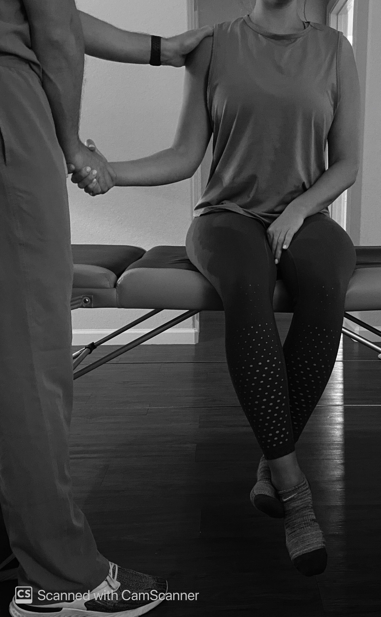
Muscle Energy Technique for an Internally Rotated AC Joint:
- Have the patient in a seated position, with the arm in slight abduction and elbow slightly flexed
- While one hand monitors AC joint, use the other to "shake hands" with the patient and guide the AC joint into external rotation until the restrictive barrier is met
- The patient internally rotates their shoulder against isometric resistance from the provider for 3 to 5 seconds.
- The provider instructs the patient to relax and takes shoulder further into the restrictive barrier (thus, further into external rotation)
- Repeat steps 3 and 4 multiple times (typically 3 to 5 times)
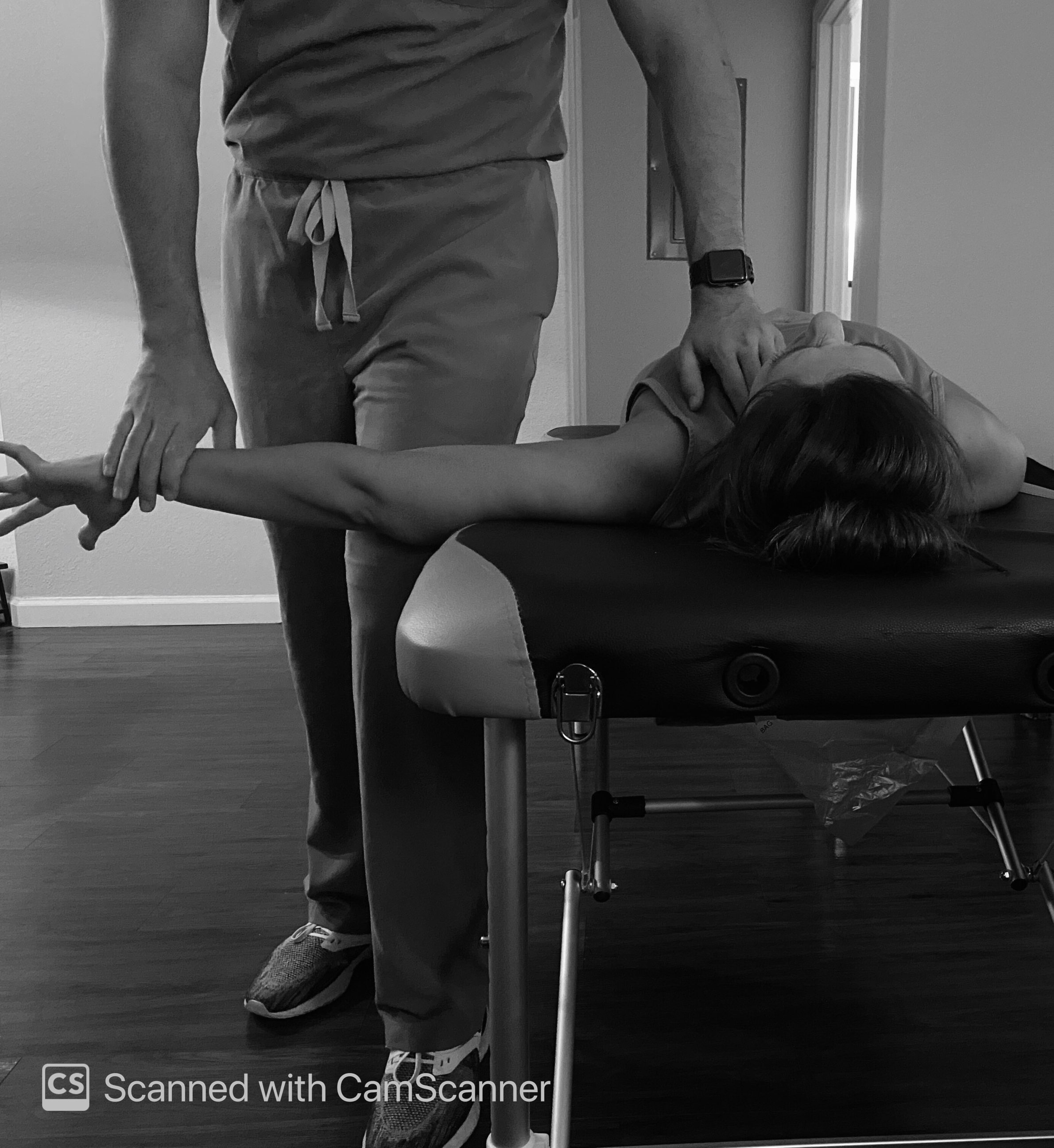
Muscle Energy Technique for an Adducted Clavicle:
- Provider standing on the side of the table/patient (on the side of the adducted clavicle) and facing cephalad relative to the patient
- The patient abducts arm on the side of dysfunction to 90 degrees with the thumb facing down.
- The provider positions his/her thigh against the patient's abducted arm. The hand closest to the patient contacts the superior part of the SC joint.
- Patient instructed to push arm into the provider's thigh, while the provider isometrically resists (for 3 to 5 seconds)
- The provider instructs the patient to relax.
- The provider abducts the shoulder further to the restrictive barrier while maintaining an inferior force on the SC joint.
- Repeat steps 4 to 6 multiple times (typically 3 to 5 times)
- Recheck
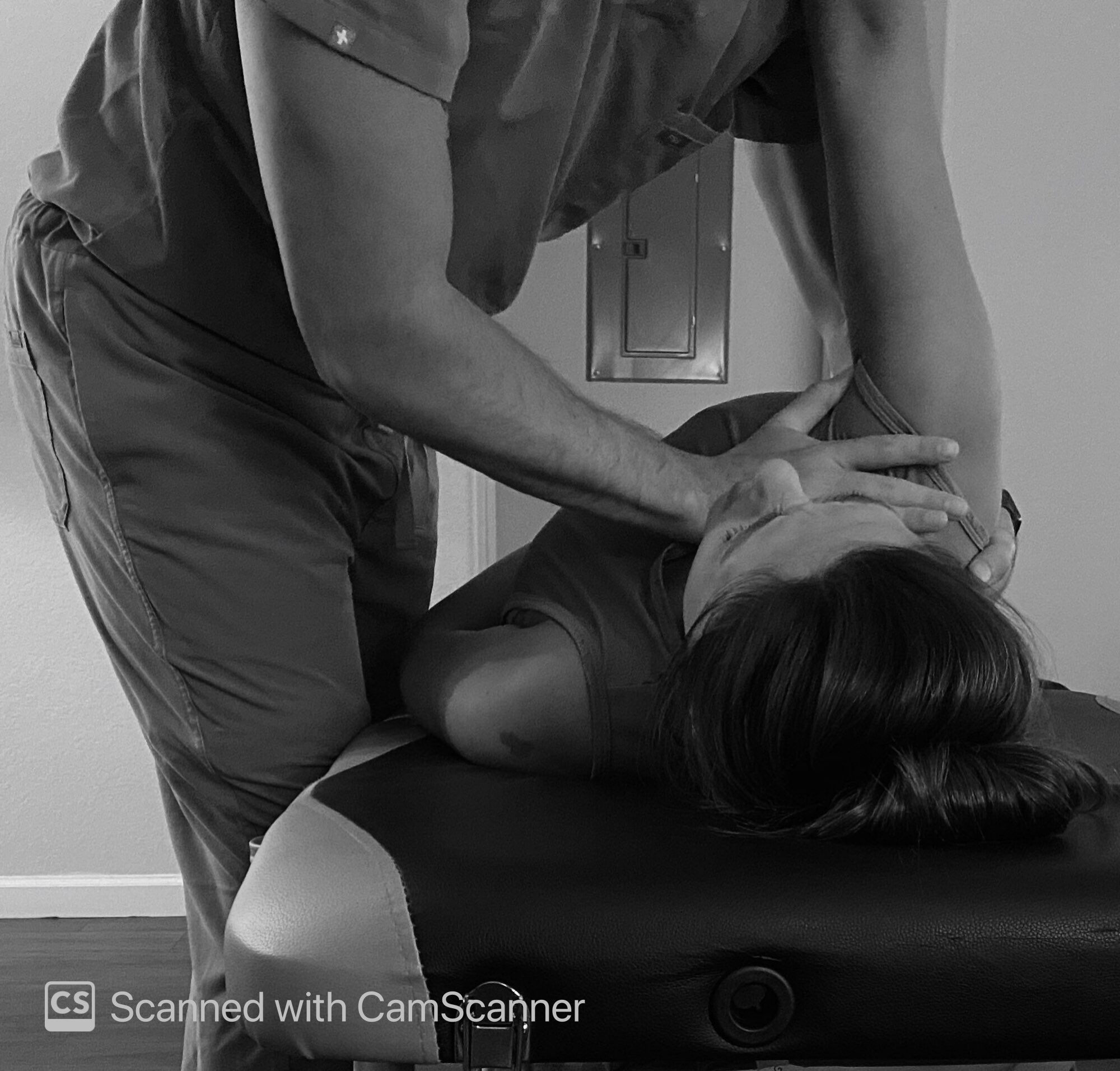
Muscle Energy Technique for a Horizontally Extended Clavicle:
- Provider standing on the side of the table/patient (opposite the side of the dysfunction)
- Provider contacts the anterior surface of the dysfunctional SC joint with the palm of the hand
- Instruct patient to bring the hand to the ceiling (flexion of the shoulder) and wrap an arm around the provider's trapezius area
- The provider should then contact the scapular region with their free-hand.
- The patient is instructed to try to bring scapula down towards the table, while the provider resists (for 3 to 5 seconds)
- The provider instructs the patient to relax.
- While relaxed, provider further pushes SC joint posterior while simultaneously bringing the patient's shoulder/scapula region anterior
- Repeat steps 5 to 7 multiple times (typically 3 to 5 times)
- Recheck
Complications
As one study states, significant adverse events are not well described or correlated with manual therapy use. For instance, there is no correlation to a musculoskeletal treatment with resulting gastrointestinal complications that we are aware of, although they rarely coincidentally occur. However, minor adverse events that require little to no follow-up are known to occur with manual therapy, such as headache, muscle soreness, and light-headedness.[12] Patients should refrain from strenuous activity after treatment and drink ample amounts of water.
Clinical Significance
Osteopathic manipulative treatment is an underutilized non-surgical alternative that can provide significant benefits to patients with somatic dysfunctions of the AC and SC Joints. As mentioned before, non-surgical approaches are best for AC injuries type I and II, and there is still dispute over types III and V. All non-surgical approaches should be exhausted prior to surgery, and muscle energy is a viable option to treat these non-surgical injuries while benefiting the surrounding stabilizing structures as well.
Enhancing Healthcare Team Outcomes
Interprofessional communication is something that lacks in terms of osteopathic manipulative treatment. As an osteopathic treatment, only DOs are familiar with the process (unless an MD has taken a separate course). With a large number of patient complaints stemming from the musculoskeletal origin, we need to educate physicians and mid-level practitioners about osteopathic manipulative treatment so patients can be directed to the proper physician to enhance patient care. [Level 5]
Nursing, Allied Health, and Interprofessional Team Interventions
Interprofessional education of osteopathic manipulative treatment and awareness of which physicians in their area utilize this treatment modality is critical. These are simple techniques that can be taught and utilized by health care professionals.
Nursing, Allied Health, and Interprofessional Team Monitoring
Communication about patient progression is essential. Treatment by an osteopathic physician requires continual monitoring and progress communicated to the patient's primary physician or orthopedist.