Continuing Education Activity
Xanthelasma palpebrarum is a benign condition characterized by the development of soft, semisolid, yellow papules or plaques containing cholesterol. These deposits are typically found on the inner aspect of the eyes, most commonly along the corners of the upper and lower eyelids. Xanthelasma palpebrarum has been linked to various medical conditions, such as hyperlipidemia, diabetes, and thyroid dysfunction. This activity elucidates the situations when xanthelasma palpebrarum should be considered a potential diagnosis and outlines the appropriate evaluation and management of this condition. This activity also discusses the etiology, pathophysiology, evaluation, and treatment of xanthelasma palpebrarum, as well as the role of the interprofessional team in caring for patients with this condition.
Although xanthelasma palpebrarum poses minimal health risks, its cosmetic impact can prompt patient concern due to its visible appearance. Linked to lipid abnormalities in approximately half of adult cases, its occurrence may signify underlying dyslipidemia, such as familial hypercholesterolemia or hyperapobetalipoproteinemia, especially in younger individuals. The course discussion explores treatment modalities, including surgical excision, laser therapy, and topical trichloroacetic acid, emphasizing the importance of lipid level management in addressing this condition. However, recurrence rates following treatment remain a concern, underlining the need for a comprehensive understanding of the condition's pathophysiology and systemic associations to enhance patient outcomes and mitigate associated risks.
Objectives:
Differentiate xanthelasma palpebrarum from other periorbital skin conditions, considering color, texture, and distribution characteristics.
Screen patients with xanthelasma palpebrarum for underlying lipid disorders, diabetes, and thyroid dysfunction through appropriate laboratory tests.
Select the most suitable treatment option for patients with xanthelasma palpebrarum based on the patient's preferences, clinical presentation, and underlying health conditions.
Coordinate with the interprofessional healthcare team and follow-up to ensure patients receive appropriate evaluation, treatment, and long-term management, addressing both cosmetic and systemic aspects of xanthelasma.
Introduction
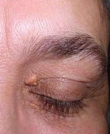
The medical term xanthelasma palpebrarum consists of 2 words—"xanthelasma" originates from ancient Greece and combines "xanthos," meaning yellow, with "elesma," meaning plate; palpebrarum is a Latin term indicating proximity to or association with the eyelid. Xanthelasma palpebrarum is primarily characterized by soft, lipid-rich deposits, especially cholesterol, manifesting as semisolid, yellowish papules or plaques (see Image. Xanthelasma palpebrarum). These deposits are typically found on the inner aspect of the eyes and are most commonly located along the corners of the upper and lower eyelids.[1]
Xanthelasma palpebrarum is the most prevalent cutaneous presentation of a xanthoma that primarily occurs on the eyelids due to cholesterol deposits in the skin. Other areas of the body may or may not be affected. Although xanthelasma palpebrarum is a benign lesion and does not pose significant health risks, this condition may be a cosmetic concern due to its appearance.
Xanthomas can be associated with hyperlipidemia, diabetes, and thyroid dysfunction. Approximately 50% of adult patients with xanthelasma have abnormal lipid levels. These deposits are also prevalent in individuals with inherited dyslipidemia, such as familial hypercholesterolemia and hyperapobetalipoproteinemia. In younger individuals, particularly children, the presence of xanthelasma should prompt consideration of an underlying inherited dyslipidemia. Xanthelasma is a distinctive feature of PBC, frequently associated with significant hypercholesterolemia.
Although xanthelasma treatment is typically not medically necessary, some patients may seek therapy for cosmetic reasons. Lowering lipid levels can be beneficial in managing this condition. Treatment options include surgical excision, laser therapy, and topical trichloroacetic acid (TCA). However, it is essential to note that recurrence rates are often high. This topic delves into the etiology, pathophysiology, assessment, and treatment of individuals with xanthelasma palpebrarum. Recognizing the connection between xanthelasma and potential underlying dyslipidemia and other conditions is crucial for improving patient outcomes and reducing morbidity and mortality.
Etiology
Xanthomas are accumulations of cholesterol-rich material found primarily in the skin, tendons, and subcutaneous tissue, but they can occur in various locations throughout the body. Approximately 50% of individuals who develop xanthelasma have either a primary or secondary lipid disorder.
Primary Lipid Disorders
- Type IIa and IIb phenotype hyperlipidemias
- Type IV phenotype hyperlipidemia
- Low levels of high-density lipoprotein (HDL)
Secondary Lipid Disorders
- Primary biliary cholangitis (PBC) characterized by the buildup of elevated levels of lipoprotein X, a carrier of substantial cholesterol in the bloodstream
- Acquired hyperlipoproteinemia resulting from systemic illnesses such as diabetes, hypothyroidism, and nephrotic syndrome
- Treatment with retinoids and estrogens
- Sarcoidosis
Epidemiology
The prevalence of xanthelasma palpebrarum is 1.1% in women and 0.3% in men. Although this condition can appear in adults between the ages of 20 and 70, it is most frequently observed between the ages of 35 and 55.[2][3] The most commonly affected area is the medial aspect of the upper lid, followed by the medial aspect of the lower lid, and the lateral canthus. The lesions are usually symmetrical.
Pathophysiology
Xanthelasmas are the most prevalent type of xanthoma, characterized by yellowish deposits rich in cholesterol that typically develop on the inner portions of the eyelids. These deposits result from the accumulation of lipids within macrophages. The primary mechanism is the compression of capillaries during each blinking cycle, mediated by the orbicularis oculi muscle. With each blinking cycle, the capillaries are squeezed by the surrounding muscle fibers. The endothelial lining tends to get weaker with age, leading to the exudation of plasma in the subcutaneous space of the eyelids. The cholesterol and lipid components are cleared out by the lymphatic system. When the lipid content exceeds the macrophage's ability to regulate lipid balance, it leads to the formation of lipid droplets, resulting in the transformation of the macrophage into a foam cell. Inflammation is believed to play a significant role in this process, as foam cells can develop in response to chronic inflammation associated with conditions such as gastric xanthoma, gastric cancer, and various metabolic, infectious, and autoimmune diseases.
Histopathology
The papillary dermis, epidermis, and subcutaneous fatty layer generally exhibit no changes.[1] In the superficial reticular dermis, there is evidence of a perivascular inflammatory infiltrate consisting of a variety of cells, including both mononucleated and multinucleated foamy histiocytes characterized by lipid-laden cytoplasmic vacuoles.
History and Physical
Evaluating individuals with xanthelasma palpebrarum involves thorough history-taking and a comprehensive physical examination. Patients may describe the lesion as having grown gradually without causing pain. Healthcare providers should inquire about the patient's personal and family history of hyperlipidemia and early ischemic heart disease.[4]
Upon examination, the lesions appear as soft, yellowish papules or plaques situated primarily along the medial canthus of the upper lid and, occasionally, the lower lid. These lesions typically have a solid or firm texture upon palpation and often display symmetry, with multiple lesions more prevalent than solitary ones.[5] However, it is noteworthy that applying pressure to the lesion will not result in the expression of lipid-like material.
Evaluation
Most individuals with xanthelasma have underlying lipid disorders, so conducting a serum lipid profile is advisable. In addition, considering a liver panel, thyroid function test, and fasting blood glucose assessment may also be warranted. In situations involving patients with diabetes, assessing the glycosylated hemoglobin level can provide valuable insights into blood sugar control over the past 3 months. Elevated cholesterol and triglyceride levels and reduced HDL levels are frequently observed in patients. Patients should adhere to 12-hour fasting before the blood draw to ensure precise lipid profile measurements.
Xanthelasma is evaluated radiologically using ultrasound biomicroscopy. The depth of involvement and echotexture are essential in deciding the appropriate management strategy. Xanthelasma is graded as follows:[6]
- Grade 1: Lesion on the upper lid only
- Grade 2: Lesion along the upper lid + medial canthus area
- Grade 3: Lesion on the medial aspect of upper and lower lids
- Garde 4: Diffuse lesions involving medial and lateral aspects of upper and lower eyelids
Treatment / Management
Treating xanthelasma entails lifestyle modifications and lipid-lowering medications. However, it is noteworthy that once xanthelasma has developed, these interventions may have a limited impact on the cosmetic appearance. Xanthelasma does not typically resolve on its own and tends to remain stable or may even increase in size. The primary reason individuals seek medical attention is often related to cosmetic concerns.[7][8] If the lesions become a cosmetic concern, potential treatment options include surgical excision, laser therapy, cryotherapy, cautery, or TCA removal.[9] Moreover, it is important to know that recurrence is common.
Laser treatments are often preferred options due to their association with minimal tissue damage and the potential to avoid surgical procedures. Among these, fractionated Erbium: YAG (Er: YAG) or fractionated CO2 lasers are the most commonly utilized for treating xanthelasma.[10][11][12] Although radiofrequency machines are considered potential options because of their lower complication rates, they are generally regarded as less effective than alternative treatments and are accompanied by higher costs.[13][14]
Laser therapy is effective for superficial lesions, with a depth of <100 µm. For deeper lesions from 100 to 1000 µm, chemical peel with trichloroacetic acid is an effective solution. For large lesions with a depth of >1 µm, surgical excision is advised. Surgical options include blepharoplasty, blepharoplasty with muscle excision, or uncapping with in-toto excision of xanthelasma[6]. The blepharoplasty is often combined with the reshaping of lid crease, removal of any epicanthal folds, and repositioning of the fat pads of the eyelids for better cosmetic results.
Differential Diagnosis
Xanthelasma palpebrarum can present with clinical similarities to necrobiotic xanthogranuloma (NXG) and syringomas, flesh-colored papules clustered on the periorbital skin. Key distinguishing features of xanthelasma palpebrarum include its bilateral occurrence, location above the medial canthus, and characteristic yellowish coloration of the lesions.
NXG is a non-Langerhans histiocytosis, usually associated with monoclonal gammopathy. In contrast to xanthelasma palpebrarum, NXG typically presents as plaques, papules, or nodules on the periorbital skin, often exhibiting violaceous, red-brown, or yellowish coloration in various areas.
Xanthelasmas are observed in approximately 30% of patients with Erdheim-Chester disease—a non-Langerhans histiocytosis subtype.[15] However, it is noteworthy that xanthelasma palpebrarum is not associated with the systemic manifestations involving the bones, heart, lungs, and kidneys, as seen in Erdheim-Chester disease.
Other potential diagnoses that may be considered in the differential include:
- Sebaceous hyperplasia
- Juvenile xanthogranuloma
- Nodular basal cell carcinoma
- Adult-onset asthma and periocular xanthogranuloma (AAPOX)
- Palpebral sarcoidosis
- Lipoid proteinosis
- Necrobiotic xanthogranuloma
Pseudo-xanthogranulomas are reported in some cases of postvitrectomy surgery, with a deposition of silicon oil in the subcutaneous plane.[16] Furthermore, if there is any uncertainty in the diagnosis of xanthelasma palpebrarum, it is recommended to undergo a skin biopsy.
Prognosis
Xanthelasma palpebrarum is a benign lesion with no malignant potential, and treatment is only necessary for cosmetic concerns. The recurrence rate of xanthelasma palpebrarum is generally less than 50%, although it may vary depending on the chosen treatment.[9][6] In addition, although recurrent cases can be retreated, it is noteworthy that the recurrence rate is around 60% after the retreatment. The deeper lesions (depth of >1 µm) have a much higher recurrence rate.[6]
In addition to managing xanthelasma, addressing underlying medical conditions such as hyperlipidemia, liver diseases, diabetes, and thyroid disorders is essential to mitigate the formation of additional lesions.
Complications
Xanthelasma itself does not cause any medical complications; however, the lesions' cosmetic appearance can be undesirable for patients. Possible complications associated with treatments include pain, erythema, scarring, and pigmentary changes. The incidence of these complications remains consistent across various treatment modalities.[17][18][19] Surgery around the eyelids can potentially lead to ectropion, eyelid retraction, ptosis, and, rarely, an injury to the eyeball. Applying cryotherapy and chemical cauterization may result in substantial scarring and skin discoloration.[20][21] As xanthomas are often associated with dyslipidemia, patients face an elevated risk of conditions such as myocardial infarction, ischemic heart disease, severe atherosclerosis, and potentially fatal outcomes.
Deterrence and Patient Education
Xanthelasma palpebrarum presents as painless, yellowish bumps that typically form near the eye or on the eyelid. This condition is the most prevalent type of xanthoma, which refers to cholesterol deposits usually found beneath the skin but can occur anywhere in the body. Xanthelasma is more frequently observed in women than men and typically manifests in adults aged 40 to 50. Approximately half of individuals with xanthelasma palpebrarum exhibit abnormal cholesterol levels, often attributable to familial hypercholesterolemia or other underlying factors. Elevated cholesterol levels heighten the risk of heart attack, stroke, peripheral vascular disease, and even death.[8] In addition, xanthelasma palpebrarum is associated with other medical conditions, including hypothyroidism and diabetes.
Although xanthelasma palpebrarum presents as painless bumps around the eyes or on the eyelids and are typically firm to the touch, occasionally, larger lesions may become uncomfortable. Notably, xanthelasma is unrelated to cancer development and does not necessitate treatment. However, this condition may be a source of cosmetic concern for some individuals. Treating high cholesterol and managing associated conditions can potentially lead to the regression of existing lesions and reduce the risk of forming new ones. Additional treatment options include laser therapy, surgical removal, and skin peels. Recurrence is a frequent occurrence. Potential complications from treatment encompass pain, scarring, infection, skin color changes, eye injury, and ectropion, where the lower lid droops away from the eye and everts.
Enhancing Healthcare Team Outcomes
The interprofessional healthcare team can optimize the treatment of patients with xanthelasma palpebrarum by recognizing the risk of underlying conditions, effective communication, and coordinated care. Xanthelasmas have diverse etiologies and are frequently linked to independent risk factors for ischemic heart disease.[22]
The interprofessional team comprises healthcare specialists from various fields. Typically, a patient's primary care, ophthalmology, or dermatology clinician is the initial evaluator of the lesion. If patients choose to proceed with removal, consultation with a dermatologist or oculoplastic surgeon is often sought. Subsequently, pathology examination verifies the diagnosis following lesion excision.
Clinicians have a multifaceted role that includes patient education, treatment, accurate maintenance of medication lists, and vigilant monitoring of drug interactions and compliance, especially when underlying conditions are involved. Conducting a comprehensive history and physical examination is essential to identify any signs of vascular disease. Nutritionists can help patients with their dietary changes or modifications to manage the condition. In addition, engaging in physical exercise can effectively lower lipid levels. Effectively managing underlying causes, such as diabetes, thyroid disorders, and obesity, can reduce mortality rates. Recognizing the risk of underlying dyslipidemia and other conditions associated with xanthelasma palpebrarum and ensuring appropriate evaluation, treatment, and follow-up can significantly reduce the morbidity and mortality associated with this condition.