Acneiform Eruptions

Earn CME/CE in your profession:


Continuing Education Activity

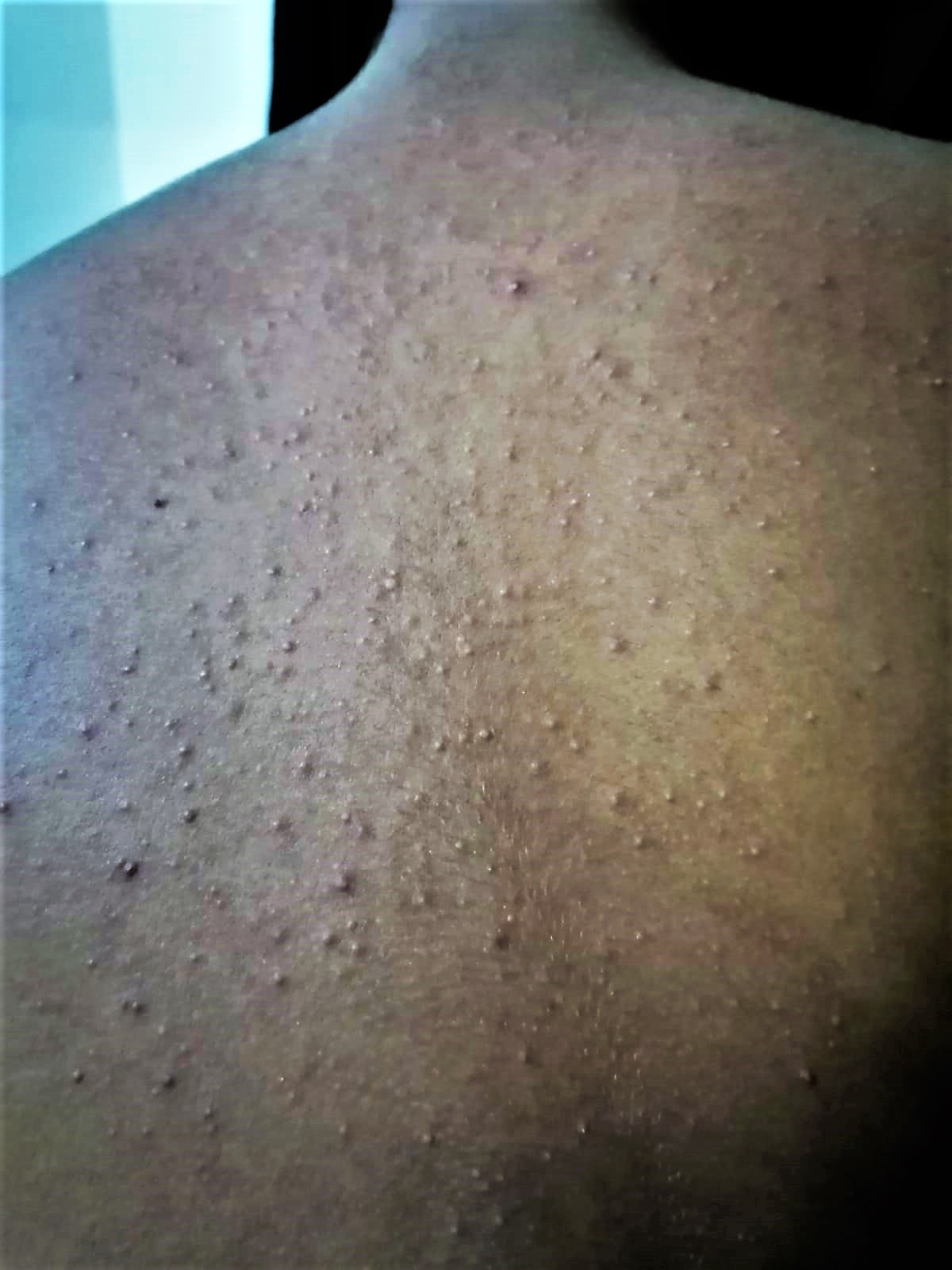
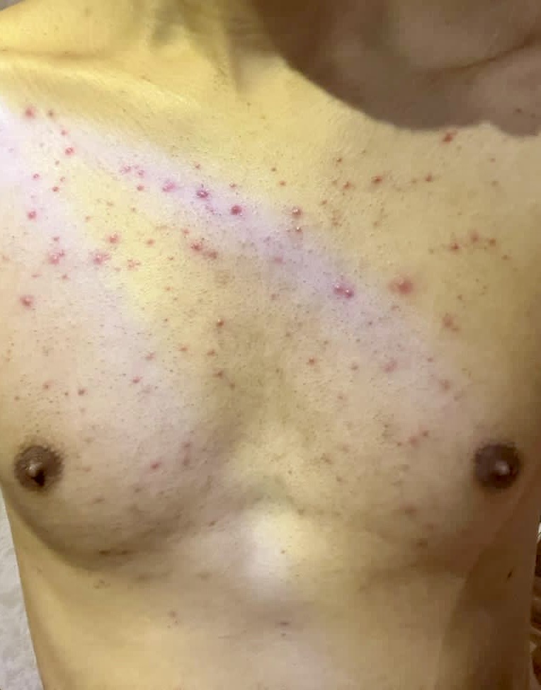
Acneiform eruptions are acne vulgaris-like dermatoses. There are papulopustular, nodular, and cystic lesions. Acne vulgaris is characterized by comedones, whereas acneiform eruptions clinically lack comedones. Acneiform eruptions demonstrate an acute onset and can affect any age group. Distinguishing between acne-like dermatoses and real acne can be difficult in clinical settings. Acneiform eruptions can display papules, pustules, nodules, and cysts, just like acne. Unlike acne, comedones are infrequent but can be seen in specific forms of acneiform eruptions.

Additionally, the sudden onset, propensity to affect people of all ages, uniformity of lesions, and dispersion outside the seborrheic regions distinguish acneiform eruptions from acne. Medications, hormonal and metabolic abnormalities, drug reactions, or genetic disorders may cause acneiform eruptions. In most cases, the diagnosis is clinical, but when there is doubt, one may obtain a biopsy or culture of any discharge. Withdrawal of the suspected medication is another way to make the diagnosis. The treatment of acneiform eruptions depends on the cause. This activity for healthcare professionals is designed to enhance the learner's competence when managing acneiform eruptions, equipping them with updated knowledge, skills, and strategies for timely identification, effective interventions, and improved coordination of care, leading to better patient outcomes.

Objectives:

  • Identify some of the etiologic factors that can contribute to the development of acneiform eruptions.

  • Differentiate acneiform eruptions from acne vulgaris.

  • Implement the treatment strategies for patients with acneiform eruptions.

  • Outline the role of a collaborative interprofessional team in improving the evaluation, management, and counseling for patients with acneiform eruptions.

Introduction

Acneiform eruptions are a group of disorders characterized by papules and pustules resembling acne vulgaris.[1][2] Distinguishing between acne-like dermatoses and real acne can be difficult in clinical settings. Acneiform eruptions can display papules, pustules, nodules, and cysts, just like acne. Unlike acne, comedones are infrequent but can be seen in specific forms of acneiform eruptions. Additionally, the sudden onset, propensity to affect people of all ages, uniformity of lesions, and dispersion outside the seborrheic regions distinguish acneiform eruptions from acne. Medications, hormonal and metabolic abnormalities, drug reactions, or genetic disorders may cause acneiform eruptions. In most cases, the diagnosis is clinical, but when there is doubt, one may obtain a biopsy or culture of any discharge. Withdrawal of the suspected medication is another way to make the diagnosis. The treatment of acneiform eruptions depends on the cause. Clinicians frequently erroneously treat patients for acne and exacerbate the condition.[1]

Etiology

Acneiform eruptions can develop due to infections, hormonal or metabolic abnormalities, genetic disorders, drug reactions, chemical contact, or friction and pressure.[3][4] Prolonged and increased excretion of causative substances might irritate the follicular epithelium and produce an inflammatory reaction. There are many causes for acneiform eruptions, including exposure to halogenated aromatic hydrocarbons and the use of antibiotics like macrolides and penicillin. Other drugs that induce acneiform eruptions include nystatin, isoniazid, corticotropin, naproxen, and hydroxychloroquine. Many organisms, like Proteus, Klebsiella, Escherichia coli, and Enterobacter infections, can also induce acneiform eruptions. Pityrosporum folliculitis, caused by Malassezia furfur, may also present on the trunk and upper extremities with pruritic eruptions. Infections that are known to cause acneiform eruptions include secondary syphilis, mycotic infections, cutaneous coccidioidomycosis, and Sporothrix schenckii.

Epidemiology

Acneiform eruptions can occur at any age and affect both genders. Individuals most prone to developing this skin disorder are those exposed to bacteria or use antibiotics. The condition often develops in hospitalized patients. Epidermal growth factor receptor inhibitors are used in the treatment of many malignant neoplasms and frequently cause drug-induced acneiform eruptions, with a reported overall incidence ranging from 60% to 80% in patients treated with them.[5]

Pathophysiology

The etiology of acneiform eruptions is multifactorial, with no singular underlying mechanism implicated. However, patient age, infections, occupation, habits, cosmetics, and medications may all contribute to the development of these eruptions. Because the observed lesions might not respond to conventional acne treatment, treatment approaches should be customized based on the identified causative agents. The following etiologies are common causes of acneiform eruptions.

Drug-induced Acne

This acne can occur due to several medications including corticosteroids, anticonvulsants (eg, phenytoin), antidepressants, antipsychotics (eg, olanzapine and lithium), antituberculosis drugs (eg, INH, thiourea, thiouracil, disulfiram, and corticotropin), antifungals (eg, nystatin and itraconazole), hydroxychloroquine, naproxen, mercury, amineptine, and chemotherapy. Acneiform eruptions are most frequently reported in individuals undergoing treatment with epidermal growth factor receptor inhibitors and mitogen-activated protein kinase inhibitors.[6] Antibiotics (eg, penicillins and macrolides) cause acute generalized pustular eruptions without comedones, and patients are often febrile with leukocytosis. Other antibiotic causes include co-trimoxazole, doxycycline, ofloxacin, and chloramphenicol. Acneiform eruption was found to be an adverse event associated with the recently approved 3 cystic fibrosis transmembrane conductance modulators (ie, elexacaftor, tezacaftor, and ivacaftor).[7] Compared to other cancer treatments, acneiform eruptions are less frequently observed with CTLA-4 inhibitors. However, healthcare professionals should know that this side effect may still occur in clinical practice.[8] There have been few reports in the literature regarding acneiform outbreaks caused by vitamin B12, with more cases reported in females than males. Possible causative variables of acneiform eruptions were postulated to include high-dose supplementation, prolonged periods of supplementing, or the concurrent use of vitamin B12 with vitamins B1, B2, or B6.[9][10][11]

Steroid acne presents as monomorphous papulopustules located mainly on the trunk and extremities, with less face involvement. Characteristically, lesions appear after the administration of systemic corticosteroids. Topical corticosteroids may also cause acneiform eruption over the skin under which the topical preparation is applied or around the nose or mouth in the case of inhaled steroids. This eruption typically presents as skin erythema along the distribution of corticosteroid application with pustules and inflamed papules. Ultimately, the lesions resolve once the corticosteroid is discontinued; however, prolonged and severe flares may result from "steroid dependency" after withdrawal. Athletes and bodybuilders ought to be questioned regarding their use of anabolic steroids.

Occupational Acne, Acne Cosmetica, and Pomade Acne

Occupational acne is caused by contact with insoluble, follicle-occluding substances in the place of employment. Among the harmful substances are derivatives of coal tar, cutting oils, petroleum-based products, and chlorinated aromatic hydrocarbons. The clinical presentation primarily consists of comedones, although a few papules, pustules, and cystic lesions may be dispersed throughout exposed and normally covered regions. Facial acne that is predominantly comedonal, characterized by closed comedones, may also manifest in areas consistently exposed to hair products or follicle-occluding cosmetics (ie, acne cosmetica). The latter condition, pomade acne, is more prevalent on the forehead and temples.[12]

Chloracne

Chloracne formation is the result of halogenated aromatic hydrocarbon exposure. Typically, the onset of symptoms occurs multiple weeks following systemic exposure, which may occur through ingestion, percutaneous absorption, or inhalation. Insecticides, fungicides, herbicides, electrical conductors and insulators, polychlorinated naphthalenes and biphenyls, tetrachloroazobenzene, and tetrachloroazoxybenzene have all been implicated. Additionally, dibenzofurans, dibenzodioxins, polybrominated naphthalenes, and biphenyls have been identified. Comedo-like lesions and yellowish cysts, characterized by minimal inflammation, predominantly manifest in the retroauricular and malar regions of the head and neck, axillae, and scrotum. To varying degrees, the extremities, buttocks, and trunk are implicated. Significant scarring may accompany the healing of cystic lesions, and the condition may persist for years after exposure has ceased. Further observations might include hypertrichosis and a grayish discoloration of the skin.[13]

Acne Mechanica

Acne mechanica is caused by repetitive frictional and mechanical obstruction of the pilosebaceous orifice, resulting in comedo formation. Robust mechanical factors thoroughly described include chin straps, suspenders, helmets, and collars. Acne mechanica caused by orthopedic factors consists of eruptions in the axillae secondary to the use of crutches and amputee stumps due to friction from prostheses. A typical instance of acne mechanica is fiddler's neck, characterized by a distinct, hyperpigmented plaque that is lichenified and interspersed with comedones, resulting from repeated trauma caused by the positioning of a violin on the lateral neck. The primary clinical feature of acne mechanica is the geometrically distributed eruptions and linear areas of involvement.

Tropical Acne

Tropical acne is an acneiform follicular eruption caused by prolonged exposure to extremely high temperatures. This may manifest in tropical regions or due to working in extremely hot environments, such as among furnace workers. Tropical acne has historically resulted in substantial morbidity among troops. Manifestations often include trunk and pelvic involvement in nodulocystic acne that is significantly inflamed; secondary staphylococcal infection is a common complication.

Radiation Acne

Radiation acne is identified by comedo-like papules that develop in areas previously exposed to therapeutic ionizing radiation.[14] The lesions manifest as the acute phase of radiation dermatitis begins to subside. The ionizing rays induce epithelial metaplasia within the follicle, creating adherent hyperkeratotic plugs resistant to expression.[15]

Pseudoacne of the Nasal Crease

The transverse nasal crease is a horizontal line located in the lower part of the nose, marking the boundary between the alar cartilage and the triangular cartilage. Milia, cysts, and comedones can be found in a row along this fold. These acne-like lesions, or pseudoacne, are not influenced by hormones and develop in childhood shortly before puberty begins.

Idiopathic Facial Aseptic Granuloma

Idiopathic facial aseptic granuloma is a painless nodule that resembles acne and usually forms on the cheeks of young children, with an average age of 3.5 years. Multiple lesions are infrequent. The histopathologic examination shows the presence of a lymphohistiocytic infiltration in the dermis, characterized by the presence of foreign body-type giant cells. Cultures are usually negative, and the lesions are unresponsive to antibiotic treatment. Typically, within a year, the lesions resolve spontaneously. Idiopathic facial aseptic granuloma has been suggested to be a type of childhood rosacea, as more than 40% of children exhibit at least 2 other clinical indications of rosacea (eg, recurring chalazions, facial flushing, telangiectasias, or papulopustules).

Childhood Flexural Comedones

This condition is identified by distinct, double-opening comedones found specifically in the axillae and, less frequently, in the groin area. Most patients exhibit a solitary lesion, and the average age of diagnosis is 6 years, with an equal prevalence among boys and girls. The condition may also be familial. No association with hidradenitis suppurativa, acne vulgaris, or precocious puberty has been identified.[16]

Histopathology

The lesions of acneiform eruptions look similar to acne but lack comedones. Biopsies, though rarely performed, may reveal the presence of fungi or bacteria. The presence of both neutrophils and eosinophils may suggest mild inflammation. The identification of fungal spores within the follicular lumen strongly supports a diagnosis of Pityrosporum folliculitis. Nevertheless, inflammation within the hair follicles, abnormal patterns of keratin plugging, and the accumulation of nuclear dust in the follicular lumen strongly support a diagnosis of acneiform eruption. The observed variations within the follicles and the inflammatory disparities are thought to result from necrotic keratinocytes, which induce vacuolar alterations in the follicular wall.[17]

History and Physical

Acneiform eruptions are primarily clinically diagnosed. Clinical features include papules and pustules without true comedones, which are present mainly over the trunk and back (see Image. Acneiform Eruptions). Rarely, nodulocystic lesions can be seen. Unlike acne, these lesions may occur in other parts of the body besides the face. When the underlying etiology is drug-induced, the patient will usually state that the lesions disappear with medication discontinuation.

Evaluation

Acneiform eruptions can be distinguished from acne vulgaris by a history of sudden onset, monomorphic morphology, the development of the eruption at any age, affecting the trunk more commonly than the face, not necessarily affecting sebaceous areas of the body, and a rarity of cyst formation (see Image. Acneiform Eruption Papules). The diagnosis is usually clinical, but clinicians may obtain a biopsy or culture of any discharge for diagnostic uncertainty. Withdrawal of the suspected medication is another way to make the diagnosis.[18][19]

Treatment / Management

The treatment of acneiform eruptions is tailored to the underlying etiology. Exposure should be discontinued for acneiform eruptions due to organisms or medications. Minimizing contacts or friction will prevent occupational and mechanical acne. Protective clothing and the removal of the worker from unsuitable environments also help.[20] The traditional agents used to treat acne vulgaris seldom work in patients with acneiform eruptions, but using skin cleaners like salicylic acid or benzoyl peroxide to reduce oily skin may be suggested. Itching is a prevalent symptom in patients with acneiform eruptions so that these patients may benefit from the use of antihistamines. For nocturnal itching, first-generation antihistamines are recommended because they also induce sleep.

Over the years, the use of retinoids to treat acneiform eruptions has increased due to successful treatment. Both oral and topical retinoids have been used. These agents are known to decrease sebum production and rapidly resolve eruptions. These agents should not be prescribed to women of childbearing age because of their teratogenic potential. Some cases of acneiform eruptions may benefit from dapsone, as anecdotal reports have demonstrated that eosinophilic pustular eruptions have responded to a short course of dapsone.[21] Patients who have gram-positive organisms causing skin lesions may also benefit from doxycycline. 

Oral tetracycline antibiotics are the most effective preventive treatment for acne-like skin rashes caused by EGFR inhibitors. These should be provided to appropriate individuals at the start of therapy and utilized with a comprehensive skincare regimen that includes moisturizers and the avoidance of irritants.[22] For fungal infections (eg, Pityrosporum folliculitis), topical antifungal agents such as ciclopirox, econazole, and ketoconazole can be helpful. Acneiform eruptions secondary to chloracne treatment are difficult as they may persist for years, even without further exposure. Some patients with eosinophilic pustular folliculitis may benefit from a short course of oral indomethacin. Lesions that fail to respond to indomethacin may be treated with cyclosporine.

Differential Diagnosis

Various lesions share common features with acneiform eruptions; therefore, identifying the specific and unique clinical aspects of these eruptions can help make an accurate diagnosis.

  • Demodicosis can be categorized into 4 distinct clinical forms, each of which derives its name from the corresponding skin condition it resembles: acne type, rosacea type, perioral type, and pityriasis folliculorum. Acne vulgaris is clinically mimicked by the latter, manifesting as non-scaly, localized follicular pustules.[23]
  • Milia, which are little white bumps, are commonly seen on the cheeks and noses of newborns. However, they typically disappear after a few months.
  • In contrast to acne vulgaris, rosacea does not exhibit comedones or seborrhea. Rosacea is a recurring condition that primarily affects the face symmetrically. Additionally, it may be accompanied by secondary symptoms such as stinging, burning, swelling, dryness, and ocular indications.
  • Eruptive vellus hair cysts have the potential to become inflammatory, and, as their name suggests, they contain numerous vellus hairs that can be readily observed under microscopic examination.
  • Steatocystoma multiplex is characterized by non-inflammatory, closed cystic papules and nodules on the central chest and back.
  • Favre-Racouchot syndrome has multiple open comedones clustered in the lateral malar region of older individuals.
  • Perioral dermatitis (PD), in its classic form, is characterized by numerous pustules and papules measuring 1 to 2 mm in diameter situated on observable erythema in the perioral region. Generally, the narrow zone encompassing the vermilion border remains unaffected.[24]
  • Hidradenitis suppurativa manifests with painful and profound papular, pustular, or nodular lesions or abscesses, which have the potential to advance and develop sinus tracts and scars.[25]
  • Pseudofolliculitis barbae is an inflammatory disorder of the hair follicle characterized by red papules and pus-filled lesions. It can occur in any area where hair is removed by shaving or plucking. The lesions primarily manifest on males' facial and neck regions due to shaving.[26]
  • Eruptive syringoma distributed on the trunk can clinically resemble acneiform eruptions. Histopathological examination can be helpful in these cases.
  • Angiofibromas and appendageal tumors derived from hair follicles, such as trichoepitheliomas, trichodiscomas, and fibrofolliculomas, frequently manifest as numerous papules on the face.
  • Eosinophilic pustular folliculitis is an uncommon skin disorder characterized by papulopustules. This condition is not caused by an infection and is associated with inflammation. One of its unique histological features is the presence of eosinophilic infiltrates around the pilosebaceous unit.
  • Sarcoidosis is linked to many skin lesions, which can be categorized as either specific or nonspecific, depending on whether or not characteristic sarcoidosis granulomas are present. Maculopapular eruptions are the most common type of particular lesions. The monomorphic lesions are usually found on the face and appear as tiny papules that may merge into annular lesions or plaques. Sarcoid lesions differ from acne vulgaris because they do not exhibit comedones or pustules.
  • Papular granuloma annulare does not exhibit the normal ring shape, which might lead to misinterpretation as an acne outbreak. Nevertheless, acne can be ruled out based on the dissimilarity in affected areas, the lack of changes in the epidermis, the absence of comedones and pustules, and the consistent appearance of granuloma annulare lesions compared to acne.[27]
  • Periorificial granulomatous dermatitis (PGD) manifests as solitary, uniform, dome-shaped papules of 1 to 3 mm in size, which usually do not cause any symptoms. The color of the lesions might range from pink-red to flesh-colored to yellow-brown. The inclusion of the vermillion border in PGD sets it apart from other conditions, offering a helpful distinction from PD. Furthermore, PGD can be distinguished from acne based on its monomorphous papules, lack of pustules or comedones, lesions around the mouth, self-limiting nature, and unique histology.
  • Bacterial folliculitis is referred to as superficial bacterial folliculitis or Bockhart impetigo. It impacts the outer layer of the hair follicle, resulting in small pus-filled lesions surrounded by redness that later become crusty. Pruritus and discomfort frequently are accompanying symptoms. Conversely, profound folliculitis occurs when the hair follicle is completely blocked. The condition is distinguished by furuncles, which initially manifest as painful inflammatory nodules and progress into necrotic lesions within a few days.
  • Gram-negative folliculitis should be considered in patients with acne who see a deterioration of pustular or cystic lesions while undergoing antibiotic treatment and in those who do not observe a substantial improvement in their acne after 3 to 6 months of antibiotic medication. In most cases, the lesions manifest in the inferonasal region and may potentially spread to the chin and cheeks.[28]
  • Malassezia folliculitis, formerly known as Pityrosporum folliculitis, affects young to middle-aged adults and is characterized by an infectious follicular papulopustular eruption on the face, torso, and upper extremities. The disorder is caused by yeasts belonging to the genus Malassezia, which are common commensal organisms found on the epidermis.
  • Hot tub folliculitis, or Pseudomonas folliculitis, is a follicular infection brought on by the Gram-negative opportunistic pathogen Pseudomonas aeruginosa. The lesions typically manifest after 1 to 4 days of exposure to contaminated swimming pools, hot tubs, or whirlpools. The characteristic pruritic dermatitis develops from follicular macules to pustules and papules over time.
  • Sporotrichosis induces lymphocutaneous infections in the majority of instances. However, it can manifest as a fixed cutaneous manifestation. When papulonodular eruptions occur, they can potentially be misdiagnosed as acne.
  • Secondary syphilis is a systemic condition that results from the hematogenous dissemination of Treponema pallidum. Typical clinical manifestations comprise crusted papules, pustules, and nodules found on the face, extremities, and trunk. Frequent concurrent systemic symptoms with the lesions include fever, lymphadenopathy, and general malaise.[29]
  • Sebaceous hyperplasia manifests as lobulated, yellowish papules originating predominantly on the forehead and cheekbones.
  • Psychogenic (ie, neurotic) excoriations and factitial dermatitis mainly occur on the face, chest, and back and can imitate acne excoriée. Linearity and the absence of clinically detectable primary lesions serve as indications.[16]

Prognosis

Most patients recover within a few weeks. For any residual lesions, treatments that have been used include laser ablation, excision, topical or oral antibiotics, drug withdrawal, or topical or oral retinoids.[16]

Complications

Complications of acneiform eruptions include:

  • Psychological distress from the cosmetic appearance
  • Irritation from itching
  • Secondary bacterial infection
  • Post-inflammatory hyperpigmentation
  • Scarring, although rare
  • Keloid formation [16]

Deterrence and Patient Education

Patients should be counseled that acneiform eruptions are temporary skin conditions that resemble acne and can arise from various factors, such as infections, medication reactions, hormonal or metabolic imbalances, or genetic disorders. Therefore, keeping a symptom diary or removing possible causative factors may help find the underlying etiology. Patients should also be instructed to avoid scratching the lesions to prevent the development of scarring and keloid.

Enhancing Healthcare Team Outcomes

Nurse practitioners, pharmacists, and primary care clinicians must know that not all facial lesions are due to acne. Acneiform eruptions have many causes and, in many cases, are caused by occupational exposure or certain medications. Erroneously treating these patients for acne often exacerbates the condition. Consult with a dermatologist is recommended for uncertain diagnoses. Clinicians should educate the public that acneiform eruptions are transient skin disorders with many causes. In many cases, simply removing the offending agent or limiting exposure to the organism can resolve the disorder. The prognosis for most patients is excellent, and there is usually no residual scarring. Patients should be warned not to scratch the lesions, which can lead to scars and keloid formation.


(Click Image to Enlarge)
<p>Acneiform Eruptions</p>

Acneiform Eruptions


Contributed by Hasnain Ali Syed, MD 


(Click Image to Enlarge)
<p>Acneiform Eruption Papules</p>

Acneiform Eruption Papules


Contributed by Haitham Saleh, MSc

Details

Updated:

1/11/2024 3:02:26 AM

References


[1]

Gupta M, Aggarwal M, Bhari N. Acneiform eruptions: An unusual dermatological side effect of ribavirin. Dermatologic therapy. 2018 Sep:31(5):e12679. doi: 10.1111/dth.12679. Epub 2018 Sep 19     [PubMed PMID: 30230152]


[2]

Ayanlowo O, Puddicombe O, Gold-Olufadi S. Pattern of skin diseases amongst children attending a dermatology clinic in Lagos, Nigeria. The Pan African medical journal. 2018:29():162. doi: 10.11604/pamj.2018.29.162.14503. Epub 2018 Mar 19     [PubMed PMID: 30050626]


[3]

Musthaq S, Mazuy A, Jakus J. The microbiome in dermatology. Clinics in dermatology. 2018 May-Jun:36(3):390-398. doi: 10.1016/j.clindermatol.2018.03.012. Epub 2018 Mar 10     [PubMed PMID: 29908581]


[4]

Sibaud V. Dermatologic Reactions to Immune Checkpoint Inhibitors : Skin Toxicities and Immunotherapy. American journal of clinical dermatology. 2018 Jun:19(3):345-361. doi: 10.1007/s40257-017-0336-3. Epub     [PubMed PMID: 29256113]


[5]

Recuero JK, Fitz JR, Pereira AA, Bonamigo RR. EGFR inhibitors: clinical aspects, risk factors and biomarkers for acneiform eruptions and other mucosal and cutaneous adverse effects. Anais brasileiros de dermatologia. 2023 Jul-Aug:98(4):429-439. doi: 10.1016/j.abd.2022.10.004. Epub 2023 Mar 27     [PubMed PMID: 36990917]


[6]

Yuan C, Wang B. Acneiform eruption induced by molecularly targeted agents in antineoplastic therapy: A review. Journal of cosmetic dermatology. 2023 Aug:22(8):2150-2157. doi: 10.1111/jocd.15704. Epub 2023 Mar 16     [PubMed PMID: 36924348]


[7]

Okroglic L, Sohier P, Martin C, Lheure C, Franck N, Honoré I, Kanaan R, Burgel PR, Carlotti A, Dupin N, Oulès B. Acneiform Eruption Following Elexacaftor-Tezacaftor-Ivacaftor Treatment in Patients With Cystic Fibrosis. JAMA dermatology. 2023 Jan 1:159(1):68-72. doi: 10.1001/jamadermatol.2022.5208. Epub     [PubMed PMID: 36449298]


[8]

Welborn M, Kubicki SL, Garg N, Patel AB. Twelve cases of acneiform eruptions while on anti-CTLA4 therapy. Supportive care in cancer : official journal of the Multinational Association of Supportive Care in Cancer. 2020 Jun:28(6):2499-2502. doi: 10.1007/s00520-020-05381-5. Epub 2020 Mar 9     [PubMed PMID: 32147760]

Level 3 (low-level) evidence

[9]

Bahbouhi I, Aboudourib M, Hocar O, Amal S. Vitamin B12 induced acneiform eruption. Heliyon. 2023 May:9(5):e16120. doi: 10.1016/j.heliyon.2023.e16120. Epub 2023 May 20     [PubMed PMID: 37305488]


[10]

Bowden A, Ekeh O, Brownstone ND, Hsu S. Acneiform Eruption Secondary to Over-the-Counter Vitamin B12. Cureus. 2023 Aug:15(8):e43275. doi: 10.7759/cureus.43275. Epub 2023 Aug 10     [PubMed PMID: 37692729]


[11]

Elmas ÖF, Metin MS, Özyurt K, Atasoy M. Vitamin B(12) : An underestimated cause of acneiform drug eruption. Dermatologic therapy. 2020 Jul:33(4):e13531. doi: 10.1111/dth.13531. Epub 2020 May 25     [PubMed PMID: 32379373]


[12]

Kligman AM, Mills OH Jr. "Acne cosmetica". Archives of dermatology. 1972 Dec:106(6):843-50     [PubMed PMID: 4264346]


[13]

Panteleyev AA, Bickers DR. Dioxin-induced chloracne--reconstructing the cellular and molecular mechanisms of a classic environmental disease. Experimental dermatology. 2006 Sep:15(9):705-30     [PubMed PMID: 16881967]


[14]

Liu W, Liu JW, Ma DL. Acneiform eruption secondary to radiotherapy. Indian journal of dermatology, venereology and leprology. 2021 [SEASON]:87(4):587-588. doi: 10.4103/ijdvl.IJDVL_421_19. Epub     [PubMed PMID: 31929232]


[15]

Martin WM, Bardsley AF. The comedo skin reaction to radiotherapy. The British journal of radiology. 2002 May:75(893):478-81     [PubMed PMID: 12036846]


[16]

Plewig G, Jansen T. Acneiform dermatoses. Dermatology (Basel, Switzerland). 1998:196(1):102-7     [PubMed PMID: 9557242]


[17]

An MK, Hong EH, Cho EB, Park EJ, Kim KH, Kim KJ. Clinicopathological differentiation between Pityrosporum folliculitis and acneiform eruption. The Journal of dermatology. 2019 Nov:46(11):978-984. doi: 10.1111/1346-8138.15070. Epub 2019 Sep 5     [PubMed PMID: 31489692]


[18]

Del Rosso JQ. Who Is Accountable When Patients Do Not Achieve Successful Treatment for Their Acne? Journal of drugs in dermatology : JDD. 2018 Jun 1:17(6):599-600     [PubMed PMID: 29879246]


[19]

Kallis PJ, Price A, Dosal JR, Nichols AJ, Keri J. A Biologically Based Approach to Acne and Rosacea. Journal of drugs in dermatology : JDD. 2018 Jun 1:17(6):611-617     [PubMed PMID: 29879248]


[20]

Truitt JM, Reichenberg JS, Sharghi KG, Sampson SM, Roenigk RK, Magid M. Isotretinoin: the ups are just as troubling as the downs. Giornale italiano di dermatologia e venereologia : organo ufficiale, Societa italiana di dermatologia e sifilografia. 2018 Aug:153(4):535-539. doi: 10.23736/S0392-0488.18.05979-5. Epub 2018 Apr 18     [PubMed PMID: 29667796]


[21]

Beshay A, Petersen M, Rhoads JLW. Severe EGFR inhibitor-induced acneiform eruption responding to dapsone. Dermatology online journal. 2021 Jul 15:27(7):. doi: 10.5070/D327754366. Epub 2021 Jul 15     [PubMed PMID: 34391331]


[22]

Gorji M, Joseph J, Pavlakis N, Smith SD. Prevention and management of acneiform rash associated with EGFR inhibitor therapy: A systematic review and meta-analysis. Asia-Pacific journal of clinical oncology. 2022 Dec:18(6):526-539. doi: 10.1111/ajco.13740. Epub 2022 Mar 29     [PubMed PMID: 35352492]

Level 1 (high-level) evidence

[23]

Li J, Wei E, Reisinger A, French LE, Clanner-Engelshofen BM, Reinholz M. Comparison of Different Anti-Demodex Strategies: A Systematic Review and Meta-Analysis. Dermatology (Basel, Switzerland). 2023:239(1):12-31. doi: 10.1159/000526296. Epub 2022 Oct 28     [PubMed PMID: 36310014]

Level 1 (high-level) evidence

[24]

Mokos ZB, Kummer A, Mosler EL, Čeović R, Basta-Juzbašić A. PERIORAL DERMATITIS: STILL A THERAPEUTIC CHALLENGE. Acta clinica Croatica. 2015 Jun:54(2):179-85     [PubMed PMID: 26415314]


[25]

Pescitelli L, Ricceri F, Prignano F. Hidradenitis suppurativa and associated diseases. Giornale italiano di dermatologia e venereologia : organo ufficiale, Societa italiana di dermatologia e sifilografia. 2018 Jun:153(3 Suppl 2):8-17. doi: 10.23736/S0392-0488.17.05772-8. Epub     [PubMed PMID: 30468375]


[26]

Ogunbiyi A. Pseudofolliculitis barbae; current treatment options. Clinical, cosmetic and investigational dermatology. 2019:12():241-247. doi: 10.2147/CCID.S149250. Epub 2019 Apr 16     [PubMed PMID: 31354326]


[27]

Xue J, Lam JM. Granuloma annulare. Paediatrics & child health. 2019 Sep:24(6):366-367. doi: 10.1093/pch/pxz003. Epub 2019 Mar 7     [PubMed PMID: 31528105]


[28]

Böni R, Nehrhoff B. Treatment of gram-negative folliculitis in patients with acne. American journal of clinical dermatology. 2003:4(4):273-6     [PubMed PMID: 12680804]


[29]

Lambert WC, Bagley MP, Khan Y, Schwartz RA. Pustular acneiform secondary syphilis. Cutis. 1986 Jan:37(1):69-70     [PubMed PMID: 2936581]