Arbovirus Encephalitides

Earn CME/CE in your profession:


Continuing Education Activity

Arboviruses refer to a diverse group of viruses that are transmitted via mosquitos, ticks, or sandflies and are capable of causing a wide range of diseases. It is important to understand the disease processes caused by these infectious agents, given the increasing frequency of infection and the potential for additional emerging diseases. This activity describes the evaluation and management of arbovirus encephalitides and reviews the role of the interprofessional team in managing patients with this condition.

Objectives:

  • Review the etiology of arbovirus encephalitides.
  • Outline the typical presentation of arbovirus encephalitides.
  • Summarize management of arbovirus encephalitides.
  • Describe interprofessional team strategies for improving care coordination and communication to improve outcomes in arbovirus encephalitides.

Introduction

Arboviruses, also known as arthropod-borne viruses, refer to a diverse group of viruses that are transmitted via mosquitos, ticks, or sandflies.[1] This group of viruses belongs to the families Togaviridae, Flaviviridae, Bunyaviridae, and Reoviridae.[1] These families have similar RNA genomes that permit mutations that allow them to acclimate to changing environments or host conditions.[2] These viruses are unique as they require vectors for biological transmission to humans. It is important to understand the disease processes caused by these infectious agents, given the increasing frequency of infection and the potential for additional emerging diseases.[3] 

Arboviruses can cause multiple neurological diseases, including myelitis, neuritis, myositis, meningitis, and encephalitis.[4] Mortality rates related to these infections increase with the diagnosis of encephalitis. Thus arboviruses are important differentials to consider when evaluating a febrile patient with neurological symptoms. Although arboviruses can cause a wide range of infections, this article will cover major features of the more common arthropod-borne neurotropic viruses that cause endemic and travel-associated encephalitis.

Etiology

Togaviridae family:

  • Chikungunya virus, an alphavirus, can be transmitted to humans by infected Aedes mosquitoes, maternal-fetal transmission, and blood products.[5][6][7] The virus, known to cause the clinical entity known as chikungunya fever, can cause meningoencephalitis in severe infections.[8] These infections have only been noted since its re-emergence in La Réunion Island and the Caribbean.[4] 
  • Eastern equine encephalitis (EEE) virus is transmitted via infected Culiseta melanura as well as some Aedes, Coquillettidia, and Culex mosquitoes. The incubation period is 4 to 10 days after the mosquito bite. Peak incidence for infection is during the months of August and September. However, infections can occur year-round.[9] About 2% of adults develop neuroinvasive diseases resulting in rapid neurological deterioration. EEE has the highest rate of mortality among arthropod-borne encephalitides, with a mortality of at least 30%.[10] There are currently no vaccinations available for humans, but there is one available for horses.
  • Western equine encephalitis (WEE) virus is transmitted by infected Culex mosquitoes. The virus can be found in North and South America. Less than 1% of infected adults develop encephalitis.[11] There are currently no vaccinations available for humans, but there is one available for horses.
  • Venezuelan equine encephalitis (VEE) virus has both epizootic subtypes transmitted by many mosquitoes and enzootic subtypes transmitted by infected Culex mosquitoes[11]. The incubation period is 1-6 days, followed by a febrile illness. Less than 0.5 percent of infected individuals develop neuroinvasive disease. This virus has been linked to several outbreaks in North and South America.[4]

Flaviviridae family:

  • Dengue virus is transmitted by Aedes aegypti or Aedes albopictus mosquitoes. Clinically, this virus can cause disease, which can be classified into 3 categories: dengue without warning signs, dengue with warning signs, and severe dengue. Studies have shown that these viruses are directly neurotropic and can cause encephalitis.[12][13][14] The frequency of neurological manifestations has been shown to be between 0.5% to 21% in case series.[15] One vaccine is currently available in Latin America and Southeast Asia. 
  • Japanese encephalitis virus (JEV) is an important cause of arthropod-borne virus encephalitis in Asia. The virus is transmitted via Culex species, predominantly by Culex tritaeniorhynchus, and is endemic in Asia.[16] This virus infects about 68,000 individuals each year. Less than one percent of infections progress to neurological disease.[17] Although there is a low risk of infection in travelers, there is a vaccine available in the US for individuals traveling to high-risk areas.[17]
  • Murray valley encephalitis (MVE) virus is transmitted via Culex annulirostris mosquitoes. Geographically, this virus is restricted to Australia, New Guinea, and Indonesia. The incubation period is between 1-4 weeks, and most patients will initially have symptoms of a flu-like illness. Mortality rates in individuals with severe infection are between 15%-30%.[18]
  • St. Louis encephalitis (SLE) virus is an important flavivirus in the US. It is transmitted via Culex mosquitoes in the Americas. Transmission occurs in the summer months when mosquitos are active. Patients typically have a flu-like illness prior to developing any neurological manifestations. Mortality rates are higher in those patients who are over the age of 60. Neurological disease has also been described in the postinfectious period.[19] 
  • West Nile virus (WNV), a mosquito-borne flavivirus, is an important cause of endemic encephalitis in the US. Cases should be suspected in individuals who have had recent mosquito exposure, organ transplantation, or blood transfusion. Additional risk factors for symptomatic infection include immunosuppressed individuals, patients with multiple co-morbidities, and the elderly. This virus is transmitted via infected Culex mosquitos, and individuals become viremic when the virus replicates in dendritic cells and macrophages, resulting in dissemination within the human host. Most infections related to WNV are asymptomatic, and less than 1% of individuals develop neurological disease. There are currently no vaccines available for use in humans.
  • Powassan virus is an RNA virus that can be transmitted by Ixodes scapularis, Ixodes cookei, and Ixodes marxi. Most cases occur in the Northeast and Great Lakes regions in the US and follow a similar distribution as Lyme disease. There are a limited number of case reports of this virus in the medical literature, however, most recognized cases present with neurological findings.[20] About 10% of Powassan virus encephalitis cases are fatal.
  • Tick-borne encephalitis (TBE) virus is transmitted via Ixodes persulcatus and Ixodes ricinus in Europe and Russia as well as Ixodes ovatus in Japan. The incubation period lasts from 7 to 14 days, and the illness is known to be biphasic with neurological manifestations occurring during the second phase. Mortality outcomes differ with varying subtypes, with outcomes being worse in the Far Eastern group.[21] Vaccines are available in Europe and Canada. 
  • Zika virus, another mosquito-borne flavivirus, is primarily transmitted via Aedes aegypti, although it can also be transmitted by Aedes albopictus. Transmission can also occur via maternal-fetal transmission, sexual transmission, blood products, organ transplantation, and laboratory exposures.[22] This virus can cause multiple neurological complications, including encephalitis.[22] 

Bunyaviridae family:

  • La Crosse encephalitis virus (LACV) is transmitted via Aedes triseriatus and is considered to be the most pathogenic member of the California encephalitis serogroup. Transmission occurs from July-September. Although many infected individuals are asymptomatic, 89% of reported cases in 2016 were neuroinvasive in the US.[23]
  • California encephalitis virus (CEV) is transmitted via Aedes mosquitoes. Although the California serogroup was named after this virus, it has rarely been implicated in CNS disease.[23] The mortality rate has been reported as <1%.[4]

Reoviridae family:

  • Colorado tick fever virus is transmitted via Dermacentor andersoni, a Rocky Mountain wood tick. Geographically, this virus is limited to the western US and Canada. 5% to 10% of infected children can develop neuroinvasive disease, however, mortality rates of this disease remain low.[20]

Epidemiology

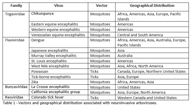
There are many epidemiologic features to consider when evaluating patients for potential etiologic agents of encephalitis. Exposure history, including place of residence, recent travel, insect contact, animal contact, occupation, recreational activities, and diet, can help guide initial diagnostic testing. Additional factors such as age, vaccination history, immune status, and seasonal variation can also help narrow one’s differential. See table 1 for vectors and geographical distribution associated with each virus.

Pathophysiology

The degree to which arboviruses can cause neuroinvasive disease depends on both host and virus factors. Although several mechanisms have been described, the exact pathway by which arboviruses can enter the central nervous system (CNS) remains unknown. The infection can then both directly and indirectly cause neuronal injury and apoptosis.[4]

History and Physical

Arbovirus infections can vary in clinical presentation. As patients can present with nonspecific symptoms, one's initial differential diagnosis must remain broad. Fevers, headaches, altered mental status, seizures, and focal neurological deficits are common findings in individuals who present with encephalitis. Some infected patients may experience a flu-like illness prior to having any neurological symptoms. Importantly, the combination of fever, headache, and altered mental status can symbolize acute encephalitis syndrome. 

Physical exam findings can also assist in determining the cause of a patient's encephalitis. Skin rashes, for example, can be associated with WNV or zika virus. Ocular findings such as chorioretinitis, retinal hemorrhages, and vitreitis can be seen with WNV.[24] Furthermore, certain neurological findings can aid in determining a specific etiology of viral encephalitis. For example, certain flaviviruses such as WNV, SLE virus, and JEV can cause signs and symptoms of parkinsonism. Additionally, these viruses can also cause seizures, cerebellitis, and symptoms of brain stem involvement.[25]

Evaluation

Patients should undergo a lumbar puncture if there are no contraindications to the procedure. Cerebrospinal fluid (CSF) should be sent for cell count and differential, glucose concentration, protein concentration, culture, and gram stain. CSF analysis will typically reveal lymphocytic pleocytosis, normal glucose, and elevated protein with a negative gram stain. 

Virus-specific IgM and PCR of CSF and serum are helpful in obtaining a diagnosis when available. It is important to note that cross-reactivity has been demonstrated among flaviviruses.[10] Plaque reduction neutralization testing can be beneficial in these instances.

Neuroimaging studies, preferably magnetic resonance imaging (MRI), should also be considered when evaluating a patient for encephalitis as distinct MRI findings can provide additional guidance in diagnostic testing. For example, MRI findings of diffusion restriction and abnormal signal intensities on T2 and FLAIR imaging can be indicative of WNV.[26] Some studies have reported similar MRI abnormalities with JEV and SLE virus.  

Electroencephalograms (EEG) are typically abnormal in patients with viral encephalitis and typically show generalized slowing. In patients with WNV encephalitis, the most common EEG abnormality include diffuse irregular slow waves.[27]

Brain biopsy can be considered in patients with encephalitis with neurological deterioration if other non-invasive test is nonrevealing.

Treatment / Management

If neuroinvasive disease is suspected or confirmed to be secondary to an arbovirus, management involves supportive care as there are no current antiviral treatment options. 

Infection prevention is important to consider in endemic regions and include vector control, personal protective measures, and vaccination when available. The approach for travelers includes avoiding infected areas, reducing exposure to vectors by wearing long clothing and using insect repellant, and considering vaccine if available if traveling to high-risk areas.

Differential Diagnosis

  • Aseptic meningitis
  • Autoimmune encephalitis
  • Bacterial meningitis
  • Herpes simplex virus (HSV) encephalitis
  • Tuberculous meningitis
  • Fungal meningitis 
  • Brain abscess
  • Syphilis
  • Malaria

Prognosis

Prognosis varies for each virus. Although most infected individuals are asymptomatic and have a complete recovery, mortality rates have been reported from <1% up to 30% with neuroinvasive disease. Patients that recover from neurological manifestations of the infection often have long-term neurological sequelae.

Complications

Many patients require hospitalization for supportive care, including respiratory support. Long-term neurological sequelae are common in individuals diagnosed with arthropod-borne encephalitis. For example, previous studies of patients hospitalized with WNV encephalitis have demonstrated neurocognitive deficits and self-reported symptoms ranging from fatigue to neurological symptoms lasting from months to years after the onset of illness.[28][29][30][31]

Deterrence and Patient Education

It is important to be aware of the infectious complications related to arboviruses that can be transmitted via mosquitos and ticks. As no treatment options are available, educating patients on the geographical distribution of these viruses to practice infection prevention is important when traveling to endemic areas. Resources for such information can be found online through the Center for Disease Control (CDC) and the World Health Organization. Using insect repellant, wearing long-sleeved clothing, and checking for ticks can help prevent diseases related to these viruses.

Enhancing Healthcare Team Outcomes

An interprofessional team approach in evaluating patients with symptoms concerning for encephalitis can help facilitate an early diagnosis as appropriate diagnostic testing can be ordered upon the first presentation. Although there are no current treatment options for arbovirus encephalitides, early diagnosis can help identify the need for supportive treatment sooner and prevent any additional invasive testing. 

Additionally, it is important to note that many endemic arboviruses are nationally notifiable diseases and should be reported to the CDC via ArboNET, a national surveillance system. This will ensure that appropriate public health measures are taken to identify outbreaks and prevent additional cases of the disease. Furthermore, it provides additional information regarding these viruses in terms of their epidemiology, seasonality, and geographical distribution.[23]


(Click Image to Enlarge)
Vectors and geographical distribution associated with neuroinvasive arboviruses.
Contributed by Rupinder Mangat, MD
Details

Editor:

Ted Louie

Updated:

8/28/2023 9:20:28 PM

References


[1]

Davis LE, Beckham JD, Tyler KL. North American encephalitic arboviruses. Neurologic clinics. 2008 Aug:26(3):727-57, ix. doi: 10.1016/j.ncl.2008.03.012. Epub     [PubMed PMID: 18657724]


[2]

Beckham JD, Tyler KL. Arbovirus Infections. Continuum (Minneapolis, Minn.). 2015 Dec:21(6 Neuroinfectious Disease):1599-611. doi: 10.1212/CON.0000000000000240. Epub     [PubMed PMID: 26633778]


[3]

Kuno G, Chang GJ. Biological transmission of arboviruses: reexamination of and new insights into components, mechanisms, and unique traits as well as their evolutionary trends. Clinical microbiology reviews. 2005 Oct:18(4):608-37     [PubMed PMID: 16223950]


[4]

Salimi H, Cain MD, Klein RS. Encephalitic Arboviruses: Emergence, Clinical Presentation, and Neuropathogenesis. Neurotherapeutics : the journal of the American Society for Experimental NeuroTherapeutics. 2016 Jul:13(3):514-34. doi: 10.1007/s13311-016-0443-5. Epub     [PubMed PMID: 27220616]


[5]

Caron M, Paupy C, Grard G, Becquart P, Mombo I, Nso BB, Kassa Kassa F, Nkoghe D, Leroy EM. Recent introduction and rapid dissemination of Chikungunya virus and Dengue virus serotype 2 associated with human and mosquito coinfections in Gabon, central Africa. Clinical infectious diseases : an official publication of the Infectious Diseases Society of America. 2012 Sep:55(6):e45-53. doi: 10.1093/cid/cis530. Epub 2012 Jun 5     [PubMed PMID: 22670036]


[6]

Panning M, Grywna K, van Esbroeck M, Emmerich P, Drosten C. Chikungunya fever in travelers returning to Europe from the Indian Ocean region, 2006. Emerging infectious diseases. 2008 Mar:14(3):416-22. doi: 10.3201/eid1403.070906. Epub     [PubMed PMID: 18325256]


[7]

Gérardin P, Barau G, Michault A, Bintner M, Randrianaivo H, Choker G, Lenglet Y, Touret Y, Bouveret A, Grivard P, Le Roux K, Blanc S, Schuffenecker I, Couderc T, Arenzana-Seisdedos F, Lecuit M, Robillard PY. Multidisciplinary prospective study of mother-to-child chikungunya virus infections on the island of La Réunion. PLoS medicine. 2008 Mar 18:5(3):e60. doi: 10.1371/journal.pmed.0050060. Epub     [PubMed PMID: 18351797]


[8]

Gérardin P, Couderc T, Bintner M, Tournebize P, Renouil M, Lémant J, Boisson V, Borgherini G, Staikowsky F, Schramm F, Lecuit M, Michault A, Encephalchik Study Group. Chikungunya virus-associated encephalitis: A cohort study on La Réunion Island, 2005-2009. Neurology. 2016 Jan 5:86(1):94-102. doi: 10.1212/WNL.0000000000002234. Epub 2015 Nov 25     [PubMed PMID: 26609145]


[9]

Centers for Disease Control and Prevention (CDC). Eastern equine encephalitis--New Hampshire and Massachusetts, August-September 2005. MMWR. Morbidity and mortality weekly report. 2006 Jun 30:55(25):697-700     [PubMed PMID: 16810146]


[10]

Johnson BW, Kosoy O, Martin DA, Noga AJ, Russell BJ, Johnson AA, Petersen LR. West Nile virus infection and serologic response among persons previously vaccinated against yellow fever and Japanese encephalitis viruses. Vector borne and zoonotic diseases (Larchmont, N.Y.). 2005 Summer:5(2):137-45     [PubMed PMID: 16011430]


[11]

Calisher CH. Medically important arboviruses of the United States and Canada. Clinical microbiology reviews. 1994 Jan:7(1):89-116     [PubMed PMID: 8118792]


[12]

Varatharaj A. Encephalitis in the clinical spectrum of dengue infection. Neurology India. 2010 Jul-Aug:58(4):585-91. doi: 10.4103/0028-3886.68655. Epub     [PubMed PMID: 20739797]


[13]

Verma R, Sharma P, Garg RK, Atam V, Singh MK, Mehrotra HS. Neurological complications of dengue fever: Experience from a tertiary center of north India. Annals of Indian Academy of Neurology. 2011 Oct:14(4):272-8. doi: 10.4103/0972-2327.91946. Epub     [PubMed PMID: 22346016]


[14]

Madi D, Achappa B, Ramapuram JT, Chowta N, Laxman M, Mahalingam S. Dengue encephalitis-A rare manifestation of dengue fever. Asian Pacific journal of tropical biomedicine. 2014 May:4(Suppl 1):S70-2. doi: 10.12980/APJTB.4.2014C1006. Epub     [PubMed PMID: 25183150]


[15]

Carod-Artal FJ, Wichmann O, Farrar J, Gascón J. Neurological complications of dengue virus infection. The Lancet. Neurology. 2013 Sep:12(9):906-919. doi: 10.1016/S1474-4422(13)70150-9. Epub     [PubMed PMID: 23948177]


[16]

van den Hurk AF, Ritchie SA, Mackenzie JS. Ecology and geographical expansion of Japanese encephalitis virus. Annual review of entomology. 2009:54():17-35. doi: 10.1146/annurev.ento.54.110807.090510. Epub     [PubMed PMID: 19067628]


[17]

Hills SL, Walter EB, Atmar RL, Fischer M, ACIP Japanese Encephalitis Vaccine Work Group. Japanese Encephalitis Vaccine: Recommendations of the Advisory Committee on Immunization Practices. MMWR. Recommendations and reports : Morbidity and mortality weekly report. Recommendations and reports. 2019 Jul 19:68(2):1-33. doi: 10.15585/mmwr.rr6802a1. Epub 2019 Jul 19     [PubMed PMID: 31518342]


[18]

Selvey LA, Speers DJ, Smith DW. Long-term outcomes of Murray Valley encephalitis cases in Western Australia: what have we learnt? Internal medicine journal. 2016 Feb:46(2):193-201. doi: 10.1111/imj.12962. Epub     [PubMed PMID: 26601912]

Level 3 (low-level) evidence

[19]

Sejvar JJ, Bode AV, Curiel M, Marfin AA. Post-infectious encephalomyelitis associated with St. Louis encephalitis virus infection. Neurology. 2004 Nov 9:63(9):1719-21     [PubMed PMID: 15534266]


[20]

Romero JR, Simonsen KA. Powassan encephalitis and Colorado tick fever. Infectious disease clinics of North America. 2008 Sep:22(3):545-59, x. doi: 10.1016/j.idc.2008.03.001. Epub     [PubMed PMID: 18755390]


[21]

Kaiser R. Tick-borne encephalitis. Infectious disease clinics of North America. 2008 Sep:22(3):561-75, x. doi: 10.1016/j.idc.2008.03.013. Epub     [PubMed PMID: 18755391]


[22]

White MK, Wollebo HS, David Beckham J, Tyler KL, Khalili K. Zika virus: An emergent neuropathological agent. Annals of neurology. 2016 Oct:80(4):479-89. doi: 10.1002/ana.24748. Epub 2016 Aug 10     [PubMed PMID: 27464346]


[23]

Burakoff A, Lehman J, Fischer M, Staples JE, Lindsey NP. West Nile Virus and Other Nationally Notifiable Arboviral Diseases - United States, 2016. MMWR. Morbidity and mortality weekly report. 2018 Jan 12:67(1):13-17. doi: 10.15585/mmwr.mm6701a3. Epub 2018 Jan 12     [PubMed PMID: 29324725]


[24]

Garg S, Jampol LM. Systemic and intraocular manifestations of West Nile virus infection. Survey of ophthalmology. 2005 Jan-Feb:50(1):3-13     [PubMed PMID: 15621074]

Level 3 (low-level) evidence

[25]

Jang H, Boltz DA, Webster RG, Smeyne RJ. Viral parkinsonism. Biochimica et biophysica acta. 2009 Jul:1792(7):714-21. doi: 10.1016/j.bbadis.2008.08.001. Epub 2008 Aug 12     [PubMed PMID: 18760350]


[26]

Ali M, Safriel Y, Sohi J, Llave A, Weathers S. West Nile virus infection: MR imaging findings in the nervous system. AJNR. American journal of neuroradiology. 2005 Feb:26(2):289-97     [PubMed PMID: 15709126]


[27]

Rodriguez AJ, Westmoreland BF. Electroencephalographic characteristics of patients infected with west nile virus. Journal of clinical neurophysiology : official publication of the American Electroencephalographic Society. 2007 Oct:24(5):386-9     [PubMed PMID: 17912061]


[28]

Klee AL, Maidin B, Edwin B, Poshni I, Mostashari F, Fine A, Layton M, Nash D. Long-term prognosis for clinical West Nile virus infection. Emerging infectious diseases. 2004 Aug:10(8):1405-11     [PubMed PMID: 15496241]


[29]

Haaland KY, Sadek J, Pergam S, Echevarria LA, Davis LE, Goade D, Harnar J, Nofchissey RA, Sewel CM, Ettestad P. Mental status after West Nile virus infection. Emerging infectious diseases. 2006 Aug:12(8):1260-2     [PubMed PMID: 16965710]


[30]

Carson PJ, Konewko P, Wold KS, Mariani P, Goli S, Bergloff P, Crosby RD. Long-term clinical and neuropsychological outcomes of West Nile virus infection. Clinical infectious diseases : an official publication of the Infectious Diseases Society of America. 2006 Sep 15:43(6):723-30     [PubMed PMID: 16912946]


[31]

Loeb M, Hanna S, Nicolle L, Eyles J, Elliott S, Rathbone M, Drebot M, Neupane B, Fearon M, Mahony J. Prognosis after West Nile virus infection. Annals of internal medicine. 2008 Aug 19:149(4):232-41     [PubMed PMID: 18711153]