Continuing Education Activity
Ocular dermal melanosis, also known as nevus of Ota or oculodermal melanocyte, is benign melanosis that involves the distribution of the trigeminal nerve, mainly the ophthalmic and the maxillary divisions with associated hyperpigmentation of the eye and its adnexa. It occurs most commonly unilaterally. There is an increased risk of uveal melanoma and glaucoma in these cases. This activity reviews the evaluation and clinical diagnosis of nevus of Ota and highlights the role of the interprofessional team in evaluating and treating patients with this condition.
Objectives:
- Review histopathology of nevus of Ota.
- Describe the appropriate evaluation steps for a possible nevus of Ota.
- Outline treatment options for nevus of Ota.
- Summarize the importance of improving care coordination amongst the interprofessional team to enhance the delivery of care for patients with nevus of Ota.
Introduction
Nevus of Ota is a benign melanosis that primarily involves the region of the trigeminal nerve distribution. The first and second divisions of the trigeminal nerve, namely the ophthalmic V1 and the maxillary V2 are most commonly involved. There is associated hyperpigmentation of the eye. Nevus of Ota is also known as ocular dermal melanosis.[1][2]
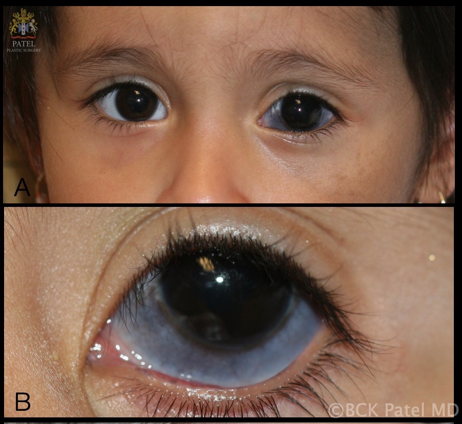
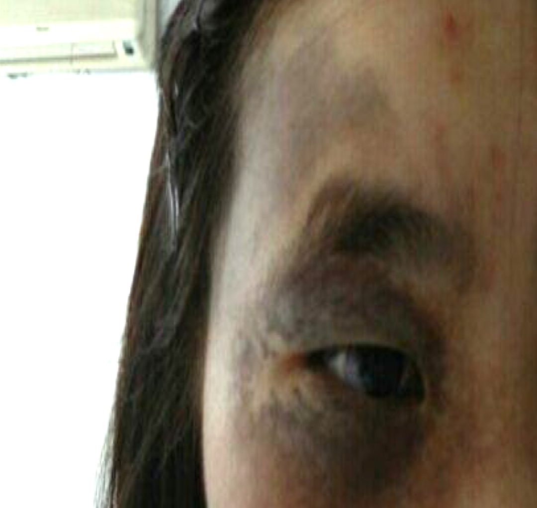
The characteristic gray-blue hyperpigmentation occurs due to entrapped melanocytes. Unilateral presentation is more common. The melanocytes are entrapped leading to gray-blue hyperpigmentation of the conjunctiva and sclera along with ipsilateral facial skin. There is an increased risk of uveal melanoma and glaucoma in these cases.[3] Palatal involvement may also occur.[4]
Nevus of Ito is very similar to nevus of Ota except it differs in the territory of distribution. It was described by Minor Ota in 1954. It involves the distribution territory of lateral cutaneous brachial nerves of the shoulder and posterior supraclavicular nerves. Both of these diseases share similar pathophysiology.
Etiology
The exact etiology is unknown, and various hypotheses have been proposed:
- Melanocytes do not migrate from neural crest cells to the basal layer of the epidermis
- History of previous radiotherapy/radiation exposure/hormonal factors
- The genes BRAF and NRAS of the MAP kinase pathway have been implicated. G-coupled protein, GNAQ is found to be mutated causing the G-coupled protein to be constitutively turned on
- During embryogenesis, the melanoblasts migrate to skin and uvea causing the typical bluish-gray pigmentation in these sites
- Monosomy of chromosome 3 and gain of the long arm of chromosome 8q is a significant risk factor for uveal melanoma and poor outcome.[5]
Epidemiology
The Asian population is more commonly involved affecting 0.014% to 0.034% of the population. Most commonly, they present at birth, but lesions can also appear in puberty or during pregnancy due to hormonal changes. Females are more commonly affected than males at a ratio of 5:1.[6] It is more common in people of Asian and African descent and less common in Whites.[7] Though the risk of malignant melanoma is much more in Whites than in other races.
Nevus of Ito also presents during infancy or at the time of puberty. Asian populations and dark skin races are affected more commonly. Nevus of Ota is also sometimes found to be associated with nevus of Ito.[8]
Pathophysiology
There is hyperpigmentation of the eye and surrounding adnexa along the distribution of the first and second divisions of the trigeminal nerve, namely the ophthalmic V1 and the maxillary V2 divisions. The hyperpigmentation is visible as a bluish pigmentation of the eyes and periocular region. The dermis contains excessive melanocytes, which can transform into malignant melanoma.[9]
Histopathology
Pathology shows numerous melanocytes in the dermis. These pigmented melanocytes are spindle-shaped or dendritic in shape. The Mongolian spot differs from the nevus of Ota in having melanocytes primarily concentrated in the deeper layer of the dermis, whereas in the nevus of Ota, the melanocytes are more superficially concentrated. Nevus of Ota can be classified into 5 types based on the location of dermal melanocytes, namely:
- Superficial
- Superficial dominant
- Diffuse
- Deep dominant
- Deep
History and Physical
Nevus of Ota is usually asymptomatic though rare cases with sensory loss have also been reported. It is characterized by bluish hyperpigmentation along the ophthalmic and maxillary divisions (V1 and V2) of the trigeminal nerve. The nevus of Ota is usually unilateral or sometimes bilateral. Morphology can be macular (rarely papular or nodular), patchy brown, slate-blue or with grey-black pigmentation, and deeper lesions appearing blue. Macules are confluent, non-hairy, and flat with poorly defined margins. Pigmentation may affect extracutaneous sites like the eyes (conjunctiva, sclera, cornea, and uvea). Pigmentation may also affect the oral cavity and nasal mucosa.
A complete ophthalmic examination, including visual acuity testing, intraocular pressure measurement, slit-lamp examination of the anterior segment, and dilated fundus examination with an indirect ophthalmoscope, should be performed. Subconjunctival melanosis and episcleral pigmentation are well appreciated on slit-lamp examination. Gonioscopy should be performed to note the hyperpigmentation of trabecular meshwork. A dilated peripheral retinal examination should be performed to rule out any choroidal mass/choroidal melanoma. Stereoscopic examination of the optic nerve head should be done to document any optic nerve head cupping. An oral examination is performed to look for palate lesions.
Tanino classifies nevus of Ota into 4 types:[10]
- Type I (mild): Type A is periocular. Type B involves the zygomatic region. Type C involves the forehead. Type D involves the only nose.
- Type II (moderate): Similar to type I but worse in severity.
- Type III (intensive): Periocular, nose, and scalp involvement.
- Type IV: Bilateral involvement.
The only difference between the nevus of Ota and the nevus of Ito is the site of involvement and distribution. Ocular complications are not seen in the nevus of Ito. The intensity of pigmentation in both of these nevi may vary with menstruation, fatigue, or weather. Pigmentation becomes more intense after puberty and remains permanent with no spontaneous regression.
Evaluation
There is no definitive diagnostic test to confirm the nevus of Ota and Ito. They are diagnosed mainly by clinical examination and history. Dermatoscopy shows bluish to slate grey homogeneous pigmentation. Skin biopsy is mandated if the overlying skin develops ulceration, or there is a variation in pigmentation with the development of papules.
Superficial variants tend to occur on the cheeks, while deeper variants affect periorbital areas, the temple, and the forehead. Periodic re-evaluation with an ophthalmologist should also be performed annually to rule out ocular complications.
Treatment / Management
Ophthalmic
Ophthalmological screening and recording of vision, intraocular pressures, gonioscopy, comprehensive slit-lamp examination, and dilated fundus examination should be done to rule out any uveal melanomas. Management of uveal melanomas will depend on the size and location and will involve surgical resection, radiotherapy, transpupillary thermotherapy, or enucleation.[9]
Dermatologic
Treatment is required for cosmetic purposes or if there is any malignant transformation. Treatment depends on the size of the lesion and the extent of invasion. Mohs micrographic surgery can be employed to remove the lesions and reconstruct the periocular tissues. For more extensive lesions or metastasis, a combination of surgical excision along with radiotherapy, transpupillary thermotherapy, or chemotherapy may be tried.
Lasers can be employed for the cosmetic treatment of skin lesions by destroying the melanocytes. Pulsed Q-switched laser surgery is the treatment of choice for nevi of Ota and Ito. It targets dermal melanocytes and melanophages by their selective photothermal and photos mechanical destruction, thereby decreasing the pigmentation. Chemical peels, dermabrasion, electrocautery, or cryotherapy are other modalities of treatment for cosmesis.[11]
Differential Diagnosis
Diagnosis of nevus of Ota and Ito can be made based on its clinical morphology and sites of involvement.[12] Dermatosis that need to be considered in the differential diagnosis are:
- Mongolian spots – mostly, the lesions are present in the lumbosacral area and rarely present over the face. They spontaneously resolve by the age of 3 to 6 years.
- Acquired bilateral nevus of Ota-like macules (ABNOM)- sclera and oral mucosa are not involved in ABNOM or Sun’s nevus
- Melanoma metastasis
- Ecchymoses
- Vascular malformations
- Drug-induced hyperpigmentation is usually after the ingestion of drugs like minocycline, amiodarone, and gold
- Melasma - typically associated with pregnancy
- Oral melanotic macule - present over the palate, smaller in size with no involvement of sclera
Prognosis
Malignant degeneration is rare. The prognosis is good. Patients usually present for cosmetic reasons due to pigmentation of the sclera and iris. Rarely it may be associated with cavernous hemangioma of the optic disc.
There is a 10.3% risk of glaucoma, and 1 in 400 patients developed malignant melanoma. For these reasons, annual ophthalmological screening is mandated for the measurement of intraocular pressures and dilated indirect ophthalmoscopy.
A skin biopsy should be done if clinical changes are suspected of malignant transformation. Ophthalmologic follow-up care is necessary for patients with increased intraocular pressure.
Overall, these lesions are typically benign with an excellent prognosis with or without treatment. Yearly screening for ruling out glaucoma and malignant melanoma by an ophthalmologist and dermatologist is recommended.
Complications
The main social impact of nevi of Ota and Ito is the cosmetic concern. In nevus of Ota, where the sclera is commonly affected, ocular complications like increased intraocular pressure and glaucoma can occur. Malignant melanoma has also been reported, which occurs mainly on the skin, but meninges and ocular areas can also be involved.
Deterrence and Patient Education
Patients should be taught that nevus of Ota and Ito are benign lesions that may present as ipsilateral grayish-blue pigmentation along the periocular region. The patient should be advised that transformation to malignancy is rare. The patients are counseled to seek immediate medical consultation if there is any change in the size and shape of the lesions. Instruct patients to schedule periodic follow-up visits with an ophthalmologist for the measurement of intraocular pressures.
Enhancing Healthcare Team Outcomes
Nevus of Ota commonly presents initially to ophthalmologists or dermatologists as a cosmetic concern for the bluish discoloration of periocular skin and the sclera. It is primarily a clinical diagnosis, but there is an increased risk of glaucoma and uveal melanoma in these cases (10% and 1 in 400, respectively). It is important to have baseline measurements of the intraocular pressures, perform gonioscopy, and indirect ophthalmoscopy on the first visit. Serial monitoring of intraocular pressures is mandated to detect early glaucoma. Close interprofessional team coordination with ophthalmologists, dermatologists, and primary clinicians is indicated for the management of these patients.