Continuing Education Activity
Pemphigus herpetiformis is a rare subtype of intraepidermal autoimmune bullous diseases that shares clinical characteristics with dermatitis herpetiformis and immunologic features with pemphigus. It usually has an indolent course and is less life-threatening than many forms of pemphigus. However, some cases may progress into pemphigus vulgaris or pemphigus foliaceous. This activity describes the etiology, pathophysiology, and evaluation of pemphigus herpetiformis and highlights the role of the interprofessional team in the management of this disorder.
Objectives:
- Identify the etiology of pemphigus herpetiformis.
- Review the presentation of pemphigus herpetiformis.
- Outline the treatment and management options available for pemphigus herpetiformis.
- Explain interprofessional team strategies for enhancing care coordination and communication to advance the treatment of pemphigus herpetiformis and improve outcomes.
Introduction
Pemphigus herpetiformis is a rare subtype of intraepidermal autoimmune bullous diseases that associate the clinical characteristics of dermatitis herpetiformis and the immunologic features of pemphigus. Although Jablonska and others proposed the name of this entity in 1975, similar clinical presentations were reported earlier and named dermatitis herpetiformis with acantholysis, mixed bullous disease, and pemphigus controlled by sulfapyridine.[1][2][3][4]
Etiology
Pemphigus herpetiformis appears to be an IgG-mediated autoantibody that mainly targets the desmoglein proteins of the epidermis. The mechanism by which autoantibodies produce characteristic skin lesions in pemphigus herpetiformis is still debated. Some cases of drug-induced pemphigus herpetiformis have otherwise been reported.[5][6][7][8]
Epidemiology
Pemphigus herpetiformis accounts for 6% to 7.3% among patients with pemphigus. Since the first description in 1975 by Jablonska and others, approximatively 100 cases have been reported in the literature. It equally affects men and women. The age of onset ranges from 5 to 92 years. Only four pediatric patients have been reported. Pemphigus herpetiformis does not appear to have a specific ethnic predominance since it has been reported from different continents including America, Europe, Asia, and Africa.
Pathophysiology
Target antigens for pemphigus herpetiformis are usually Desmoglein 1 (Dsg1) and less commonly Desmoglein 3 (Dsg3). Recently, several cases of pemphigus herpetiformis without anti-Dsg1 or anti-Dsg3 autoantibodies have been reported, and some patients show reactivity against other antigens such as desmocollin (Dsc). In contrast to pemphigus vulgaris and pemphigus foliaceous, pemphigus herpetiformis is characterized by an intense inflammation that may not be associated with acantholysis. In fact, pemphigus herpetiformis autoantibodies may recognize functionally less important epitopes of Dsg1 or Dsg3, and therefore, do not induce acantholysis. A recent study supports this hypothesis through its discovery of a broader epitope distribution compared to pemphigus vulgaris or pemphigus foliaceous. It is also held that autoantibodies in pemphigus herpetiformis lead to the secretion of proinflammatory cytokines by keratinocytes, especially interleukin-8. This, in turn, consecutively leads to the recruitment and stimulation of inflammatory cells, resulting in eosinophils and/or neutrophils spongiosis and focal intercellular edema that are responsible for the blistering process.[9][10]
Histopathology
Histopathological features of pemphigus herpetiformis differ from pemphigus vulgaris and pemphigus foliaceous because of the presence of spongiosis, mostly without acantholysis. The histological findings may vary among patients, and one patient can have various histological features at different times. Therefore, more than one biopsy may be necessary for the diagnosis of pemphigus herpetiformis. The main histopathological finding of pemphigus herpetiformis is eosinophilic spongiosis. Other reported features include neutrophilic spongiosis, mixed eosinophilic-neutrophilic spongiosis, subcorneal pustules and/or intraepidermal vesicles filled with neutrophils and/or eosinophils. Acantholysis is usually absent but may appear later in the disease process. Dermal papillary neutrophilic microabscesses also can be present. The pattern of immune cell infiltration may vary, being mixed eosinophil neutrophil in 60% of cases, eosinophil dominant in approximately 20%, and neutrophil-dominant in 20%.
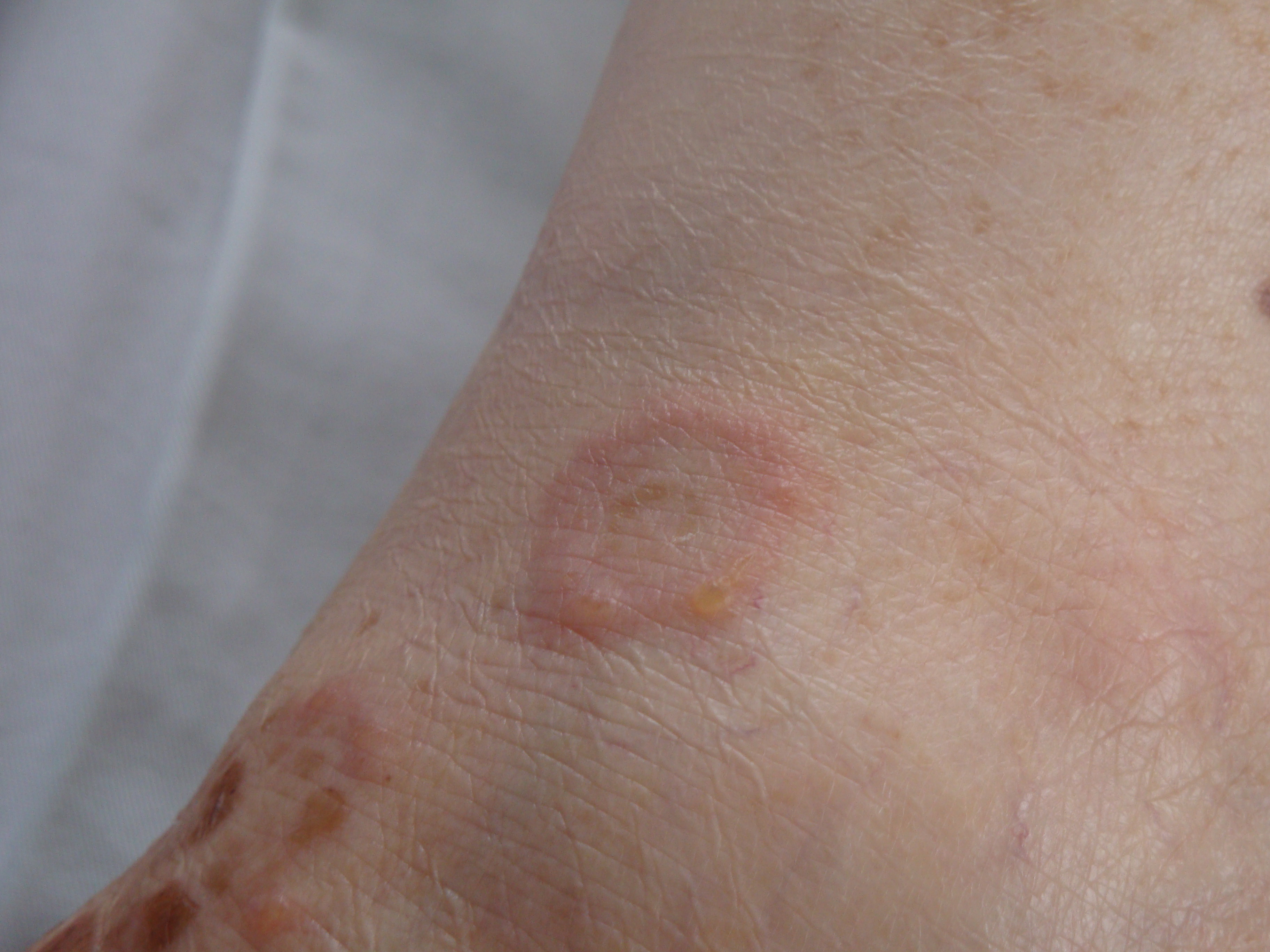
History and Physical
Patients affected with pemphigus herpetiformis typically have a subacute onset of disease. The diagnosis of pemphigus herpetiformis is rarely established at the first consultation, as the clinical presentation is usually atypical, and other diseases can be hypothesized such as dermatitis herpetiformis, bullous pemphigoid, and linear IgA bullous dermatosis. Patients affected with pemphigus herpetiformis usually show erythematous, vesicular, bullous, pustular or papular lesions, often in a "herpetiform" pattern and with severe pruritus. Skin lesions tend to present with annular-shaped distribution, probably due to the centrifugal spread of inflammatory processes. In some cases, the main lesions can be urticarial itching papules and plaques. These features are not frequently seen in pemphigus vulgaris and pemphigus foliaceous. The affected sites for pemphigus herpetiformis are mostly the trunk and proximal extremities. Although pemphigus herpetiformis usually manifests a widespread distribution, some cases of atypical and localized lesions have been reported. However, mucous membrane involvement is not a frequent issue. During the clinical course of the disease, pemphigus herpetiformis may evolve into pemphigus vulgaris or pemphigus foliaceous. The opposite has also been reported.
Evaluation
Peripheral blood eosinophilia may be found in some patients. Direct immunofluorescence staining on skin biopsy specimen is similar to the other forms of pemphigus: interkeratinocytic deposits of IgG and C3 most often seen in the superficial layers of the epidermis. Less commonly, these immunoreactants can be seen in the lower layers of the epidermis, mainly when circulating autoantibodies against Dsg3 are present. Indirect immunofluorescence with healthy human skin, the monkey/guinea pig esophagus or rat bladder as a substrate may reveal intercellular binding of IgG antibodies. Enzyme-linked immunosorbent assay and/or immunoblotting can reveal circulating antibodies targeting epidermal proteins, mainly Dsg1, less commonly Dsg3. Recently, other target antigens have been reported such as Dsc1, Dsc3, 178-Kd antigen, BP 180 C-terminus, and laminin 332 gamma2 subunit.
Treatment / Management
Pemphigus herpetiformis usually has an indolent course, and it is less life-threatening than other forms of pemphigus. Some cases, however, may progress into pemphigus vulgaris or pemphigus foliaceous. The small sample sizes limit the evidence base for management of pemphigus herpetiformis. Based on a review of the literature, dapsone and corticosteroids are the drugs mainly prescribed. Pemphigus herpetiformis usually respond well to dapsone which is considered by many authors as the first drug of choice due to its ability to decrease neutrophil migration. Corticosteroids are also frequently reported to be effective in PH, and the daily doses required for complete remission are much lower than those required in pemphigus vulgaris or pemphigus foliaceous. Dapsone and steroids may be given as monotherapy or in combination. More intense therapy modalities have been applied in cases of pemphigus herpetiformis evolving to the classical forms of pemphigus in patients with recalcitrant pemphigus herpetiformis, including cyclophosphamide, mycophenolate mofetil, sulfapyridine, azathioprine, methotrexate, intravenous immunoglobulin, rituximab, and plasmapheresis.[11]
Differential Diagnosis
- Familial benign pemphigus
Pearls and Other Issues
Pemphigus herpetiformis is an uncommon subtype of pemphigus with atypical clinical, histological and immunopathological findings. The exact mechanisms underlying pathogenesis remain unclear. The diagnosis of pemphigus herpetiformis is challenging since it is a rare disease with unusual presentation. A significant delay in diagnosis is, therefore, often reported. Intense pruritus associated with urticarial plaques, vesiculobullous eruption in a herpetiform pattern and the absence of mucosal involvement should prompt consideration of the diagnosis. Pemphigus herpetiformis has a good prognosis and responds well to dapsone and/or low doses of steroids; however, some cases may progress into pemphigus vulgaris or pemphigus foliaceous.
Enhancing Healthcare Team Outcomes
Pemphigus herpetiformis is a rare blistering skin disorder that is best managed by an interprofessional team that includes clinicians, specialists, nursing staff, and pharmacists. pharmacists. There is no universal treatment for the disorder but many patients do respond to dapsone and corticosteroids. However, many other immunosuppressive agents and biological drugs are now used to treat this disorder with variable success. The patient needs to be educated about the adverse effects of the drugs; which often reduces compliance. Both nursing and the pharmacist can fulfill this role and should report any concerns back to the prescribing clinician. The pharmacist will also verify all dosing and perform medication reconciliation to rule out drug interactions. This interprofessional methodology will lead to improved patient outcomes for patients with pemphigus herpetiformis.