Continuing Education Activity
Lichen sclerosus is an uncommon autoimmune condition characterized by skin atrophy and hypopigmentation. It mostly affects the genital skin and occurs in women more than men. This activity outlines the evaluation and treatment of lichen sclerous and reviews the role of the interprofessional team in managing patients with this condition.
Objectives:
- Review the role of T cells, autoantibodies and cytokines in the pathophysiology of lichen sclerosus.
- Describe the typical presentation of patients with lichen sclerosus.
- Identify the use of biopsy in the evaluation of lichen sclerosus.
- Explain the importance of collaboration and communication among the interprofessional team for the long-term follow up of patients with lichen sclerosus.
Introduction
Lichen sclerosus (LS) is a chronic inflammatory disease. It was first described by Hallopeau in 1881. Since then, multiple names have been used to describe this condition such as leukoplakia, kraurosis vulvae, balanitis xerotica obliterans, and lichen sclerosis et atrophicus. In 1976, the International Society for the Study of Vulvovaginal Disease adopted the term of lichen sclerosus.
LS is a mucocutaneous autoimmune disorder characterized by hypopigmentation and skin atrophy. It involves most commonly the genital skin, less often the extragenital sites. LS is more common in women than in men. It may cause phimosis or scarring of the vaginal introitus. The diagnosis is based on the clinical features, but it is often confirmed by biopsy. The lesions can evolve towards the destruction of anatomic structures, functional impairment and a potential risk for malignant evolution. Thus, treatment and long term follow-up are mandatory.
Etiology
LS is considered to be an autoimmune condition; however, its etiology remains unclear. Given its association with other autoimmune diseases such as alopecia areata, vitiligo, autoimmune thyroiditis, and pernicious anemia, its etiology is probably multifactorial. Further etiologic factors have been implicated such as genetic susceptibility, infectious agents such as spirochetes, sex hormone, and the Koebner phenomenon. Furthermore, recent data demonstrated a high prevalence of lichen sclerosis associated with morphea. [1]
Epidemiology
LS is relatively rare. Although reports indicate that it affects between 1 in 1000 and 1 in 300 individuals in the general population, the exact prevalence is unknown. [2] It still seems unknown and underestimated because many patients are asymptomatic, and it is frequently misdiagnosed. Both sexes are affected, but it is more common in women than men with a female to male ratio varies from 1:1 to 10:1. There is no racial predilection. It occurs at all ages. Moreover, its incidence in women has two peaks, the first one occurs between eight and thirteen-year-old girls and the second one is during the fifth and the sixth decades. The mean age at diagnosis ranges between 52 and 60 years. LS occurs in the genital skin in 85 to 98 %, and on the extragenital skin in only 15 to 20 %. [2] Extragenital lichen sclerosus is uncommon during childhood. LS can involve the oral mucosa, known as oral lichen sclerosus, and has been rarely reported.
Pathophysiology
- The underlying pathogenesis of LS includes an infiltrate of activated T cells releasing interleukin 4 (IL 4) and transforming growth factor β (TGF β). So, these cytokines activate fibroblasts producing significantly altered collagen leading to fibrosis. Besides, the pathogenesis includes vascular damage by the decrease in the number of capillaries.
- Interleukin 1 (IL 1) and interleukin one receptor antagonist (IL 1ra) may also be included in the pathogenesis of lichen sclerosus as well as increased number of monoclonal T lymphocyte CD4 +, lymphocyte T dendritic CD1a + cells, macrophages, mast cells, and decreased number of T lymphocyte CD3 +. [3]
- Another hypothesis has been suggested such as an increased number of circulating IgG autoantibodies targeting extracellular matrix 1 (ECM 1) protein leading to widespread deposition of hyaline material in the dermis. [4]
Histopathology
LS has a specific histologic pattern characterized by a band-like lymphocytic infiltrate below a zone of dermal edema and orthokeratotic hyperkeratosis. Histopathology findings vary depending on disease duration. In earlier stages, it shows vacuolar degeneration of the basal layer, hyalinization of subepithelial collagen, decreased elastic fibers in the upper dermis and dilated blood vessels under the basement membrane. In older lesions, histology shows a reduced number of mononuclear cells and dispersed patchy islands of mononuclear cells within the hyalinized dermis. [3][4]
History and Physical
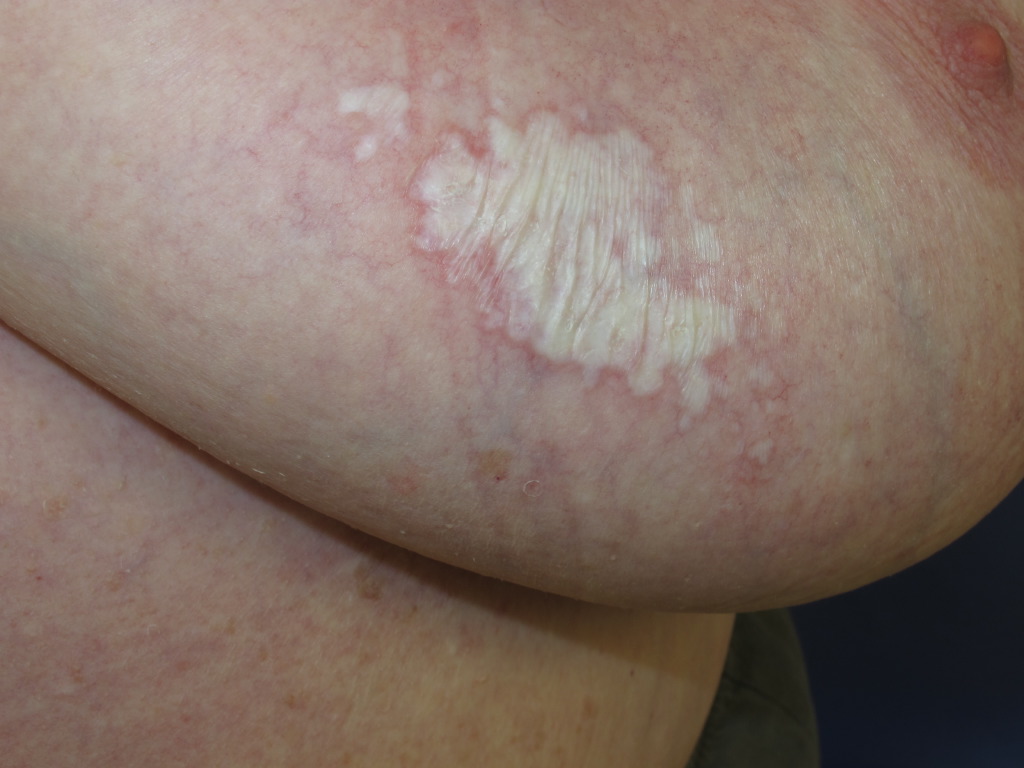
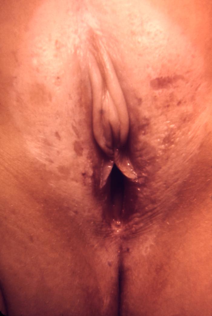
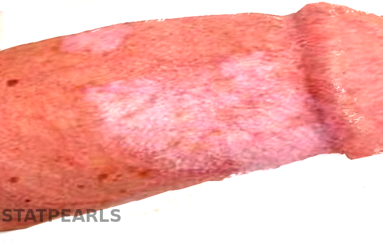
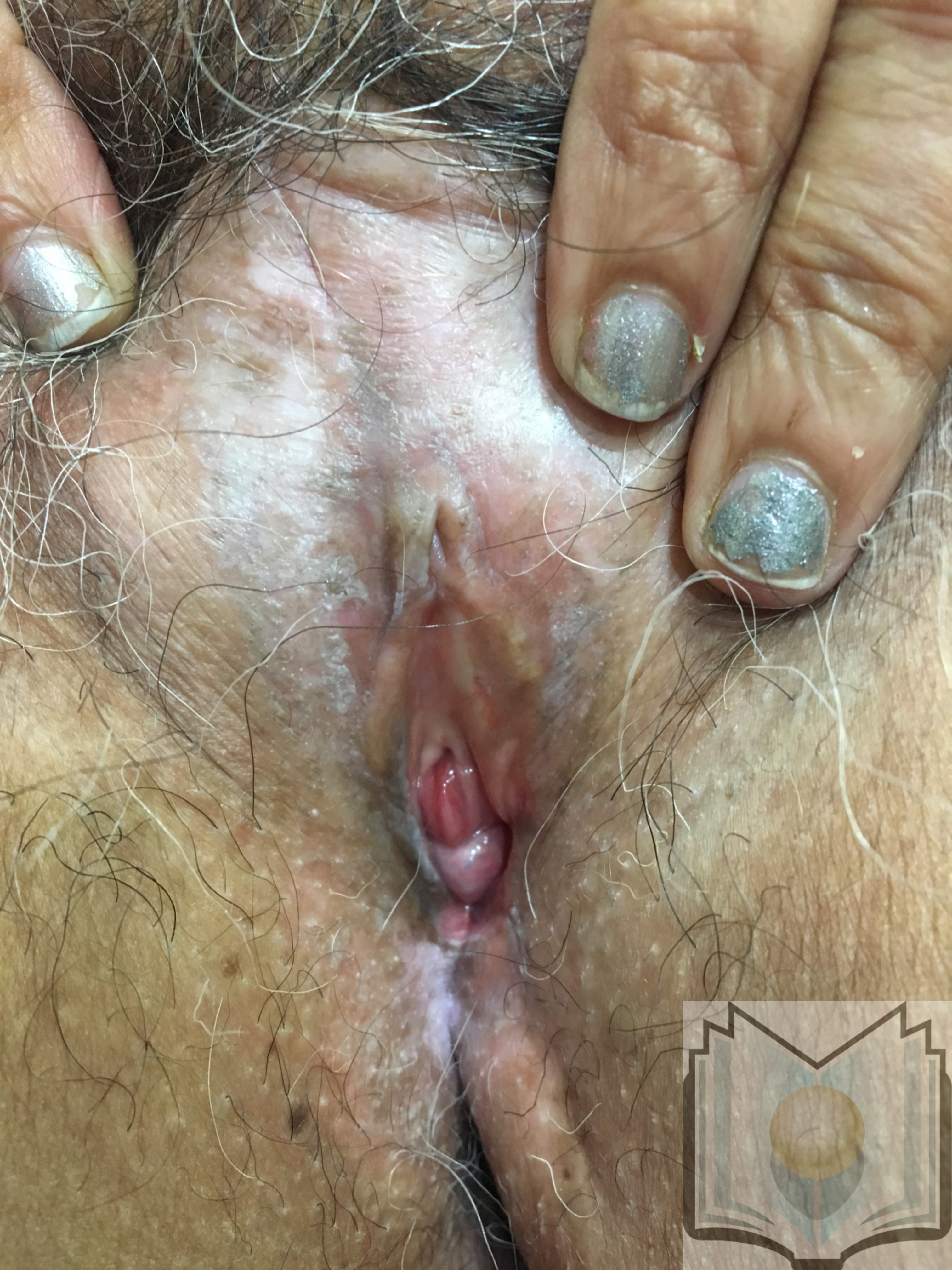
LS affects most commonly the genital site and less often the extragenital area. Typical lesions begin as a sharply demarcated erythema that becomes thin, hypopigmented, ivory-white, porcelain-like, and sclerotic plaques. Plaques may become later thickened due to repeated excoriations. Itch is the main symptom and is often worse at night. Other lesions may include telangiectasias, purpura, fissures, ulcerations, and edema. Typical complaints commonly include significant pruritus, local burning sensation, pain, painful defecation. Constipation is frequent in children but rarely seen in adults. However, lesions can be asymptomatic. Genital lesions begin around the periclitoral hood. The affected area varies from a small and single area to large area involving the entire region of the vulva, perineum, and perianus assuming a typical aspect of ‘keyhole sign.’
However, LS usually spares the vagina and cervix. In girls, LS presents commonly with irritation and soreness although it can mimic sexual abuse. However, LS and sexual abuse can coexist. Visual lichen sclerosus exhibits the Koebner phenomenon at sites of trauma; sexual abuse can aggravate lesions of LS. [5] Male genital LS (boys and men) occurs in the foreskin, glans penis, and the coronal sulcus penis. Extragenital lesions occur on any part of the skin and usually asymptomatic. The most commonly involved areas are inframammary areas, neck, wrists, thighs, upper back, and shoulders. The involvement of the oral mucosa appears clinically as bluish-white papules on the buccal mucosa or under the tongue.
Evaluation
The diagnosis is based on taking a careful history including the autoimmune diseases in the patient and family, examination of the mucosas, extragenital skin and completed by a gynecological exam. Thus, the diagnosis of LS is usually clinical. But in some cases, a biopsy can be performed. However, atypical histology does not rule out the diagnosis.
Biopsies should be performed in case of:
- Atypical clinical presentation
- Suspected malignancy
- Nonresponse, after an appropriate duration, to recommended first-line treatment
The workup of the condition should include investigation of thyroid function, and according to symptoms investigation of the other autoimmune diseases.
Treatment / Management
The aims of the treatment are relief of the symptoms, stopping the atrophy, prevention scar formation, and anatomical distortion, as well as malignant transformation. The therapy includes general care, topical treatments, systemic treatments, and surgical procedures. It is fundamental to inform the patient to avoid the use of irritating products such as soap and to prefer emollients to break the itch-stretch cycle.
For genital LS, the gold standard treatment is three months application of high potency topical steroids (clobetasol propionate). Second-line therapies include topical calcineurin inhibitors and imiquimod. In men, early circumcision may be recommended. Surgery is indicated only for the treatment of complications associated with lichen sclerosus. For extragenital LS, therapeutic modalities are limited and include phototherapy, ultrapotent topical steroids, tacrolimus ointment 0, 1%, and systemic steroids or methotrexate. Follow-up examinations must be kept indefinitely. [6]
Differential Diagnosis
- Lichen planus
- Lichen simplex chronicus
- Vitiligo
- Morphea
- Eczema
- Contact dermatitis
- Psoriasis
- Child sexual abuse
Prognosis
The prognosis of genital LS in children is good. Often the disease may resolve itself spontaneously. LS responds mostly well to local corticosteroid treatment. The forecast is not as positive for extragenital LS and chronic atrophic genital disease.
When untreated, LS leads to disease complications making early diagnosis and treatment crucial. Timely diagnosis and treatment may prevent disease complications.
Although LS is not a precancerous condition, patients with genital LS have an increased risk of squamous cell carcinoma.
Complications
LS is a chronically relapsing disease and if untreated leads to a potential scarring process that may evolve to a complete loss of standard vulvar architecture in women including introital stenosis, fusion, and resorption of the labia minora, and urethral strictures in males. Tissue adhesion and sclerosis lead to tearing and then the loss of sexual function, in addition to dysuria, constipation, itching, and soreness. Furthermore, vulvar lichen sclerosis may evolve to vulvar squamous cell carcinoma in the affected area with an estimated risk up to 5 %; however, its association with penile SCC is not clear. Extragenital lesions are not associated with this risk of transformation. Melanoma and basal cell carcinoma have been reported. [7]
Deterrence and Patient Education
LS may lead to scarring and loss of standard genital architecture. Hence, education of the patient relating to sexual dysfunction may be required.
Given its potential for malignant transformation to squamous cell carcinoma, the patients with anogenital LS should receive long-term follow-up.
Enhancing Healthcare Team Outcomes
It is essential that dermatologists, gynecologists, urologists, surgeons, and pediatricians, respectively, do not hesitate to cooperate. LS may have a negative impact on sexual health. Care coordination by different specialists is necessary to improve outcomes.