Etiology
The consequences of arthropod bites are generally due to traumatic injury or local inflammation and hypersensitivity to arthropod saliva. Even though some arthropods are capable of injecting venom when biting, most envenomation occurs via a stinger connected to a venom gland. Notable arthropods possessing stingers include bees, wasps, hornets, fire ants, and scorpions. Both bites and stings create tissue injury which can serve as a portal of entry for secondary bacterial infection.
The four classes medically significant arthropods are the chilopods, diplopods, insects, and arachnids. Members belonging to each class are as follows:
- Chilopoda: centipedes
- Diplopoda: millipedes
- Insecta: Hymenoptera (bees, wasps, hornets and fire ants), mosquitoes, bedbugs, fleas, lice, beetles, caterpillars and moths, and kissing bugs
- Arachnids: spiders, scorpions, mites, and ticks
Chilopods
Centipedes are chilopods characterized by a cephalad biting structure connected to a venom gland. Bites often produce two hemorrhagic punctures accompanied by surrounding erythema and swelling. The venom consists of biochemical mediators, including metalloproteases, which result in immediate localized pain.[1] Treatment is generally supportive and includes cleaning the area with soap and water, local application of ice, topical steroids, and pain management with acetaminophen or NSAIDs.[2] Cases manifesting with severe localized pain are treatable with the local infiltration of lidocaine.[2]
Diplopods
Millipedes are members of the class Diplopoda, characterized by the presence of two pairs of legs per body segment. Millipedes inflict damage through secretion of a toxic liquid from glands on the sides of their body segments which produces a localized caustic-like effect to tissues. Clinically this may present with an intense burning sensation accompanied by erythema and occasionally vesicle formation. The toxic liquid often causes the development of a localized area of hyperpigmentation, usually brown or black, which may last for months.[1][3] More significant injury can occur with ocular exposure which can cause chemical conjunctivitis or corneal ulceration. Treatment of topical exposure is similar to that of a second-degree burn and includes topical antibiotics and analgesics. Washing the area immediately with soapy water following exposure may help reduce the effects of the toxin. Treatment of ocular exposure requires copious irrigation, fluorescein staining to evaluate for ulceration, topical antibiotics, cycloplegics, and ophthalmology referral.
Insecta
Members of the class Insecta, which comprise approximately 60% of all arthropod species, include Hymenoptera (bees, wasps, hornets and fire ants), mosquitoes, bed bugs, fleas, lice, beetles, caterpillars and moths, and kissing bugs.[2]
Caterpillars and Moths
While most caterpillars and moths are harmless to humans, some cause cutaneous disease when contact with their protective hairs or spines occurs. The most common clinical manifestations of exposure are erythema, pruritus, and the development of papules or urticaria at the site of contact. In the United States, the most serious clinical reactions occur in contact with the Pus Caterpillar, primarily found in the Southeastern United States. Rather than pruritus, exposure often results in local burning pain and the development of hemorrhagic, purpuric papules organized in a grid-like pattern.[3] Treatment of caterpillar and moth induced cutaneous reactions consists primarily of symptomatic care including local ice application, pruritus control with topical corticosteroids and systemic antihistamines, and pain control with NSAID’s or acetaminophen.[2] If hairs or spines are present on the skin, they can be removed with adhesive tape.[3]
Bedbugs
Cimex lectularius, the human bedbug, is an obligate blood feeder with a worldwide distribution. It is oval shaped, flat, red-brown in color and about 3 to 6 mm in length. Bedbugs usually live in crevices in walls, floors, mattresses, cushions, bed frames, and other structures. Bites sometimes occur linearly and most commonly manifest as a small papule or punctum usually without a surrounding reaction.[3]Treatment of bites consists primarily of symptomatic care with the use of topical glucocorticoids and systemic antihistamines to control pruritus.[2] Secondary infections should be treated with the appropriate antibiotics. Elimination of infestations can be difficult and requires a combination of professionally applied insecticide and nonchemical controls.
Kissing Bugs
Kissing bugs, or triatome bugs, are a type of reduviid bug that can carry the parasite Trypanosoma cruzi, the etiologic agent of Chagas disease. Kissing bugs are 1.5 to 2.5 cm long, brown or black and typically have red or yellow stripes on their abdomen. Triatomes are blood-sucking insects and nocturnal feeders which transmit Trypanosoma cruzi through their feces. Their range runs in the southern United States, Mexico, Central America and South America where they typically live in thatched roofs or cracks and holes of substandard housing. Victims may sometimes inadvertently scratch or rub the feces into the bite wound or the mucous membranes, especially the eyes or mouth. Bites are usually painless and present as papules with hemorrhagic puncta or vesiculobullous lesions.[3] An area of localized swelling, erythema, and induration corresponding to the site of trypanosome entry is known as a chagoma. The classic finding in acute Chagas disease is the presence of a chagoma on the eyelid, known as Romana’s sign. Following the acute phase of the infection, which can last for several months, patients enter the chronic phase, during which the infection may remain asymptomatic for decades. Approximately 20 to 30% of infected patients will develop cardiac or gastrointestinal complications.
Lice
Lice are parasitic insects that feed on human blood. They have six legs, are tan to grayish-white, 2.5 to 3.5 cm in length, have no wings, and are flat in shape. The three types of lice that feed on humans are the head louse, Pediculus humanus capitus, the body louse, Pediculus humanus corporis, and the pubic or crab louse, Pthirus pubis. While head and pubic lice do not have an association with disease transmission, body lice can transmit typhus and louse-borne relapsing fever.
Head lice
Head lice occur most frequently in younger children in daycare or elementary school and typically present on the head, eyebrows, and eyelashes. Head lice exist in one of three forms: the adult, the nymph, and the egg. Eggs, also known as nits, are yellowish and are cemented to the hair shafts. The nymph, an immature louse, hatches from the egg in about 8 days, and as the adult form, is an obligate blood feeder. Common symptoms of head lice infestation include a sensation of something moving in the hair, itching, or the visualization of nits or lice. Diagnosis is by identification of nits, nymphs or the adult louse on the scalp or hair. Head lice infestation treatment is with pediculicides, many of which are available without a prescription. Common over-the-counter treatments include pyrethrins and permethrin 1% lotion. Oral ivermectin at an initial dose of 200mcg/kg and repeated 10 days later is often used to treat lice infections but is not FDA approved for this purpose. Nits removal should be with a fine comb. Since most treatments do not entirely eradicate the eggs, retreatment is usually necessary seven to ten days later.
Pubic lice
Pubic lice are smaller than head or body lice, measuring around 1 to 2 mm in length. They have six legs with two large front legs which resemble the pinchers of a crab. The distribution of pubic lice is generally limited to areas where the hair is short, such as pubic hair, but may occasionally be found on eyelashes, eyebrows, and axillary and beard hair. As with head lice, pubic lice have an egg, nymph and adult form. Transmission is usually through sexual contact, so infestations are most frequently found in adults. If pubic lice are found, clinicians should consider evaluating for the presence of other sexually transmitted infections. The symptoms of infestation are similar to that of head lice, and the finding of lice or nits in the pubic hair establishes the diagnosis. The treatment of pubic lice infestation is the same as that of head lice. Treatment of lice or nits found on the eyelashes involves the application of ophthalmic-grade petroleum ointment to the eyelid margins for 10 days.[3]
Body Lice
Unlike head and pubic lice, body lice do not live on the skin but rather live and lay their eggs in seams of clothing or bedding, moving to the skin only to feed. Body lice usually spread by direct contact but can also be transmitted through clothing, bed linens and towels. In the United States, body lice infestations are most commonly found in homeless populations. While body lice do share some symptoms with other lice, they produce more severe pruritus secondary to an allergic response to lice saliva. Areas of pyoderma may be found in areas covered by clothing such the axilla, trunk, and groin. Unlike head and pubic lice, diagnosis of an infestation is typically by finding eggs and lice in seams of clothing rather than on the skin. Treatment of body lice does not usually require the use of a pediculicide because improvements in hygiene, including showering and access to regular laundered clothing, will often eradicate the infestation. Wash all clothing, bed linens and towels in hot water and machine dry on the hot cycle. Itching can be treated with systemic antihistamines and antibiotics may be required for secondary infection. If pharmacologic treatment is deemed appropriate, it consists of the same agents utilized for pubic and head lice. Body lice pose significant health risks because of their ability to transmit the diseases of epidemic typhus, trench fever, and epidemic relapsing fever.[3]
Mosquitoes
Mosquitoes belong to the family Culicidae and are characterized by compound eyes, delicate wings, long thin legs, and proboscises which allow for biting and obtaining blood meals. Bites from mosquitoes produce minimal trauma and are often not felt by the host. The most common symptom is pruritus, and the most common cutaneous finding is the presence of urticarial wheals. Treatment of mosquito bites is symptomatic and includes ice application, topical corticosteroids, and systemic antihistamines. The greatest danger from mosquitos is their ability to transmit several serious diseases including malaria, filariasis, yellow fever, dengue fever, West Nile virus, Zika virus, and chikungunya.
Flies
There are a number of medically significant flies that bite humans, most notably the deer fly, horse fly, sand fly, and tsetse fly. When obtaining a blood meal, flies use their specially designed mouthparts to lacerate the skin, inject its anticoagulant-containing saliva and then suck up the resulting bleeding. Some flies can transmit serious diseases such as the Tsetse fly which transmits trypanosomiasis, the sand fly which transmits bartonellosis and leishmaniasis, and the deer fly which transmits tularemia. Some flies such as the black fly and horse fly can produce severe allergic and even anaphylactic reactions. Fly bites are usually quite painful and may produce significant cutaneous inflammation and the development of large urticarial wheals and papules. Treatment consists of symptomatic care including ice application, pain control with acetaminophen or NSAIDs, thorough washing of the wound, and systemic antihistamines for itching.
Myiasis is a condition that occurs when there is an infestation of fly larvae. Wound myiasis occurs when flies deposit larvae on or near a wound where they feed on the surrounding tissue. Copious irrigation will usually remove the larvae, but sometimes debridement is required.[4] Furuncular myiasis occurs when fly larvae burrow into the skin. The most notable cause of furuncular myiasis is the human botfly, found in the Caribbean and Central and South America. Transmission usually occurs when a female fly lays eggs directly on a blood-sucking insect such as a mosquito.[2] When the insect lands and bites its victim, the eggs get deposited on the skin, hatch, and the larvae quickly burrow under the skin and emerge fully mature after 5 to 10 weeks. Clinically, botfly larvae produce an erythematous papule or nodule with a central punctum representing the larva’s breathing tube [4]While the larvae will emerge spontaneously in 5 to 10 weeks, surgical incision and extraction are usually the treatment. Another treatment option is to cover the site with a thick layer of petroleum jelly or pork fat which occludes the larva’s breathing tube and causes it to emerge within 24 hours.[4]Additional treatment includes updating tetanus immunization, administering antibiotics if secondarily infected, and acetaminophen or NSAIDs for pain control.
Hymenoptera
The order Hymenoptera includes wasps, bees, yellow jackets, hornets and fire ants. These insects all have a painful sting delivered by their venom-injecting caudal stinger. The composition of these venoms are complex, and some have the potential for cross-sensitization. Local reactions to stings are most common and present with an immediate onset of localized pain, erythema and edema. Anaphylactic reactions may present with the initial symptoms of pruritus, facial flushing, and urticaria which can rapidly progress to wheezing, dyspnea, angioedema and stridor, vomiting, abdominal cramping, and syncope. The Apoidea family of Hymenoptera, consisting of honeybees and bumblebees, possess a stinger with curved barbs which remains in the victim following a sting. Africanized, or “killer” bees, are an extremely aggressive bee imported to the United States from Brazil in the late 1950s. They are most widely distributed in the southwestern United States, particularly Texas, Arizona, and California but have continued to migrate north and eastward. Wasps and yellow jackets comprise the Vespidae family which, unlike members of the Apoidea family, do not lose their stinger in an attack and have the ability to sting multiple times.
Treatment of uncomplicated stings includes manual remover of the stinger, application of ice compresses, and analgesics for pain. Short courses of systemic antihistamines and corticosteroids are effective for more severe localized reactions.[5]
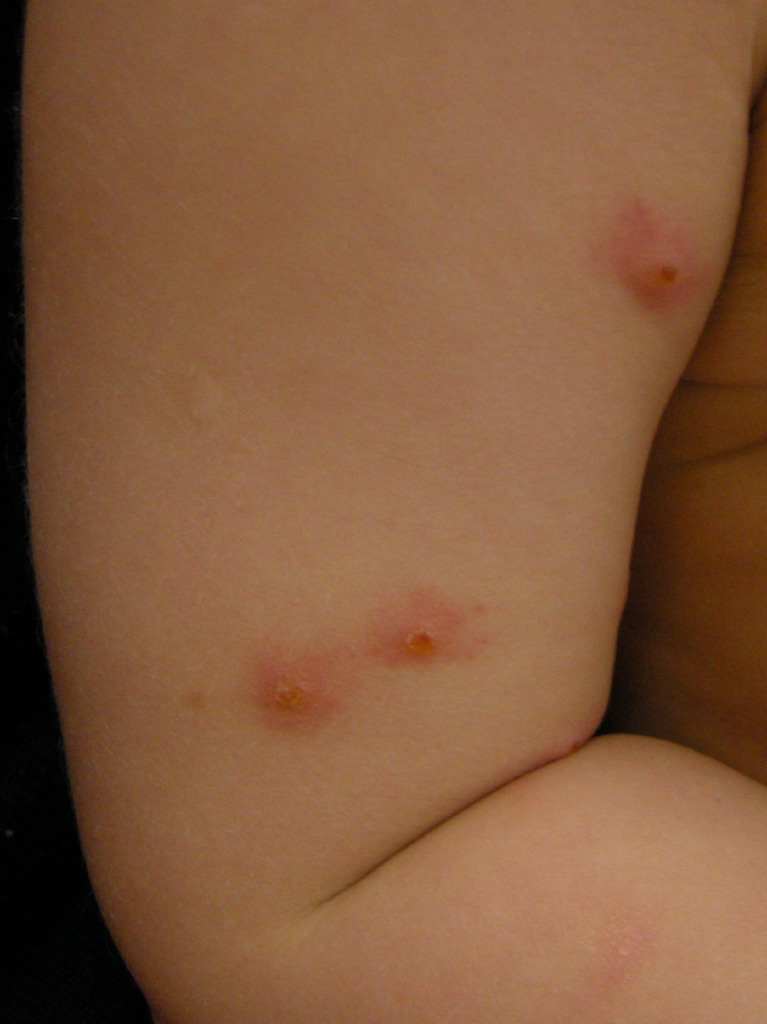
Fire ants are Hymenoptera belonging to the Formicidae family which live in large ground colonies in the southeastern United States. They may be red or black, are very aggressive, and often attack in swarms. Fire ants begin their attack by latching on to their victim with powerful jaws and then deliver up to 10 stings with their ovipositor. Their venom is composed of alkaloids and causes intense, burning-like pain. Their venom may produce anaphylaxis and has potential crossover sensitization with other Hymenoptera venom. As with the Vespidae and Apoidea families, most reactions consist of localized dermatologic findings. The presence of two central hemorrhagic puncta from the bite of the ant, surrounded by a ring of erythematous papules caused by the sting, is a distinctive characteristic.[5] These papules develop into vesicles and then sterile, pruritic pustules over 6 to 24 hours. Treatment is similar to that of other Hymenoptera stings.[2]
Fleas
Fleas are wingless ectoparasites that feed on mammals and birds. They are 2 to 4 mm long, thin, and are red to brown. Flea bites typically present as erythematous papules, often with a hemorrhagic appearing center. Bites may also manifest as urticarial lesions, vesicles or bullae. The pruritus can be severe, and scratching of the lesions can result in skin excoriation and secondary bacterial infection. The primary goal in treating flea bites is to control the intense itching through the use of topical calamine lotion or corticosteroids, and systemic antihistamines. The most significant medical impact of fleas is their ability to serve as vectors for several serious, and potentially fatal diseases including tularemia, endemic typhus, and bubonic plague.
Arachnids
Arachnids are the class of arthropods that include ticks, mites, scorpions, and spiders.
Ticks
Nymphal and adult ticks are characterized by the presence of eight legs tipped with a pair of claws and an oval-shaped body which becomes engorged during feeding. Most ticks are categorized as hard ticks, belong to the Ixodidae family, or soft ticks belonging to the Argasidae family. Ticks feed by cutting a hole in the epidermis and injecting anticoagulants or compounds which inhibit platelet aggregation. Tick bites are usually painless and can present with a wide variety of rashes and other dermatologic findings, making diagnosis challenging. Bites often appear as an erythematous papule with surrounding erythema while others may present as pruritic urticarial lesions. Tick-borne infectious diseases such as Lyme disease and Rocky Mountain spotted fever present with characteristic rashes; however, these are not always identified.
The most significant impact of ticks on humans is their ability to serve as vectors for significant diseases including Rocky Mountain spotted fever, endemic typhus, ehrlichiosis, Q-fever, encephalitis, hemorrhagic fever, Lyme disease, relapsing fever, tularemia, babesiosis.
Simple, uncomplicated tick bites are treated with routine wound care, topical corticosteroids and systemic antihistamines for pruritic lesions and antibiotics if secondary infection is present. Ticks should be removed with fine-tipped tweezers, grasping the tick as close to the skin as possible and pulling upward with steady, gentle pressure. Other commonly touted methods of tick removal such as applying fingernail polish, alcohol or a hot extinguished match are not recommended as they do not affect detachment and may cause the tick to regurgitate into the wound, increasing the risk of disease transmission.[6]
Scabies
Scabies is an infestation of the skin caused by the 8-legged human itch mite, Sarcoptes scabiei. The mite is small, around 0.4mm, and burrows into the stratum corneum layer of skin where it lays its eggs. Scabies has a worldwide distribution and usually spread through prolonged direct contact, often sexual. Initial infections are often asymptomatic for 2 to 6 weeks, and the development of symptoms is the result of an allergic response to mite proteins deposited under the skin.[4] The most common manifestations of scabies infestation include intense itching, often worse at night or with exposure to warm temperatures, and the presence of erythematous papules or nodules. Tiny burrows created by the tunneling of the female mite under the skin may appear as raised, linear or curved grayish-white lines on the skin surface, and sometimes contain a black speck representing the female mite. Cutaneous vesicles may be present but are a more common presentation of scabies infestation in infants and young children. The dermatologic findings in scabies infestations most commonly present in the web spaces of the fingers and toes, ventral surface of the wrist, elbows, back, buttocks and external genitals. The diagnostic basis is on symptoms of intense pruritis accompanied by the appearance of a corresponding rash or burrows. Confirmation of the diagnosis can is by microscopic evaluation of a skin scraping revealing the mite, its eggs or fecal material.[3]
Permethrin, 5% cream, is the drug of choice to treat scabies and is applied from the neck down at bedtime and then washed off in the morning. The recommendation is for two treatments administered a week apart.[3] While not FDA approved for the treatment of scabies, the oral antiparasitic agent Ivermectin is often used, especially in those who have failed other treatments or cannot tolerate topical medications.[4] Pruritus treatment from scabies infestation is with systemic antihistamines.
Norwegian scabies, also known as crusted scabies, is a severe form of scabies infestation that can occur in patients who are elderly, debilitated or immunocompromised. Unlike patients with typical scabies infection, those with Norwegian scabies manifest with thick hyperkeratotic skin, sometimes described as “dirty” in appearance. A treatment protocol for crusted scabies is published by the Centers for Disease Control (CDC) consisting of permethrin cream applied daily for 1 week then twice-weekly coupled with ivermectin 200 µg/kg on days 1, 2, 8, 9, and 15.[4]
Scorpions
Scorpions are large arachnids with a pair of anterior legs possessing pinchers. Their tail-like structure containing a stinger and two venom glands. In the United States, only the bark scorpion, Centruroides exilicauda, possesses venom with the potential to cause systemic toxicity.[7]Most stings produce only localized pain similar to that of Hymenoptera stings, and a diagnostic clue is increased sensitivity to touch or tapping on the area.[5] While systemic symptoms are uncommon, the venom from the bark scorpion can cause several adverse autonomic and motor effects such as hypertension, tachycardia, tachydysrhythmias, myoclonus, and fasciculations.[5] The clinical impacts of these effects can be especially severe in children.
The diagnosis of scorpion envenomation generally relies on a history of a scorpion sting, presence in a scorpion endemic region and characteristic findings of envenomation. In mild envenomation, laboratory studies are usually not needed. In patients with moderate to severe symptoms, serum electrolytes, liver enzymes, creatine kinase, and urinalysis should are necessary. Additional studies such as a serum lipase, complete blood count, coagulation studies and an EKG may be required depending on patient presentation.
Except in the case of children, most stings manifest similarly to Hymenoptera stings and can be managed with supportive care including removing the stinger if present, cleaning the site with soap and water, ice application to the area and acetaminophen for pain. Agitation, muscle spasms and myoclonus should be managed with benzodiazepines while tachyarrhythmias and hypertension treatment is with intravenous beta-blockers. An FDA approved centruroides-specific antivenom is available but is only for cases of severe systemic toxicity.[1]
Spiders
Spiders are carnivorous arthropods which use venom to immobilize, and in some cases, digest their prey. In North America, the two spiders that have the greatest potential to cause significant morbidity are the Black Widow and Brown Recluse.
Brown Recluse spiders (Loxosceles reclusa) are approximately 1 to 1.5 cm in length with a leg span of greater than 2.5 cm. They have a yellow to brown cephalothorax, a tan abdomen, and possess a violin-shaped marking on their dorsal cephalothorax which accounts for its nickname, the “fiddle-back” spider. They are predominantly found in the south and the central United States and reside in dark, dry places such as woodpiles, sheds, closets, and garages. Bites typically occur on the extremities when the spider’s dwelling is disturbed, or it feels threatened. Bites may be perceived as a sharp, stinging sensation but are often painless and cause only minor, inconsequential reactions, usually presenting as small erythematous lesions. Some bites will develop an area of cyanosis or pallor, sometimes with the appearance of hemorrhagic blisters, due to tissue ischemia. The most common complication in serious envenomations is full-thickness skin necrosis which may require significant debridement and skin grafting.[1]
Brown recluse venom is extremely complex and contains hemolytic enzymes which can cause tissue destruction and necrosis. Diagnosis of uncomplicated brown recluse bites may be difficult as the initial bite is often painless and the spider may go unseen. Diagnosis is usually based on history, especially if the spider was seen, in conjunction with the presence characteristic dermatologic findings. Treatment of brown recluse envenomation depends on the clinical presentation. In uncomplicated bites, treatment consists of routine wound care, evaluation of tetanus status, and the local application of ice which may decrease the activity of damaging enzymes found in the venom. In cases of necrotic ulceration, early excision is not a recommendation as it can result in recurrent wound breakdown, delayed healing, scarring, and long-term distal extremity dysfunction.[5]
Black Widows are spiders from the genus Latrodectus, with the most well-known being the North American black widow, Latrodectus mactans. There are five species of Latrodectus spiders found in North America, and only three of the five are black. They are approximately 1.5 cm in length and have up to a 4 cm leg span. Widow spiders are dark brown or black with a rounded, shiny abdomen and are most widely recognized for the presence of a red or orange hourglass on the ventral surface of their abdomen. They reside throughout the United States and prefer to spin their webs in dark, close quarters such as woodpiles, basements, crawl spaces, attics, and stored boxes. Most bites are defensive, occurring when the female spider perceives a threat to herself or her eggs, or when the spider is unintentionally disturbed. While the black widow has potentially dangerous venom, many bites result in only minimal symptoms and produce no severe damage.
Perception of the bite is usually as a sharp pinprick-like sensation which may develop into a dull ache or numbness at the site. Two red puncta may be visible, and surrounding erythema may appear within 60 minutes of the bite. Serious reactions may manifest as severe muscle spasms and pain in the chest, abdomen and lower back. Other clinical manifestations may include hypertension, sweating, salivation, restlessness, fasciculation, ptosis, nausea, vomiting, and dyspnea. Severe symptoms usually occur within 1 to 6 hours and last anywhere from 12 to 48 hours.
The venom of the black widow spider is most notable for the potent neurotoxin, alpha-latrotoxin, which unlike the brown recluse, does not cause local necrosis. Management for those without systemic symptoms is with supportive care including washing the bite site, application of an ice pack to the area, updating tetanus immunization, and treatment of pain with acetaminophen.[5] Muscle spasms, cramping, and pain are usually manageable with benzodiazepines and opiates.[7] A latrodectus antivenom, derived from horse serum, is available but reserved for those with significant systemic involvement.
Chiggers
Chiggers are tiny red mite larvae, measuring 0.3 to 1.0 mm in length, belonging to the Thrombiculidae family. Encounters with chiggers tend to be in tall grasses, weeds, and in woodlands. Infestations occur when mite larvae feed on human skin, predominantly in areas where they reach a constricting area of clothing such as at the ankles, thigh or waist. Bites are usually not felt initially, but an allergic reaction to the mite saliva causes the development of extremely pruritic red papules 3 to 14 hours later. Mites are visible on the skin as tiny red dots that will often crawl until reaching an area of clothing-skin interfaces such as the top of a sock or the belt-line of pants. If present, chiggers can easily be removed in the shower by scrubbing the skin with soap and water. The primary treatment is to control the intense itching with topical calamine lotion or corticosteroids and systemic antihistamines. Chiggers do not transmit any diseases in the U.S. but are vectors for Scrub Typhus in parts of Asia, Russia, and islands of the Indian and the Pacific Oceans.