Incisional Hernia

Earn CME/CE in your profession:


Continuing Education Activity

Incisional hernias are a common complication of laparotomy incisions and a major healthcare burden. The management of incisional hernias are challenging and complex. This activity reviews preoperative optimization, techniques for repair, mesh use, and complications of incisional hernia repair and highlights some strategies to reduce the rate of incisional hernia.

Objectives:

  • Outline risk factors leading to incisional hernia and potential strategies to reduce the incidence.
  • Describe the workup and diagnosis of patients with incisional hernia.
  • Review the use and rationale behind the use of mesh for repair of incisional hernias.
  • Explain the importance of collaboration and communication among the interprofessional team to ensure appropriate management of incisional hernias.

Introduction

Incisional hernia refers to abdominal wall hernia at the site of a previous surgical incision. It is a type of ventral hernia. Midline incisional hernias are more common than other sites. It can be a definite hernia with all the hernia components of the defect, sac, and content. Or, it can be a weakness of the wall with shallow sac and occasional bulge of content. It is a common surgical problem. Surgeons are often asked to evaluate patients with incisional hernias as they can often be symptomatic to patients. The classical presentation is a bulge with a positive cough impulse at the site of the incision. Complicated incisional. Patients with incisional hernias are also at risk for incarceration, obstruction (if the content is bowel), or strangulation. This activity reviews the assessment and management principles of incisional hernia.

Etiology

Incisional hernias can occur after any abdominal surgical procedure where the abdominal wall is incised. Incisional hernia has also been reported after traumatic abdominal wall injuries[1]. Incisional hernias develop because of the failure of the abdominal wall to close properly[2]. Despite advancements in techniques for abdominal wall closure the incisional hernia rate following laparotomy is as high 15% to 20%. Although research is ongoing for the ideal closure methods to prevent incisional hernias, and recent guidelines have been published, surgeons are still often faced with incisional hernias. Reasons for failure to close properly include: patients related factors, disease-related factors, and technical factors[3][4][5].

Patient-related factors that impair proper wound healing and affect the strength of the new tissue to support the abdominal wall increase the incidence of incisional hernia. Systemic chronic diseases like DM, renal failure, obesity, smoking, and malnutrition conditions; or systemic long term medications like steroids and immunosuppressants increase the likelihood of developing an incisional hernia. Morbid obesity is a common associated risk factor[6].

Disease-related factors including incision site, timing, and urgency of procedure, complications, and the underlying disease play an important role in the occurrence of incisional hernia. Emergency surgeries[7], midline incisions, infection[8], and acute abdominal surgeries are associated with a higher incidence of incisional hernia development. Wound infection, in particular, is a commonly associated risk factor with incisional hernia.

Technical factors related to the surgical technique or suture materials used for closure. Poor surgical technique may result in acute wound dehiscence or delayed healing failure in the form of incisional hernia[9]. When the fascial edges are not approximated side to side appropriately with using the appropriate strength and length of suture material incisional hernia is more likely to occur. Despite advancements in techniques for abdominal wall closure the incisional hernia rate following laparotomy is as high 15% to 20%[10]. Although research is ongoing for the ideal closure methods to prevent incisional hernias, and recent guidelines have been published, surgeons are still often faced with incisional hernias.

Epidemiology

Millions of abdominal surgeries are done in the USA and globally. It is estimated that wound healing problems occur in at least 20% of cases, many of which eventually result in an incisional hernia. Initially, the hernia may not be obvious but in almost all cases, it progresses and the patient may complain of a bulge. The condition can occur in individuals of all ages, both genders, and all ethnicities.

Pathophysiology

The exact pathophysiologic mechanism for development for an incisional hernia is not clearly known[11]. It is believed to be multifactorial. Associated factors like technical factors, including those relating to suturing techniques of the fascia, disease factors, and patient factors contribute to the occurrence of an incisional hernia. Intimate edge to edge fascial healing is necessary to provide the strength of fascia against the development of hernia[12]. The presence of gaps between the two healing edges that will be filled with healing scar predisposes for incisional hernia. These gaps or poor healing sites can be caused by the above risk factors in variable combinations. Chronically increased intraabdominal pressure predisposes more weak areas to develop hernias.

Non-midline incisional hernias are less common. Pathophysiologically, improper healing in more than one layer of the abdominal wall is required before hernia can fully develop. Less obvious or not well-defined hernias may develop from a weakness in one layer of the three abdominal muscles. Recognizing and treating this entity might be challenging. In addition, a ventral hernia can  predispose the patient to the classical complications of incarceration, obstruction or strangulation.

History and Physical

Incisional hernias are often noticed as a protrusion of the abdominal wall at the site of the incision from previous abdominal surgery[13]. This is typically more pronounced with increased intraabdominal pressure. Symptoms can range from no symptoms to discomfort, pain, or symptoms of complications like bowel obstruction and or strangulation. In most people, hernias limit patients' physical activities either due to the associated symptoms or as a precaution to avoid worsening. 

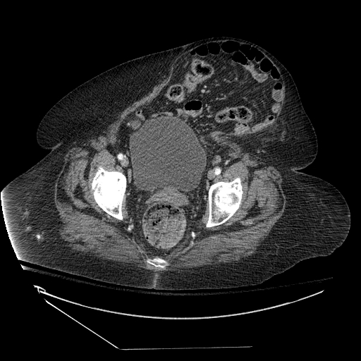
The physical exam involves abdominal palpation. In most of the cases, hernial content can be palpated. Occasional the edges of hernial defect can be felt and the size can be estimated. Planning for a surgical approach can be discussed with the patient on that visit. Although most cases of an incisional hernia are diagnosed with a history and physical, imaging is sometimes warranted like in early stages, obese patients, or complex cases[14]. Computed Tomography (CT) scanning focused on the abdominal wall is not only helpful to confirm the diagnosis when it is not clear but helps in planning the surgical approach and extent of repair[15].

Occasionally small incisional hernias are identified intraoperatively when the patients undergo another abdominal surgery. Incisions made at the same scar site or laparoscopic approach may show the fascial defects with occasional fat incarceration.

Evaluation

History and physical exam will allow for diagnosis of incisional hernias in the vast majority of cases. In cases of occult hernias, either small incisional hernias or hernias in obese patients, further workup with imaging is warranted. The computed tomography (CT) scan is the most commonly used method to diagnose an incisional hernia and can be useful in complex cases as well in helping plan operative management[15][16]. Ultrasound techniques have also been described for the evaluation of abdominal wall hernias (Dynamic Ultrasonography Assessment for Hernia-DASH). MRI can also be used to assess abdominal wall hernias but are less commonly used and is institution dependent.

Complete evaluation of incisional hernias includes confirming the diagnosis, sizing the defect, identifying the herniated content, and assessing the abdominal cavity to plan the surgical treatment in complex hernias. CT scan imaging is useful for obtaining these details.

Treatment / Management

Treatment strategies for an incisional hernia involve surgical repair or conservative nonoperative treatment[17]. In this section, a general review of the treatment principles will be discussed.  The decision to choose between the two options depends on a few factors like symptoms, the size of a hernia, complications and patients' preference. Small and asymptomatic hernias can be observed safely with a low risk of complication, 2.6% annually in some studies[7]. Unless contraindicated, large or symptomatic hernias should be surgically repaired to avoid complications, relieve symptoms, and improve quality of life. 

Open, laparoscopic, and robotic techniques have been commonly used to repair incisional hernias and must be tailored to the patient and hernia characteristics[18].  In general, the mesh should be used to repair the majority of incisional hernias as they reduce the rate of recurrence compared to primary suture repair. Mesh provides the strength for the repair and scaffold for the healing tissue. There are different options for mesh placement. Onlay (above the fascial defect), inlay (between the fascial edges), sublay (below the fascial defect but above the posterior rectus sheet), and underlay or Intraperitoneal onlay (intraperitoneally below the fascial defect). 

Several different types of meshes are available.  Meshes are characterized as permanent vs. absorbable, and synthetic vs. biologic. Permanent synthetic meshes are commonly used[19]. Absorbable meshes are used in the contaminated or infected field. Biologic meshes can be used for the same reasons when long-standing support is needed and for other special indications.

In general, the fascial closure should be performed when the defect is 10 cm or smaller, and the closure augmented with mesh. If fascial closure cannot be achieved secondary to tension, other techniques such as component separation have been used. Details of the surgical repair are discussed in other articles.

Differential Diagnosis

  • Cancer
  • Cyst
  • Lipoma
  • Wound infection
  • Rectus diastasis

Prognosis

The prognosis for patients with incisional hernias is guarded. Despite the use of mesh and minimally invasive techniques, incisional hernias have high recurrence rates. Infection, pain, and recurrence are common problems. If the patient does not eliminate the risk factors the risk of recurrence remains high. Bowel obstruction, strangulation, and incarceration are also serious complications of the incisional hernias.

Complications

The natural history of incisional hernia is progressive enlargement over a variable time period. It can be asymptomatic or produce one or a combination of symptoms as discussed above. Possible complications include:

- Incarceration. Incarceration can be chronic nonsymptomatic as in large defect hernias, or acute and likely symptomatic incarceration[20]. It can be reducible or irreducible, associated with other complications like obstruction or with no complication.  In chronically incarcerated hernias, adhesions develop between the hernial content and the sac. While in acutely incarcerated hernias the small defect size the cause of the incarceration.

- Obstruction. Luminal obstruction of the bowel is an acute complication that may lead to more serious complications like strangulation or perforation[21].

- Strangulation. Incarceration or obstruction can lead to strangulation, a more serious and acute complication. Strangulation should be relieved emergently.

Enhancing Healthcare Team Outcomes

Managing incisional hernia requires an interprofessional team. The condition has many risk factors and the key is to prevent the problem in the first place.

Prevention of an incisional hernia is an essential concept in abdominal surgery. There are several principles and recommendations that can minimize the risk of incisional hernias. Some of these recommendations are: using slowly absorbing monofilament suture, suture to wound length ratio not less than 4:1 has been shown to decrease incisional hernia rates, prevention of infections, less tissue traumatic techniques, and optimizing postoperative patient care[9].

In some high-risk patients and cases, the incisional hernia rate is exceptionally high despite the adequate closure. Open abdominal aortic aneurysm (AAA) repair and formation of permanent colostomies are associated with high incisional/ parastomal hernia rates.  Prophylactic mesh augmentation has been used in these cases. 

The dietitian should encourage the patient to lose weight as obesity is a major risk factor for incisional hernia. The pharmacist should monitor patients on corticosteroids as they may affect wound healing. If possible, alternative drugs should be used or the steroid use should be shortened. The nurse should educate the patient on adequate control of blood glucose as diabetes is a risk factor for wound infections. Close communication between the team is vital if one wants to improve outcomes.


(Click Image to Enlarge)
<p>Abdomen CT,&nbsp;Ventral Hernia</p>

Abdomen CT, Ventral Hernia


Contributed by Scott Dulebohn, MD

Details

Editor:

Faiz Tuma

Updated:

6/12/2023 8:11:00 PM

References


[1]

Yagnik VD, Joshipura V. Non-incisional traumatic lateral abdominal wall hernia. ANZ journal of surgery. 2017 Nov:87(11):952-953. doi: 10.1111/ans.14052. Epub     [PubMed PMID: 29098778]


[2]

Berrevoet F. Prevention of Incisional Hernias after Open Abdomen Treatment. Frontiers in surgery. 2018:5():11. doi: 10.3389/fsurg.2018.00011. Epub 2018 Feb 26     [PubMed PMID: 29536013]


[3]

Kaneko T,Funahashi K,Ushigome M,Kagami S,Goto M,Koda T,Nagashima Y,Shiokawa H,Koike J, Incidence of and risk factors for incisional hernia after closure of temporary ileostomy for colorectal malignancy. Hernia : the journal of hernias and abdominal wall surgery. 2018 Nov 13     [PubMed PMID: 30426253]


[4]

Doussot A, Abo-Alhassan F, Derbal S, Fournel I, Kasereka-Kisenge F, Codjia T, Khalil H, Dubuisson V, Najah H, Laurent A, Romain B, Barrat C, Trésallet C, Mathonnet M, Ortega-Deballon P. Indications and Outcomes of a Cross-Linked Porcine Dermal Collagen Mesh (Permacol) for Complex Abdominal Wall Reconstruction: A Multicenter Audit. World journal of surgery. 2019 Mar:43(3):791-797. doi: 10.1007/s00268-018-4853-x. Epub     [PubMed PMID: 30426186]


[5]

Blatnik JA, Michael Brunt L. Controversies and Techniques in the Repair of Abdominal Wall Hernias. Journal of gastrointestinal surgery : official journal of the Society for Surgery of the Alimentary Tract. 2019 Apr:23(4):837-845. doi: 10.1007/s11605-018-3989-1. Epub 2018 Oct 18     [PubMed PMID: 30338444]


[6]

Krivan MS, Giorga A, Barreca M, Jain VK, Al-Taan OS. Concomitant ventral hernia repair and bariatric surgery: a retrospective analysis from a UK-based bariatric center. Surgical endoscopy. 2019 Mar:33(3):705-710. doi: 10.1007/s00464-018-6492-6. Epub 2018 Oct 19     [PubMed PMID: 30341658]

Level 2 (mid-level) evidence

[7]

Dai W, Chen Z, Zuo J, Tan J, Tan M, Yuan Y. Risk factors of postoperative complications after emergency repair of incarcerated groin hernia for adult patients: a retrospective cohort study. Hernia : the journal of hernias and abdominal wall surgery. 2019 Apr:23(2):267-276. doi: 10.1007/s10029-018-1854-5. Epub 2018 Nov 12     [PubMed PMID: 30421299]

Level 2 (mid-level) evidence

[8]

Tubre DJ, Schroeder AD, Estes J, Eisenga J, Fitzgibbons RJ Jr. Surgical site infection: the "Achilles Heel" of all types of abdominal wall hernia reconstruction. Hernia : the journal of hernias and abdominal wall surgery. 2018 Dec:22(6):1003-1013. doi: 10.1007/s10029-018-1826-9. Epub 2018 Oct 1     [PubMed PMID: 30276561]


[9]

Zucker BE, Simillis C, Tekkis P, Kontovounisios C. Suture choice to reduce occurrence of surgical site infection, hernia, wound dehiscence and sinus/fistula: a network meta-analysis. Annals of the Royal College of Surgeons of England. 2019 Mar:101(3):150-161. doi: 10.1308/rcsann.2018.0170. Epub 2018 Oct 5     [PubMed PMID: 30286645]

Level 1 (high-level) evidence

[10]

Söderbäck H, Gunnarsson U, Hellman P, Sandblom G. Incisional hernia after surgery for colorectal cancer: a population-based register study. International journal of colorectal disease. 2018 Oct:33(10):1411-1417. doi: 10.1007/s00384-018-3124-5. Epub 2018 Jul 17     [PubMed PMID: 30019246]


[11]

van den Hil LCL, Vogels RRM, van Barneveld KWY, Gijbels MJJ, Peutz-Kootstra CJ, Cleutjens JPM, Schreinemacher MHF, Bouvy ND. Comparability of histological outcomes in rats and humans in a hernia model. The Journal of surgical research. 2018 Sep:229():271-276. doi: 10.1016/j.jss.2018.03.019. Epub 2018 May 5     [PubMed PMID: 29937000]


[12]

Belokonev VI, Pushkin SIu, Fedorina TA, Nagapetian SV. [A biomechanical concept of the pathogenesis of postoperative ventral hernias]. Vestnik khirurgii imeni I. I. Grekova. 2000:159(5):23-7     [PubMed PMID: 11188811]


[13]

Cherla DV, Bernardi K, Blair KJ, Chua SS, Hasapes JP, Kao LS, Ko TC, Matta EJ, Moses ML, Shiralkar KG, Surabhi VR, Tammisetti VS, Viso CP, Liang MK. Importance of the physical exam: double-blind randomized controlled trial of radiologic interpretation of ventral hernias after selective clinical information. Hernia : the journal of hernias and abdominal wall surgery. 2019 Oct:23(5):987-994. doi: 10.1007/s10029-018-1856-3. Epub 2018 Nov 14     [PubMed PMID: 30430273]

Level 1 (high-level) evidence

[14]

Halligan S, Parker SG, Plumb AA, Windsor ACJ. Imaging complex ventral hernias, their surgical repair, and their complications. European radiology. 2018 Aug:28(8):3560-3569. doi: 10.1007/s00330-018-5328-z. Epub 2018 Mar 12     [PubMed PMID: 29532239]


[15]

Farukhi MA, Mattingly MS, Clapp B, Tyroch AH. CT Scan Reliability in Detecting Internal Hernia after Gastric Bypass. JSLS : Journal of the Society of Laparoendoscopic Surgeons. 2017 Oct-Dec:21(4):. doi: 10.4293/JSLS.2017.00054. Epub     [PubMed PMID: 29279662]


[16]

Holihan JL, Karanjawala B, Ko A, Askenasy EP, Matta EJ, Gharbaoui L, Hasapes JP, Tammisetti VS, Thupili CR, Alawadi ZM, Bondre I, Flores-Gonzalez JR, Kao LS, Liang MK. Use of Computed Tomography in Diagnosing Ventral Hernia Recurrence: A Blinded, Prospective, Multispecialty Evaluation. JAMA surgery. 2016 Jan:151(1):7-13. doi: 10.1001/jamasurg.2015.2580. Epub     [PubMed PMID: 26398884]


[17]

Khorgami Z, Hui BY, Mushtaq N, Chow GS, Sclabas GM. Predictors of mortality after elective ventral hernia repair: an analysis of national inpatient sample. Hernia : the journal of hernias and abdominal wall surgery. 2019 Oct:23(5):979-985. doi: 10.1007/s10029-018-1841-x. Epub 2018 Nov 3     [PubMed PMID: 30392164]


[18]

de Vries HS, Smeeing D, Lourens H, Kruyt PM, Mollen RMHG. Long-term clinical experience with laparoscopic ventral hernia repair using a ParietexTM composite mesh in severely obese and non-severe obese patients: a single center cohort study. Minimally invasive therapy & allied technologies : MITAT : official journal of the Society for Minimally Invasive Therapy. 2019 Oct:28(5):304-308. doi: 10.1080/13645706.2018.1521431. Epub 2018 Oct 11     [PubMed PMID: 30307356]


[19]

Köckerling F, Lammers B. Open Intraperitoneal Onlay Mesh (IPOM) Technique for Incisional Hernia Repair. Frontiers in surgery. 2018:5():66. doi: 10.3389/fsurg.2018.00066. Epub 2018 Oct 23     [PubMed PMID: 30406110]


[20]

Azin A, Hirpara D, Jackson T, Okrainec A, Elnahas A, Chadi SA, Quereshy FA. Emergency laparoscopic and open repair of incarcerated ventral hernias: a multi-institutional comparative analysis with coarsened exact matching. Surgical endoscopy. 2019 Sep:33(9):2812-2820. doi: 10.1007/s00464-018-6573-6. Epub 2018 Nov 12     [PubMed PMID: 30421078]

Level 2 (mid-level) evidence

[21]

Petersson J, Koedam TW, Bonjer HJ, Andersson J, Angenete E, Bock D, Cuesta MA, Deijen CL, Fürst A, Lacy AM, Rosenberg J, Haglind E, COlorectal cancer Laparoscopic or Open Resection (COLOR) II Study Group. Bowel Obstruction and Ventral Hernia After Laparoscopic Versus Open Surgery for Rectal Cancer in A Randomized Trial (COLOR II). Annals of surgery. 2019 Jan:269(1):53-57. doi: 10.1097/SLA.0000000000002790. Epub     [PubMed PMID: 29746337]

Level 1 (high-level) evidence