Anatomy, Abdomen and Pelvis, Penis Dorsal Nerve


Introduction

Pudendal nerve arises from the sacral plexus in the pelvis. It is the chief nerve of the perineum and external genitalia. It derives from S2, S3, and S4. Several branches of the pudendal nerve innervate the penis, including the dorsal nerve of the penis. The pudendal nerve carries axons responsible for sensation and motor responses for defecation, micturition, and procreation.[1] It relays information from the external genitalia as well as the skin around the anus and perineum. It also carries motor axons to various pelvic muscles as well as the external urethral sphincter, the external anal sphincter, and bulbospongiosus muscle.[2] 

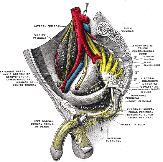
Inside the pudendal canal, the pudendal nerve divides into several branches. It first gives off the inferior rectal nerve followed by the perineal nerve and, finally, the dorsal nerve of the penis. The dorsal nerve of the penis or clitoris is a smaller terminal branch of the pudendal nerve. It runs forward first in the pudendal canal above the internal pudendal vessels and then in the deep perineal space between these vessels and the pubic arch. After branching off of the pudendal nerve, the dorsal nerve of the penis runs along the inferior ischial ramus; then, it is joined by the deep dorsal vein at the base of the penis.[1] Then it passes through the lateral part of the oval gap between the apex of the perineal membrane and arcuate pubic ligament, and runs on the dorsum of the penis and clitoris and ends in the glans penis and glans clitoris.

In addition to the efferent fibers of the dorsal nerve of the penis, this nerve includes afferent fibers that innervate the penile skin, clitoris in females, and also the glans penis. These afferents propagate sensory signals to the central nervous system that are critical to achieving an erection and sexual function.[3]

Structure and Function

The dominant function of the dorsal nerve of the penis is primarily somatosensory with a small motor component. The dorsal nerve of the penis has a direct connection to sexual function and micturition as its afferent transmit signals to the central nervous system.[4] Its small muscular branches supplies to the deep transverse perinei and the urethral sphincter.[5] 

The dorsal nerve of the penis has branching fibers that course laterally and transect the corpus spongiosum to the sinusoidal erectile tissue containing the urethra. The fibers of the dorsal nerve of the penis penetrate the corpus spongiosum to innervate the urethral lumen as afferents. These fibers receive input and relay the information during micturition and ejaculation. Semen is primarily expelled from the bulbous urethra via contraction of the bulbospongiosus muscle.[6] However, this muscle is not innervated by the dorsal nerve of the penis but by a sister branch of the pudendal nerve. Electrophysiological studies have documented that stimulation of the penis' dorsal nerve causes a reflex contraction of the bulbospongiosus muscle. During ejaculation, the penis dorsal nerve fibers provide the urethral sensation associated with the passage of seminal fluid necessary to sustain bulbospongiosus contractions until the expulsion of seminal fluid is complete.[7] 

Neural pathways are arranged in reflex arcs that provide the reflex for ejaculation and bulbospongiosus contraction. The sensory axons of the afferent perineal nerve and the dorsal and ventrolateral branches of the dorsal nerve of the penis merge at the pudendal motor neurons in the conus medullaris portion of the central nervous system. The motor portion of the arc branches from the spinal cord as the perineal nerve to terminate in muscle fibers of the bulbospongiosus muscle for the contraction of these muscles leading to ejaculation.[6]

Blood Supply and Lymphatics

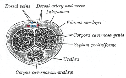
A neurovascular bundle runs along the dorsal aspect of the penis in a groove between the corpora cavernosa hemispheres. This groove includes the dorsal nerve of the penis, dorsal penile vein, and dorsal penile artery. These lay beneath the deep fascia, also known as Buck's fascia.

Nerves

The dorsal nerve of the penis is composed of multiple different collections of axons along the dorsal aspect of the erect penis. They traverse the dorsum of the penis deep to Buck's fascia and superior to the cavernous bodies.[8] 

The initial cluster consists of fibers parallel to the shaft of the penis that moves distally and ends at the glans of the penis. An additional group of fibers branches from the main body towards the lateral aspects of the penis.[9] These branches vary in position and quantity. However, they always branch laterally away from the main body of the penis. The axons do not cross the dorsal midline. The gauge of the nerve bundles varies greatly, but they consistently narrow as branching continues distally.[4]

The dorsal nerve bundles continue straight into the glans penis to form midline branches, and ventrolateral branches arise from the lateral bundles. The distal branches of the terminating nerves travel toward the meatus of the urethra.[10] As the dorsal nerve of the penis enters the glans, it immediately branches and displays a more three-dimensional pattern as opposed to a linear convention seen proximally. Each of the main glandular branches continues along the tunical surface, projecting bundles of smaller nerves that extend into the tissue of the glans and outwards toward the surface of the penis.

Physiologic Variants

The number of dorsal bundles most commonly reported are two and three, but there have been periodic reports of up to 8 variants. Most of the dorsal nerves run parallel to each other in the dorsal aspect of the penis, traveling distally. Communicating branches are commonly viewed in no specific orientation or frequency. These branches usually communicate amongst each other, but ventral nerve communications are noted in cadaver studies infrequently. Most dissection studies comment on the absence of nerves that cross the dorsal midline.

Clinical Significance

Clinicians often employ a dorsal penile nerve block for anesthesia before circumcision, ischemic priapism treatment, the release of paraphimosis, cystoscopy, and the repair of penile lacerations.[11][12] Localizing the dorsal nerve involves injecting 1 to 5 mL of 1% or 2% lidocaine superficially at the 2 and 10 o'clock positions to a depth of approximately 5 mm dorsally can provide adequate analgesia for the minor procedures. Studies have also proven that a ring block provides better anesthesia for circumcisions as well as other procedures.[13] Care is necessary to avoid lidocaine with epinephrine due to the potential for vascular compromise in this as well as other terminating anatomy.

Many advancements have been made in the transplantation of nerves to restore sexual function and sensation.[14] In a two year trial from 2014 to 2016, nearly two dozen patients underwent sural nerve bridging transplantation in an attempt to restore sensation after a dorsal nerve neurotomy. In follow up, approximately half the patients experienced a marked increase in feeling, a quarter had some improvement, and only one patient had no change. Additionally, stimulation of pudendal afferents such as the dorsal penile nerve has shown to be effective in studies of neurogenic detrusor overactivity. Previous results in laboratory testing revealed a prolonged subdual of the peripheral nervous system urination reflex arc after the stimulation of the penis's dorsal nerve via urethral afferents.[2] In individuals with multiple sclerosis or cord injury, dorsal genital nerve stimulation with an electrode stimulator inhibited detrusor contractions and increased bladder volume that provided urinary continence to those that had previously been without it.[15]


(Click Image to Enlarge)
<p>The Sacral and Coccygeal Nerves

The Sacral and Coccygeal Nerves. Sacral plexus of the right side, pelvic area, coccyx, dorsal nerve of penis, nerve to bulb, and genitofemoral nerve.


Henry Vandyke Carter, Public Domain, via Wikimedia Commons


(Click Image to Enlarge)
<p>The Penis, Transverse Section of the Penis, Corpus Cavernosum Urethrae, Urethra, Septum Pectiniforme, Corpora Cavernosa Pe

The Penis, Transverse Section of the Penis, Corpus Cavernosum Urethrae, Urethra, Septum Pectiniforme, Corpora Cavernosa Penis, Fibrous Envelope


Henry Vandyke Carter, Public Domain, via Wikimedia Commons

Details

Author

David Weech

Editor:

John V. Ashurst

Updated:

8/8/2023 12:14:09 AM

References


[1]

Nolan MW, Marolf AJ, Ehrhart EJ, Rao S, Kraft SL, Engel S, Yoshikawa H, Golden AE, Wasserman TH, LaRue SM. Pudendal nerve and internal pudendal artery damage may contribute to radiation-induced erectile dysfunction. International journal of radiation oncology, biology, physics. 2015 Mar 15:91(4):796-806. doi: 10.1016/j.ijrobp.2014.12.025. Epub     [PubMed PMID: 25752394]


[2]

Rembetski BE, Cobine CA, Drumm BT. Laboratory practical to study the differential innervation pathways of urinary tract smooth muscle. Advances in physiology education. 2018 Jun 1:42(2):295-304. doi: 10.1152/advan.00014.2018. Epub     [PubMed PMID: 29676616]

Level 3 (low-level) evidence

[3]

Alwaal A, Breyer BN, Lue TF. Normal male sexual function: emphasis on orgasm and ejaculation. Fertility and sterility. 2015 Nov:104(5):1051-60. doi: 10.1016/j.fertnstert.2015.08.033. Epub 2015 Sep 16     [PubMed PMID: 26385403]


[4]

Gosling JA, Dixon JS, Jen PY. The distribution of noradrenergic nerves in the human lower urinary tract. A review. European urology. 1999:36 Suppl 1():23-30     [PubMed PMID: 10393469]


[5]

Kinter KJ, Newton BW. Anatomy, Abdomen and Pelvis, Pudendal Nerve. StatPearls. 2023 Jan:():     [PubMed PMID: 32134612]


[6]

Giuliano F. Neurophysiology of erection and ejaculation. The journal of sexual medicine. 2011 Oct:8 Suppl 4():310-5. doi: 10.1111/j.1743-6109.2011.02450.x. Epub     [PubMed PMID: 21967393]


[7]

Alyaev YG, Akhvlediani ND. [Comparing efficacy of selective penile denervation and circumcision for primary premature ejaculation]. Urologiia (Moscow, Russia : 1999). 2016 Mar:(1 Suppl 1):60-64     [PubMed PMID: 28247749]


[8]

Manrique OJ, Adabi K, Maldonado AA, Huang TC, Martinez-Jorge J, Brassard P, Galan R, Ciudad P, Sabbagh MD. Cadaver Study of Combined Neurovascular Sensate Flaps to Create Vaginal Erogenous Sensation During Male-to-Female Genital Confirmation Surgery: The Pedicle "O" Flap. Annals of plastic surgery. 2018 Nov:81(5):571-575. doi: 10.1097/SAP.0000000000001558. Epub     [PubMed PMID: 29994881]


[9]

Jung J, Jo HW, Kwon H, Jeong NY. Clinical neuroanatomy and neurotransmitter-mediated regulation of penile erection. International neurourology journal. 2014 Jun:18(2):58-62. doi: 10.5213/inj.2014.18.2.58. Epub     [PubMed PMID: 24987557]


[10]

Chhabra A, McKenna CA, Wadhwa V, Thawait GK, Carrino JA, Lees GP, Dellon AL. 3T magnetic resonance neurography of pudendal nerve with cadaveric dissection correlation. World journal of radiology. 2016 Jul 28:8(7):700-6. doi: 10.4329/wjr.v8.i7.700. Epub     [PubMed PMID: 27551340]


[11]

Flores S, Herring AA. Ultrasound-guided dorsal penile nerve block for ED paraphimosis reduction. The American journal of emergency medicine. 2015 Jun:33(6):863.e3-5. doi: 10.1016/j.ajem.2014.12.041. Epub 2014 Dec 26     [PubMed PMID: 25605058]


[12]

Qiu Y, Hu AM, Liu J, Du GZ. Dorsal penile nerve block for rigid cystoscopy in men: study protocol for a randomized controlled trial. Trials. 2016 Mar 18:17(1):147. doi: 10.1186/s13063-016-1281-9. Epub 2016 Mar 18     [PubMed PMID: 26988368]

Level 2 (mid-level) evidence

[13]

Yang BB, Xia JD, Hong ZW, Zhang Z, Han YF, Chen Y, Dai YT. No effect of abstinence time on nerve electrophysiological test in premature ejaculation patients. Asian journal of andrology. 2018 Jul-Aug:20(4):391-395. doi: 10.4103/aja.aja_10_18. Epub     [PubMed PMID: 29600795]


[14]

Si S, Wang H, Pu Y, Cen Y, Xiao H. [Efficacy evaluation of sural nerve bridging transplantation for restoration of penis disturbance of sensation after selective dorsal nerve neurotomy]. Zhongguo xiu fu chong jian wai ke za zhi = Zhongguo xiufu chongjian waike zazhi = Chinese journal of reparative and reconstructive surgery. 2017 Jun 15:31(6):709-713. doi: 10.7507/1002-1892.201611114. Epub     [PubMed PMID: 29798653]


[15]

McGee MJ, Swan BD, Danziger ZC, Amundsen CL, Grill WM. Multiple Reflex Pathways Contribute to Bladder Activation by Intraurethral Stimulation in Persons With Spinal Cord Injury. Urology. 2017 Nov:109():210-215. doi: 10.1016/j.urology.2017.07.041. Epub 2017 Aug 8     [PubMed PMID: 28801220]