Continuing Education Activity
Axenfeld-Rieger syndrome (ARS) is a disease that encompasses anterior segment ocular dysgenesis in addition to systemic abnormalities such as dental, cardiac, craniofacial, and abdominal wall defects. Previously, this syndrome was separated into multiple entities, including Axenfeld anomaly, Rieger anomaly, and Rieger syndrome. Axenfeld anomaly has been characterized by posterior embryotoxon and peripheral iris strand attachment. Rieger anomaly denoted posterior embryotoxon, iris hypoplasia, polycoria, and corectopia. Association of systemic findings with Rieger anomaly, including dental abnormalities, changes of the facial bones such as maxillary hypoplasia, umbilical abnormalities (redundant periumbilical skin), hypospadias, and pituitary abnormalities, has been called Rieger syndrome. However, it has been recognized that all these previous entities fall under a group of diseases, which is now called ARS. This activity reviews the presentation of ARS and highlights the role of the interprofessional team in its management.
Objectives:
Identify individuals with suspected Axenfeld-Rieger syndrome based on clinical signs and symptoms, such as ocular abnormalities, dental malformations, and maxillary hypoplasia.
Implement appropriate treatment and management strategies for patients with Axenfeld-Rieger Syndrome, which may include regular monitoring of intraocular pressure, early detection and management of glaucoma, and referral to relevant specialists.
Apply evidence-based treatment approaches for managing specific complications associated with Axenfeld-Rieger syndrome, tailoring interventions to individual patient needs and characteristics.
Collaborate with other healthcare professionals, including ophthalmologists, geneticists, and dentists, to develop comprehensive management plans and ensure coordinated care for individuals with Axenfeld-Rieger Syndrome.
Introduction
In 920, the German ophthalmologist Theodor Axenfeld made an important contribution to the field by describing a prominent and anteriorly displaced Schwalbe line (posterior embryotoxon) with the adhesion of peripheral iris strands.[1] Later, these observations became known as the Axenfeld anomaly.[2] Another significant advancement came from the Austrian ophthalmologist Herwigh Rieger, who described a distinct set of ocular abnormalities known as the "Rieger anomaly." This anomaly encompasses posterior embryotoxon, iris hypoplasia, polycoria, and corectopia.[3][4]
The constellation of systemic findings in conjunction with Rieger anomaly, such as dental abnormalities, facial bone changes, umbilical abnormalities, hypospadias, and pituitary abnormalities, was called Rieger syndrome.[1][2][5] In current medical terminology, the terms Axenfeld anomaly, Rieger anomaly, and Rieger syndrome are no longer used. Instead, these ocular findings and associated systemic manifestations are now recognized under a spectrum of disorders called Axenfeld-Riger syndrome (ARS).[6]
ARS is a disease that encompasses anterior segment ocular dysgenesis and systemic abnormalities such as dental, cardiac, craniofacial, and abdominal wall defects. The mutations associated with ARS include PITX2 (chromosome 4q25), FOXC1 (chromosome 6p25), PAX6 (chromosome 11p13), FOXO1A (chromosome 13q14), and CYP1B1 (chromosome 2p22.2).[2][7][8][9]
Etiology
ARS is associated with various chromosome mutations, including mutations in chromosomes 2, 4, 6, 9, 13, 18, and 21.
ARS type 1 (RIEG 1, OMIM #180500) is an autosomal dominant (AD) disorder caused by a heterozygous mutation in the PITX2 gene located on chromosome 4q25. The PITX2 gene is a homeobox transcription factor crucial in developing teeth and the iris.
ARS type 2 (RIEG 2, OMIM %601499) is an autosomal dominant (AD) disorder linked to chromosome 13q14. In the OMIM nomenclature, the "%" sign denotes that the phenotype description or locus is known, but the molecular basis is yet to be identified.
ARS type 3 (RIEG 3, OMIM # 602482) is an autosomal dominant (AD) disorder linked to chromosome 6p25.3, specifically involving the FOXC1 gene. Heterozygous mutations in the FOXC1 gene are known to cause ARS type 3. Chromosome 6pter-p24 deletion syndrome (OMIM # 612582) is a contiguous gene deletion syndrome that shares phenotypic similarities with ARS type 3. The FOXC1 gene, also known as forkhead box 1, has been referred to as forkhead, drosophila, homolog-like 7 (FKHL7), and forkhead-related activator 3 (FREAC3).[10]
Approximately 25% to 30% of cases with ocular features suggestive of ARS correlate with mutations in FOXC1 and PITX2 on chromosomes 6p25 and 4q25, respectively.[11] Forkhead-Box C1 gene (FOXC1) and the Pituitary Homeobox 2 gene (PITX2) are the most studied transcription factor-encoding genes associated with ARS. Research has characterized FOXC1 mutations as frameshift, nonsense, missense, deletions, and duplications. PITX2 has been related to splice-site mutations, omissions, and chromosomal translocations in patients with ARS.[11]
Approximately 10% to 30% of patients with ARS present PITX2 mutations in 4q25.[1][12][13] Animal models have shown that ocular structures affected in ARS express FOXC1 and PITX2.[7][14] A distinct phenotype of ARS, referred to as De Hauwere Syndrome (OMIM 109120), has been characterized by the presence of hydrocephalus, partially absent eye muscles, hypertelorism, lax joints, psychomotor retardation, and sensorineural deafness.[15][16]
Genetic heterogeneity in conditions like ARS means that various genes can lead to similar features or phenotypes. The inheritance pattern of most cases of ARS is autosomal dominant, although sporadic cases also arise. ARS is typically characterized by high penetrance but exhibits variable expressivity.
Epidemiology
The prevalence of ARS is estimated to be around 1 in 50,000 to 100,000 to 1 in 200,000 live births.[2][17] There is no racial or gender preference. In most cases, both eyes are affected by the characteristic features of ARS.
Pathophysiology
Axenfeld-Rieger syndrome is believed to be caused by abnormal neural crest (NC) migration during early embryogenesis. Important ocular structures such as the ciliary body, cornea, and iris stroma rely on adequate NC cell migration. Transcription factors in different genes strongly regulate these processes.[18][19]
During late gestation, the primordial endothelium that covers the cornea is expected to undergo resorption; disruption in this process may result in the development of posterior embryotoxon and abnormal insertion of the iris, causing pupillary changes such as pseudopolycoria or ectropion uveae.[18][19] The previously discussed abnormalities in the development of the anterior chamber may affect the development of the Schlemm canal, impairing the outflow of aqueous humor and increasing the risk of glaucoma.[18][19]
Abnormal NC migration associated with ARS often affects extraocular tissues, such as vestibuloacoustic ganglion tissue, which may cause hearing loss in some patients with ARS.[18][19] The pathogenesis of ARS involves abnormalities in the differentiation, development, or migration of neural crest cells, contributing to the ocular and systemic features observed in affected individuals. The third wave of neural crest gives rise to the iris stroma.[20] Research suggests abnormalities or disturbances in the third wave of neural crest cell migration can result in ARS.[21] It is proposed that the developmental arrest of the tissues derived from neural crest cells in late gestation leads to the retention of the primordial endothelium over the iris and the angle of the anterior chamber.[6] This abnormal tissue continues to contract, resulting in some of the progressive changes seen in patients with ARS.[6]
Histopathology
The widely accepted definition of posterior embryotoxon is an anteriorly displaced and visibly prominent Schwalbe line. However, an immunohistopathological study revealed that in some cases, posterior embryotoxon is a direct extension of the corneal stroma ('peripheral corneal stromal nub') lined by the Descemet membrane.[22] This study noted that the locations of these nubs were anterior, posteriorly, at the Schwalbe line, or partially embedded in the trabecular meshwork.[22]
The histology of ARS shows features of abnormal anterior segment cleavage, including prominent Schwalbe line, hypoplasia of Schlemm canal and iris, hypocellular trabecular meshwork, and iridocorneal adhesion.[23] The histopathological features of ARS and iridocorneal endothelial syndrome can be similar and characterized by a Descement-like membrane and endothelial-like cells.[24] These extend from the cornea to the iris's anterior surface and the anterior chamber's angle. These changes are typically located toward the side of pupillary displacement or ectropion uveae and at the opposite quadrant of the atrophic disrupted iris.[24]
History and Physical
Axenfeld-Rieger syndrome commonly correlates with ocular, dental, facial, and abdominal abnormalities.[8][25] ARS is the preferred term for discussing this group of phenotypically similar disorders, which was historically divided into different entities, including Axenfeld anomaly, Rieger anomaly, and Rieger syndrome. The majority of ARS cases are diagnosed during infancy or childhood. Glaucoma is usually detected during middle to late childhood or even in adulthood. However, congenital or early-onset glaucoma (diagnosis within 3 years of age) may occur.[26]
Congenital glaucoma presents with a large bluish cornea (buphthalmos), watering, and photophobia. The usual presenting complaints include an abnormal eye appearance, squint, or dimness of vision. Photophobia is common and is caused by abnormalities of the iris and the pupil. Many cases are detected on routine ocular examination. All patients must receive a comprehensive ophthalmic examination.
Posterior embryotoxon is characterized by a prominent and anteriorly displaced Schwalbe line visible in the slit-lamp without gonioscopy. Peripheral iris strands are attached to this posterior embryotoxon, better visualized with gonioscopy. The prominent Schwalbe line may be missed in the slit-lamp examination if the gonioscopy is not done. Intraocular pressure should also be checked.
The historical term 'Axenfeld anomaly' included a prominent and anteriorly displaced Schwalbe line on evaluation. It is also necessary to recognize that posterior embryotoxon can be observed in various conditions, including Alagille syndrome, and can also be present in individuals without any underlying health issues. Posterior embryotoxon is incidentally found in approximately 8% to 15% of normal individuals. This finding often occurs without any ocular or systemic abnormalities, including glaucoma.[27][28]
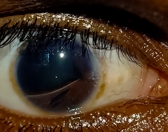
Iris abnormalities associated with ARS include stromal hypoplasia, full-thickness iris defects (iris holes) that can give rise to the appearance of multiple pupils (pseudo polycoria), a decentered pupil (corectopia), and ectropion uveae. Typically, the cornea is clear, contrary to the iridocorneal endothelial (ICE) syndrome. The previous name for these findings and posterior embryotoxon was Rieger anomaly. These features may be clinically very similar to ICE syndrome, which is usually unilateral and seen in older age groups. The iris may be normal in some cases. Other ocular findings include anterior peripheral anterior synechiae, corneal opacity, megalocornea, microcornea, and glaucoma.
The association of Rieger anomaly with extraocular conditions was previously called Rieger syndrome. An abnormality in the neural crest cell-related tissue development also causes these. Tooth-related abnormalities commonly observed include hypodontia (absence of 1-5 teeth), oligodontia (absence of ≥ 6 teeth), and microdontia (small-sized teeth). Facial features include mild craniofacial dysmorphism, telecanthus, hypertelorism, maxillary hypoplasia, and broad flat nasal bridge. Other systemic associations include hypospadias, redundant periumbilical skin, umbilical hernia, hydrocephalus, deafness, anal stenosis, renal anomalies, cardiac (valvular) abnormalities, arachnoid cyst,[29] pituitary (empty sella) abnormalities[19], endocrine abnormalities (short stature, growth retardation), and congenital hip dislocation.
Evaluation
Gonioscopy is an essential component of the evaluation process for individuals with ARS, as it allows for a detailed examination of the iridocorneal angle. Commonly observed features of ARS when performing gonioscopy include a prominent Schwalbe line, iris strands attached to the Schwalbe line, anterior insertion of the iris, and the presence of iris processes. The iris strands attached to the cornea can be delicate or broad.
In addition to gonioscopy, other ocular investigations for ARS include anterior segment photography, goniophotography, fundus (optic disc) photography, corneal pachymetry, optical coherence tomography of peripapillary retinal nerve fiber layer and macular ganglion cell complex, and Humphrey visual fields.[30]
Evaluation for patients with 6p25 microdeletion syndrome should be comprehensive and include a referral to an ophthalmologist, particularly for patients with posterior embryotoxon. Other studies should consist of neurodevelopmental evaluation, screening echocardiogram, brain imaging, hearing and vision tests, and auditory brainstem response evaluation.[31]
Childhood glaucoma is a common complication in ARS. Patients with ARS should undergo prompt evaluation and appropriate treatment to preserve their vision. In cases of congenital glaucoma, a comprehensive examination under anesthesia is often necessary to assess various parameters, including corneal diameter, intraocular pressure, optic nerve head status, and axial length measurement. While primary care providers may detect the clinical symptoms and initially diagnose, these patients will require further evaluation by specialists as they may eventually require surgical interventions or specialized care to prevent permanent disabilities or blindness.[26][32]
Treatment / Management
Treatment is individualized as the clinical presentation of patients with Axenfeld-Rieger syndrome is highly variable. Approximately half of the patients will develop glaucoma, which may eventually require surgical procedures such as trabeculectomy and trabeculotomy. These therapies have effectively controlled intraocular pressure, as described in a retrospective analysis of 24 children diagnosed with ARS and early-onset glaucoma.[26][32]
Medical management of glaucoma associated with ARS is usually ineffective in most cases. A study found that out of 46 eyes with ARS-related glaucoma, 31 (67%) needed surgery for glaucoma.[33] The options for medical management include beta-blockers, carbonic anhydrase inhibitors, and prostaglandin analogs. Alpha agonists, including brimonidine, can cause sleep apnea and central nervous system depression in children, which must be avoided in individuals younger than 2 years.
Goniotomy and trabeculotomy procedures may have low effectiveness in the long term as angle dysgenesis and other developmental abnormalities may be present.[33] In such cases, surgical options are more effective. These include trabeculectomy with anti-fibrotics, including mitomycin c,[33] combined trabeculotomy and trabeculectomy,[26] and glaucoma drainage devices.[34] However, most of these cases require more than one surgery, and maintaining the intraocular pressure (IOP) long-term is difficult in many cases, even with medical and surgical management.
Some patients may need cycloablative procedures, including cyclocryotherapy and diode laser cyclophotocoagulation. The risk of surgery may vary and includes hypotony, suprachoroidal hemorrhage, and endophthalmitis. A thorough risk-benefit analysis should be performed before planning an intervention for such cases.
Differential Diagnosis
Iridocorneal Endothelial Syndrome
- More commonly observed in middle age females
- Often affects one eye asymmetrically
- Presence of an abnormal corneal endothelium
- Three variants:
- Chandler Syndrome: Abnormal behavior of the corneal endothelium, which takes on epithelial-like characteristics. This abnormality results in distinct features, such as a beaten bronze appearance of the corneal endothelium and subsequent corneal edema.
- Essential iris atrophy: Progressive iris atrophy resulting in the formation of iris holes, pseudopolycoria, and corectopia.
- Cogan-Reese (iris nevus): Iris nodules at the anterior surface of the iris.
Iridoschisis
- There is a splitting of iris layers.[35]
- Usually, pigmented iris epithelium on the posterior surface of the iris remains intact.
- The inferior iris is generally involved.
- Associated with the shallow anterior chamber and angle closure glaucoma.
Peters Anomaly
- Congenital central corneal opacity with or without iris adhesion or cataract[36]
Aniridia (iris hypoplasia)
- Iris is absent in slit-lamp examination; however, a rudimentary iris stump is visible on careful examination or gonioscopy.[37]
- Limbal stem cell deficiency, corneal pannus
- Glaucoma
- Foveal hypoplasia
- Optic nerve head hypoplasia
Isolated Posterior Embryotoxon
- Normal iris and absence of glaucoma
Oculodentodigital Dysplasia (OMIM # 164200)
- Autosomal dominant
- Due to mutations in the connexin-43 gene (GJA1) on chromosome 6q22
- Microphthalmia
- Microcornea
- Glaucoma
- Cataract
- Narrow, pinched nose
- Enamel hypoplasia and microdontia
- Syndactyly of the 4th and 5th fingers
Congenital Ectropion Uveae
- Cryptless smooth anterior iris surface
- Visible pigmented epithelium at the pupillary margin and anterior surface of the iris[38]
- High iris insertion
- Associated glaucoma
- Other causes of congenital ectropion uveae should be ruled out
Ectopia Lentis Et Pupillae
- Autosomal recessive
- Mutation of ADAMTSL4 gene (chromosome 1q21)
- Both eyes involved
- Usually, inferotemporally displaced pupil (corectopia)
- Subluxation of the lens in the opposite side of pupillary displacement (usually superonasally subluxated lens)
- Myopia
- Retinal detachment
- Cataracts
Prognosis
Prognosis varies individually as patients with ARS have varied clinical presentations and different degrees of comorbidities. While some patients may only present anterior ocular chamber abnormalities, others may also have cardiac defects of varying severity, which may worsen their prognosis.[11] The ocular prognosis is good in the absence of glaucoma. However, glaucoma may be challenging to treat and not respond to medical therapy alone. Patients with ARS who require glaucoma surgery typically undergo multiple surgeries, with an average of approximately 2.2 surgeries performed on each eye.[33] Late diagnosis and delayed management of glaucoma can cause blindness, phthisis, or painful blind eye.[39]
Complications
The diseases linked to Axenfeld-Rieger syndrome can be associated with various complications, including ocular abnormalities such as posterior embryotoxon. Approximately 50% of patients with ARS develop glaucoma.[11] While glaucoma is usually diagnosed after middle childhood, early-onset glaucoma may occur.[26] High iris insertion is a common finding in all ARS cases, but these are more pronounced in eyes with glaucoma.[6] Developmental abnormalities, including peripheral iris strands, iris changes, and posterior embryotoxon, may not correlate with the presence or severity of glaucoma.[6] The cause of glaucoma in ARS is thought to be angle dysgenesis or incomplete development of the trabecular meshwork and the Schlemm canal. It is believed that an arrest of the development of neural crest cells leads to the persistence of primordial endothelial cells over the angle and the iris, resulting in iridogoniodysgenesis that obstructs aqueous outflow and leads to glaucoma.
Congenital glaucoma (CG) in patients is often associated with FOXC1 and PITX2 gene mutations. CG may lead to severe complications, including blindness or partial vision loss in children.[40] Amblyopia is a relatively common complication of childhood glaucoma.[32] Patients with FOXC1 mutations may also present with hearing loss and ocular and heart complications.[1] In a study involving a three-generation family with 3 cases of ARS and heart disease, FOXC1 mutations were detected, suggesting the involvement of this gene in cardiac development.[41]
Deterrence and Patient Education
Patients and their families may benefit from being informed about the type and risk of complications associated with the spectrum of ARS, including the increased risk of glaucoma.[11] Alongside ocular abnormalities, ARS carries associations with dental malformations and maxillary hypoplasia. Referral to an orofacial specialist, such as a maxillofacial surgeon, may benefit patients experiencing these complications.[1] Genetic counseling and possibly testing can be offered to patients as some variants of ARS are inherited in an autosomal dominant fashion.[1]
Enhancing Healthcare Team Outcomes
Some authors have described different variants or subtypes of ARS, leading to various terms such as Axenfeld anomaly and Rieger anomaly, among others, being used to describe different pathological presentations. Despite these variations in terminology, all these presentations share a common phenotype. It may be beneficial to use a unified terminology encompassing all these conditions to facilitate better communication among specialists involved in the care of patients with ARS, including optometrists, ophthalmologists, and ophthalmic nurses.[11]
Managing patients with ARS requires a multidisciplinary approach involving various healthcare providers, such as primary physicians, nurses, ophthalmologists of different subspecialties, dentists, endocrinologists, and geneticists.