Continuing Education Activity
Kienböck disease refers to avascular necrosis of the lunate carpal bone, known as lunatomalacia. It was first recognized and described by Austrian radiologist Robert Kienböck in 1910. Kienböck disease is the second most common type of avascular necrosis of the carpal bones, preceded only by avascular necrosis of the scaphoid. The typically affected population is males aged 20 to 40 years. This activity reviews the etiology, presentation, evaluation, and management of Keinbock disease and reviews the role of the interprofessional team in evaluating, diagnosing, and managing the condition.
Objectives:
- Describe the various factors that can lead to the presentation fo Keinbock disease in the lunate.
- Detail the clinical and raadiographic findings that present on examination and evaluation, leading to a diagnosis of Keinbock disease.
- Discuss treatment option for Keinbock disease, based on the Stage and specific etiology.
- Explain the importance of interprofessional team strategies for improving care coordination and communication to aid in prompt diagnosis of Keinbock disease and improving outcomes in patients diagnosed with the condition.
Introduction
Kienböck disease refers to avascular necrosis of the lunate carpal bone, known as lunatomalacia. It was first recognized and described by Austrian radiologist Robert Kienböck in 1910.[1]
Relevant anatomy: The proximal carpal row of the mid-carpal joint is largely responsible for wrist motion, as opposed to the relatively fixed distal carpal row. The lunate is the central bone in the proximal row, and it articulates with the scaphoid, capitate, triquetrum, and occasionally the hamate. More proximally, the lunate is a component of the radiocarpal joint, and it also articulates with the ulna via the triangular fibrocartilage complex (TFCC). Of note, nearly 10% of the axial-radial/ulnar/carpal load is transmitted through the TFCC and subsequently to the ulna, and 35% through the radiolunate articulation.[2][3]
Etiology
No consensus is present regarding the primary causative factor of Kienböck disease. It is multifactorial, related to the following variables:
Ulnar negative variance (or ulna minus): This refers to a disproportionately shortened ulna when compared to the radius. As deduced from the previous section, a shortened ulna results in excessive mechanical stress and repetitive microtrauma exerted on the lunate by the relatively long radius. In some studies, up to 78% of Kienböck cases correlate with this finding.[4]
Vascular supply to the lunate bone: The lunate receives its blood supply from a variable number of dorsal and volar penetrating arteries that branch off of the dorsal and palmar radiocarpal and intercarpal arches. Intraosseous collaterals are sparse. The lower the number of penetrating arteries (especially volar branches from the radiocarpal palmar arch), the greater the likelihood of developing the disease.[5][6]
Lunate morphology: Risk of developing Kienböck disease increases as lunate size decreases, forcing the lunate to carry a larger axial load. The lunate can either have a square/rectangular shape (type II or type III), or a more triangular shape whereby the medial articular facet is absent (type I). The latter has a weaker trabecular pattern and is a risk factor for disease development and progression.[7]
Radial inclination angle: the radial inclination angle is a measurement of the angle formed between the horizontal and a line drawn from the ulnar tip of the radial articular surface to the tip of the radial styloid. The risk of developing Kienböck disease increases as the radial inclination angle decreases.[8]
Epidemiology
Kienböck disease is the second most common type of avascular necrosis of the carpal bones, preceded only by avascular necrosis of the scaphoid. The typically affected population is males aged 20-40 years.[5]
History and Physical
Patients usually present with unilateral pain over the dorsal aspect of the wrist, limited wrist motion, weakness, or a combination of the three. Wrist extension and axial loading exacerbate pain. Symptoms range from mild to debilitating. It is rarely bilateral, and trauma is often absent.[9]
Physical examination commonly reveals wrist swelling, tenderness over the expected location of the lunate, synovitis, and loss of grip strength.[9]
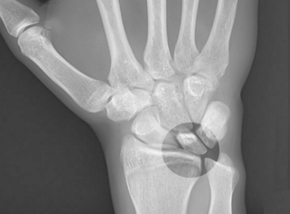
Evaluation
Kienböck disease is a clinical and imaging diagnosis. Both radiography/computed tomography and magnetic resonance imaging (MRI) are highly specific. However, MRI is the most sensitive and detects radiographically occult cases.
Magnetic Resonance Imaging: Diffuse decrease in lunate bone marrow signal on T1-weighted images is a hallmark of the disease. Signal changes on T2-weighted images or short-TI inversion recovery (STIR, which nullifies signal from fat) images vary with progression and extent of osteonecrosis. MRI also assesses the integrity of the articular cartilage.[9][6]
Radiography: Normal early in the disease. Findings, when present, depend on the morphological stage and include diffuse lunate sclerosis, cystic changes, articular surface collapse, carpal collapse, mid-carpal and/or radio-carpal secondary arthrosis. Coronal fractures may occur in lunates with a type I morphology.[9][6]
Computed Tomography: Is useful for surgical planning. It is also more sensitive than radiography for detecting subtle subchondral fractures, coronal lunate fractures, fragmentation, carpal instability, and the degree of trabecular disruption. Patients are frequently re-staged after CT imaging.[10]
Nuclear Scintigraphy: Findings are non-specific. It previously found use as an adjunct for diagnosing early-stage disease. It has fallen out of favor since introducing MRI.[6]
Treatment / Management
The goal of treating Kienböck disease is pain relief, wrist motion preservation, and preservation of grip strength.[11]
Treatment of Kienböck disease depends on the stage of the disease and its causative factors. Stage I is always treated with splinting or cast immobilization. Stage II can also be treated with immobilization if necrosis is incomplete. Stages II with complete necrosis, III, and IV require "joint-leveling" surgery possibly coupled with vascular bone grafting or transfer of branches from adjacent arteries. Later stages with lunate collapse and secondary wrist degenerative arthrosis may also require proximal row carpectomy or intercarpal arthrodesis. Radial shortening osteotomy is the most common procedure performed to unload the lunate in cases with coexistent ulnar negative variance. Treatment may improve symptoms and functionality without affecting imaging findings in the advanced stages.[6][9][11]
Differential Diagnosis
Ulnar impaction syndrome: More common with ulnar positive variance and results from repetitive microtrauma to the lunate from a relatively long ulna. Similar to Kienböck disease, ulnar impaction syndrome results in decreased T1-weighted signal coupled with increased T2 signal if hyperemia exists, or decreased T2 signal if the disease has already progressed to lunate sclerosis. The signal changes in Kienböck disease, however, are more diffuse or more severe on the radial side of the lunate. Additionally, ulnar impaction syndrome affects the ulnar head and triquetrum, which are intact in Kienböck patients.[12][6].
Lunate intraosseous ganglion: Intraosseous ganglia are true cysts which have low T1 signal and high T2 signal on MRI. Sharp, smooth margins on both MRI and radiography allow distinction from Kienböck disease.[6]
Bone contusion: May be difficult to distinguish early-stage Kienböck disease on MRI. A history of recent trauma and concomitant injuries to the wrist/hand, in this case, would aid in making the diagnosis.[6]
Arthritis: Bone marrow signal changes in inflammatory or degenerative arthritides may mimic those seen in Kienböck disease. Distinguishing features include demographics, clinical presentation, and absence of ulna minus in arthritis.[6]
Osteoid osteoma: Osteoid osteoma of the carpal bones is rare, and only a few case reports exist in the literature. Clinical presentation and the finding of a lucent nidus within a sclerotic rim on CT differentiate it from Kienböck disease.[13]
Enostosis/bone island: Has low signal intensity on all sequences, however, shows preserved bone morphology and the area of "sclerosis" is stellate and interdigitated with the normal trabeculae.[6]
Staging
Staging of Kienböck disease is essential for treatment planning and divides into morphological and functional staging.
Morphologic Staging follows the Lichtman Classification, which is based on radiographic and MR imaging findings and has low interobserver variability.[9][6][14]
Stage 1: Normal radiograph, lunate signal intensity changes on MRI.
Stage 2: Lunate sclerosis on radiography with or without fracture lines. The lunate shape is normal.
Stage 3: Collapse of the lunate articular surface.
- 3A: with preserved carpal alignment and height.
- 3B: with scaphoid flexion and loss of carpal height.
- 3C: with an associated coronal fracture
Stage 4: Stage 3B + radio-carpal or mid-carpal arthrosis.
Functional Staging relies on contrast-enhanced MRI to assess osseous perfusion and the extent of necrosis. It is helpful to guide revascularization techniques during surgery for morphologic stages 2 and 3A. The pre- and post-contrast images are acquired using a T2 fat-suppressed sequence.[6][15]
Stage 1: Intense homogeneous contrast enhancement reflects bone marrow ischemia and edema and a viable lunate (corresponds to morphologic stage 1).
Stage 2: Patchy, inhomogeneous enhancement reflects partial necrosis. The remaining viable portion is usually distal.
Stage 3: Absent contrast enhancement reflects complete necrosis.
Prognosis
Kienbock disease is invariably progressive, and joint destruction occurs within 3-5 years of onset.[6]
Prognosis depends on:
Functional Staging: The greater the extent of viable bone, the better the prognosis.[6]
Negative ulnar variance: The greater the negative variance, the more severe the disease and the more likely it is to progress.[16]
Age at diagnosis: Patients diagnosed at an older age are more likely to have advanced stage disease and are more likely to progress.[16]
It is significant to note that the severity of symptoms does not always correlate with morphologic stage.
Complications
Kienböck disease can lead to scapholunate dissociation, secondary radiocarpal and mid-carpal degenerative arthrosis, and malalignment of the triquetrum.[5]
Enhancing Healthcare Team Outcomes
Kienböck disease is the second most common type of avascular necrosis of the carpal bones, preceded only by avascular necrosis of the scaphoid. The typically affected population is males aged 20-40 years.[5]
Kienbock disease is invariably progressive, and joint destruction occurs within 3-5 years of onset.[6]
Prognosis depends on:
Functional Staging: The greater the extent of viable bone, the better the prognosis.[6]
Negative ulnar variance: The greater the negative variance, the more severe the disease and the more likely it is to progress.[16]
Age at diagnosis: Patients diagnosed at an older age are more likely to have advanced stage disease and are more likely to progress.[16]
Symptomatic severity does not always correlate with morphologic stage.