Pseudomembranous Colitis

Earn CME/CE in your profession:


Continuing Education Activity

Pseudomembranous colitis, a severe inflammation of the inner lining of the large intestine, manifests as an antibiotic-associated colonic inflammatory complication. The disease most commonly results from a serious Clostridium difficile infection, an increasing nosocomial issue over the last two decades. This activity reviews the cause, pathophysiology and presentation of pseudomembranous toxicity and highlights the role of the interprofessional team in its management.

Objectives:

  • Identify the etiology pseudomembranous toxicity.
  • Review the evaluation of a patient with pseudomembranous toxicity.
  • Summarize the treatment and management options available for pseudomembranous toxicity.
  • Describe interprofessional team strategies for improving care coordination and outcomes in patients with pseudomembranous toxicity.

Introduction

Pseudomembranous colitis, a severe inflammation of the inner lining of the large intestine, manifests as an antibiotic-associated colonic inflammatory complication. The disease most commonly results from a serious Clostridium difficile infection, an increasing nosocomial issue over the last two decades.[1][2][3]

Etiology

Pseudomembranous colitis results most commonly from C. difficile infections; however, less common etiologies include ischemic colitis, collagenous colitis, inflammatory bowel disease, cytomegalovirus-induced colitis, vasculitis, bacterial and parasitic organisms, Behcet’s Disease, chemotherapeutic medications, and toxins, such as heavy metal poisoning. Often considered as solely a nosocomial pathogen, C. difficile most commonly causes diarrheal illness in the elderly with histories of antimicrobial utilization or healthcare facility stays. Other C. difficile risk factors include the history of prior C. difficile infection, advanced age, use of gastric acid-suppressing medications, gastrointestinal surgery, feeding tubes, immunodeficiency, chemotherapy, chronic kidney disease, and inflammatory bowel disease. The absence of risk factors does not exclude the presence of C difficile infection.[4][5]

Epidemiology

C. difficile infections have increased over the last 20 years with almost 500,000 episodes and 29,000 associated deaths reported annually in the United States, which makes it among the most common nosocomial infections. C. difficile colitis in patients without antibiotic and healthcare exposure has led to the recognition of community-associated C difficile infection. The most common cause of infectious diarrhea in healthcare settings, C. difficile causes substantial morbidity and can potentially result in mortality in vulnerable inpatient populations. C. difficile colonization occurs in 13% of hospitalized patients with stays of 2 weeks and up to 50% of patients with stays more than 4 weeks. C. difficile can colonize the human colon; 2% to 5% of the healthy outpatient community will be colonized and will not manifest signs of infection.[5][6]

Pathophysiology

The administration of antibiotics, chemotherapeutic drugs, or immunosuppressive therapy disrupts the normal colonic biome, which allows for C. difficile colonization. C. difficile induces colitis via exotoxin production, toxin A, and toxin B. These toxins generate inflammation, colonic cell cytoskeleton disruption, and cellular death. The pseudomembranes of pseudomembranous colitis form as these toxins pathologically hyperstimulate the native immune system by drawing neutrophils to invade the colonic mucosa.[7]

Histopathology

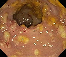
An invasive test not usually performed for the initial diagnosis of C. difficile or antibiotic-induced colitis, colonoscopy aids in confirming the diagnosis of pseudomembranous colitis in the presence of suggestive symptoms, negative testing, and failure to respond to conventional treatment. Colonoscopies of patients with pseudomembranous colitis demonstrate inflamed colonic mucosa characterized by raised yellowish, occasionally hemorrhagic nodules or plaques that congregate into widespread, pseudomembranes on the surface of the colonic mucosa. Up to 2 cm in diameter, these pseudomembranes present in scattered diffusely among areas of normal or healthy mucosa. The most severely affected have confluent pseudomembranes covering the entirety of their colonic mucosa.

History and Physical

Patients with pseudomembranous colitis most commonly report symptomatic diarrhea. C. difficile presents in various ways. Patients may be asymptomatic carriers or present with fulminant pseudomembranous colitis with toxic megacolon. Generally, the presence of pseudomembranes indicates a more severe case of C. difficile infection or antibiotic-associated diarrhea. Other notable symptoms and signs include fever, abdominal cramping, and leukocytosis. Severe pseudomembranous colitis can manifest profound leukocytosis with white blood cell counts up to 100,000/mm3, hypovolemia, hypotension, protein-losing enteropathy, reactive arthritis, and toxic megacolon. About 3% to 8% of pseudomembranous colitis will develop fulminant infection including severe ileus, toxic megacolon, hypovolemia, hypotension, renal dysfunction, colonic perforation with attendant peritonitis, and septic shock.

Evaluation

Initiate stool testing for C. difficile when patients have positive guaiac diarrhea in combination with radiographic evidence of toxic megacolon or the presence of C. difficile risk factors such as hospitalization, antibiotic or chemotherapy exposure, and marked leukemoid reactions. Screening for C. difficile as the causative agent for pseudomembranous colitis begins with screening for the C. difficile toxins. [8][9]

A common lab algorithm utilizes an enzyme immunoassay (EIA) to screen for glutamate dehydrogenase antigen, present in most C. difficile isolates followed by EIA sampling for C. difficile toxin A and toxin B.

Nucleic acid amplification tests, polymerase chain reaction, loop-mediated isothermal amplification for C. difficile have excellent sensitivity and specificity with high positive likelihood ratios; utilization of these assays aids in clarifying discrepancies between glutamate dehydrogenase and EIA toxin results. Nucleic acid amplification tests results should be interpreted with caution because they do not distinguish C. difficile colonization from active infection unless testing for concurrent toxin production. 

Consider initiating testing for C. difficile colitis on inpatients having multiple, 3, liquid consistency stools per day. Avoid sending stool samples to test for a cure because assays remain positive in 60% upon completing therapy. With its inherent hazards and monetary fees, endoscopy is not recommended unless clinical suspicion persists despite negative diagnostic testing.

Treatment / Management

Although only oral vancomycin and fidaxomicin have US Food and Drug Administration approval for treatment of C. difficile colitis, metronidazole has remained a first-line agent for four decades. Multiple studies document near the equivalent effect of metronidazole with vancomycin for the initial treatment of mild to moderate C. difficile colitis. Criteria for severe C. difficile colitis include: leukemoid reaction greater than 35,000/mm3, hypotension, admission to an intensive care unit, elevated serum lactate, and/or evidence of end-organ damage such as acute kidney injury or delirium; most manifestations of pseudomembranous colitis will signify severe disease. Because of superior cure rates for severe disease, administer oral vancomycin as first-line treatment of C. difficile colitis. With recurrent C. difficile colitis after oral vancomycin therapy, consider either fidaxomicin or rifaximin oral administration. For fulminant C. difficile pseudomembranous colitis refractory to pharmacological therapy or with complications such as megacolon or colonic perforation, surgical intervention may be warranted, including hemicolectomy. Recurrent C. difficile colitis commonly occurs following initial pharmacologic therapy; usually, the initial antibiotic therapy is reinstituted unless evidence exists of worsening disease severity. With three or more disease recurrences despite therapy with oral vancomycin, fecal microbiota transplant has shown benefit in small trials.[10][11][12]

Differential Diagnosis

Pseudomembranous colitis from C. difficile needs to be distinguished from other etiologies of infectious and noninfectious dysentery which include:

  • Diverticulitis
  • Crohn disease
  • Irritable bowel disease
  • Salmonella infection
  • Vibrio infections
  • Viral and bacterial gastroenteritis

Complications

  • Colitis
  • Bowel perforation
  • Toxic megacolon
  • Sepsis

Pearls and Other Issues

A sizeable minority of successfully treated C. difficile colitis patients will have persistent postinfectious alterations of their bowel habits, similar to irritable bowel syndrome and other functional gastrointestinal disorders. Recrudescent colitis can occur in up to 30% of cases. One challenge of managing recrudescent pseudomembranous colitis involves distinguishing failures to eliminate C. difficile, or at least transforming it into a quiescent phase, from new colonic infections.

Enhancing Healthcare Team Outcomes

C difficile is not only an annoying infection it can be associated with high morbidity and mortality. Today the emphasis is on prevention. All healthcare workers including nurses and pharmacists need to play a proactive role in preventing C.difficile colitis. When dealing with patients with C.difficile colitis, one should wear gloves and proper washing techniques. All staff and visitors need to be educated about hand hygiene. All hospital surfaces and inanimate objects need to be disinfected including thermometers, bedpans, doorknobs, and sinks. Patients who have a severe infection should be isolated. The pharmacist should regularly assess the need for antibiotics and gastric acid suppressants as these have been associated with severe C.difficile colitis.[13][14][15] (Level V)

Evidence-based Outcomes

Patients with mild C.difficile colitis usually recover and have a good prognosis. Many of these individuals will only require supportive therapy like hydration. However, those with severe colitis can develop dehydration, electrolyte deficiencies and persistent diarrhea that may last several weeks. In these patients, treatment is recommended. When treatment is initiated with metronidazole or vancomycin, the majority of these individuals will improve in 7-10 days. About 20% of patients with C.difficile colitis will relapse after treatment has been completed. Patients who relapse tend to have recurrent episodes. In severe cases of C.difficile colitis, the hospital stay is prolonged and some develop complications that require immediate surgery. Mortality rates of 3-7% have been reported in many centers.[2][16]


(Click Image to Enlarge)
Pseudomembranous colitis
image donated by sbhimji
Details

Author

Philip Salen

Updated:

8/8/2023 12:33:15 AM

References


[1]

de Curraize C, Rousseau C, Corvec S, El-Helali N, Fihman V, Barbut F, Collignon A, Le Monnier A. Variable spectrum of disease and risk factors of peripartum Clostridium difficile infection: report of 14 cases from French hospitals and literature review. European journal of clinical microbiology & infectious diseases : official publication of the European Society of Clinical Microbiology. 2018 Dec:37(12):2293-2299. doi: 10.1007/s10096-018-3372-x. Epub 2018 Sep 3     [PubMed PMID: 30178230]

Level 3 (low-level) evidence

[2]

Khanafer N, Vanhems P, Barbut F, Eckert C, Perraud M, Vandenesch F, Luxemburger C, Demont C, CDI01 Study Group. Outcomes of Clostridium difficile-suspected diarrhea in a French university hospital. European journal of clinical microbiology & infectious diseases : official publication of the European Society of Clinical Microbiology. 2018 Nov:37(11):2123-2130. doi: 10.1007/s10096-018-3348-x. Epub 2018 Aug 17     [PubMed PMID: 30120646]


[3]

Dicks LMT, Mikkelsen LS, Brandsborg E, Marcotte H. Clostridium difficile, the Difficult "Kloster" Fuelled by Antibiotics. Current microbiology. 2019 Jun:76(6):774-782. doi: 10.1007/s00284-018-1543-8. Epub 2018 Aug 6     [PubMed PMID: 30084095]


[4]

Prechter F, Stallmach A. [Clostridium difficile in the intensive care unit]. Medizinische Klinik, Intensivmedizin und Notfallmedizin. 2020 Mar:115(2):81-87. doi: 10.1007/s00063-018-0459-1. Epub 2018 Jul 11     [PubMed PMID: 29995234]


[5]

Sachu A, Dinesh K, Siyad I, Kumar A, Vasudevan A, Karim S. A prospective cross sectional study of detection of Clostridium difficile toxin in patients with antibiotic associated diarrhoea. Iranian journal of microbiology. 2018 Feb:10(1):1-6     [PubMed PMID: 29922412]


[6]

Schäffler H, Breitrück A. Clostridium difficile - From Colonization to Infection. Frontiers in microbiology. 2018:9():646. doi: 10.3389/fmicb.2018.00646. Epub 2018 Apr 10     [PubMed PMID: 29692762]


[7]

Papatheodorou P, Barth H, Minton N, Aktories K. Cellular Uptake and Mode-of-Action of Clostridium difficile Toxins. Advances in experimental medicine and biology. 2018:1050():77-96. doi: 10.1007/978-3-319-72799-8_6. Epub     [PubMed PMID: 29383665]

Level 3 (low-level) evidence

[8]

Wessling J. [Radiological imaging of acute infectious and non-infectious enterocolitis]. Der Radiologe. 2018 Apr:58(4):302-311. doi: 10.1007/s00117-018-0379-3. Epub     [PubMed PMID: 29569035]


[9]

Jessurun J. The Differential Diagnosis of Acute Colitis: Clues to a Specific Diagnosis. Surgical pathology clinics. 2017 Dec:10(4):863-885. doi: 10.1016/j.path.2017.07.008. Epub     [PubMed PMID: 29103537]


[10]

Al Momani LA, Abughanimeh O, Boonpheng B, Gabriel JG, Young M. Fidaxomicin vs Vancomycin for the Treatment of a First Episode of Clostridium Difficile Infection: A Meta-analysis and Systematic Review. Cureus. 2018 Jun 11:10(6):e2778. doi: 10.7759/cureus.2778. Epub 2018 Jun 11     [PubMed PMID: 30112254]

Level 1 (high-level) evidence

[11]

von Braun A, Lübbert C. [Treatment of acute and recurrent Clostridium difficile infections : What is new?]. Der Internist. 2018 May:59(5):505-513. doi: 10.1007/s00108-018-0401-x. Epub     [PubMed PMID: 29536125]


[12]

Shen NT, Maw A, Tmanova LL, Pino A, Ancy K, Crawford CV, Simon MS, Evans AT. Timely Use of Probiotics in Hospitalized Adults Prevents Clostridium difficile Infection: A Systematic Review With Meta-Regression Analysis. Gastroenterology. 2017 Jun:152(8):1889-1900.e9. doi: 10.1053/j.gastro.2017.02.003. Epub 2017 Feb 10     [PubMed PMID: 28192108]

Level 1 (high-level) evidence

[13]

van der Wilden GM, Velmahos GC, Chang Y, Bajwa E, O'Donnell WJ, Finn K, Harris NS, Yeh DD, King DR, de Moya MA, Fagenholz PJ. Effects of a New Hospital-Wide Surgical Consultation Protocol in Patients with Clostridium difficile Colitis. Surgical infections. 2017 Jul:18(5):563-569. doi: 10.1089/sur.2016.041. Epub 2017 May 30     [PubMed PMID: 28557651]


[14]

Cruz-Betancourt A, Cooper CD, Sposato K, Milton H, Louzon P, Pepe J, Girgis R, Patel SV, Ibrahim D, Van Horn S, Hsu V. Effects of a predictive preventive model for prevention of Clostridium difficile infection in patients in intensive care units. American journal of infection control. 2016 Apr 1:44(4):421-4. doi: 10.1016/j.ajic.2015.11.010. Epub 2016 Jan 5     [PubMed PMID: 26775936]


[15]

Vassallo A, Tran MC, Goldstein EJ. Clostridium difficile: improving the prevention paradigm in healthcare settings. Expert review of anti-infective therapy. 2014 Sep:12(9):1087-102. doi: 10.1586/14787210.2014.942284. Epub     [PubMed PMID: 25109301]


[16]

Venkat R, Pandit V, Telemi E, Trofymenko O, Pandian TK, Nfonsam VN. Frailty Predicts Morbidity and Mortality after Colectomy for Clostridium difficile Colitis. The American surgeon. 2018 May 1:84(5):628-632     [PubMed PMID: 29966560]