Midgut Volvulus

Earn CME/CE in your profession:


Continuing Education Activity

A volvulus is a medical condition where the intestines twist upon themselves. This condition can occur at any age. However, it is more frequent in children and infants. A volvulus can cause a blockage that may cut off blood flow usually due to a congenital anomaly called intestinal malrotation; this can occur in any part of the intestine without this condition present. Intestinal malrotation can make an infant more likely to develop a midgut volvulus, usually occurring in the first few weeks of life. This activity reviews the evaluation and management of midgut volvulus and highlights the role of the interprofessional team in recognizing and managing this condition.

Objectives:

  • Identify the etiology of midgut volvulus.

  • Determine the evaluation of midgut volvulus.

  • Compare the treatment and management options available for midgut volvulus.

  • Communicate interprofessional team strategies for improving care coordination and communication to advance knowledge regarding midgut volvulus and improve outcomes in pediatric patients.

Introduction

A volvulus is a medical condition where the intestines twist upon themselves. This condition can occur at any age. However, it is more frequent in children and infants.[1] A volvulus can cause a blockage that may cut off blood flow usually due to a congenital disability called intestinal malrotation; this can occur in any part of the intestine without this condition present. Intestinal malrotation can make an infant more likely to develop a midgut volvulus and occurs in the first few weeks of life. A midgut volvulus is usually part of a vascular compromise in the intestinal mesentery in intestinal malrotation.[2]

Etiology

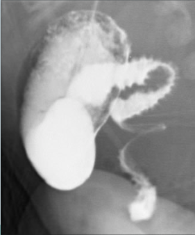
During fetal development, the intestines develop in 3 portions. These are foregut, midgut, and hindgut. The midgut portion of the intestines undergoes normal rotation during the 4 weeks and 6 weeks of gestation. The fetal midgut intestines develop so rapidly that the abdominal cavity cannot house all the intestines. Therefore, a portion of the intestines herniates into the umbilical cord. The intestines herniate back into the abdominal cavity during the eighth to tenth week of gestation with a 270-degree counter-clockwise rotation around the bloody supply to the midgut, the superior mesenteric artery. The fourth portion of the duodenum and proximal jejunum of the intestines acquires a C-loop, with the development of the ligament of Treitz, and is superior to the superior mesenteric artery (see Image. Abnormal C-Loop and Corkscrew Appearance of Jejunum). The cecal portion of the intestine becomes fixed to the retroperitoneum, superior to the superior mesenteric artery, in a diagonal axis at approximately the level of the T12 vertebra to the level of the right of the L5 vertebra.[3]

Epidemiology

Anomalies of rotation and fixation are twice as common in males as in females. They are classified as non-rotation, incomplete rotation, reverse rotation, and anomalous fixation of the mesentery.[3]

Pathophysiology

Genetic mutations likely disrupt the signaling critical for normal intestinal rotation. For instance, mutations in the gene BCL6 leading to the absence of left-sided expression of its transcript result in malrotation. In an incomplete rotation, the cecum remains in the epigastrium, and the fibrotic bands develop between the duodenum to the retroperitoneum as the cecum continues to form. These bands, also known as Ladds bands, cross over the second part of the duodenum, connecting the cecum to the lateral abdominal wall. The Ladds bands create a potential point of obstruction.

The mesenteric takeoff is then confined to the epigastrium leading to a narrow pedicle, constricting all the superior mesenteric artery branches that supply the midgut. A volvulus can occur around this narrow base of the mesentery, which then obstructs the proximal jejunum and causes ischemia to the midgut. Obstruction and bowel ischemia can then ensue if the problem is not promptly corrected surgically.[4][5]

History and Physical

Midgut volvulus can happen at any age but most commonly occurs during the first few weeks of life. Bilious emesis is usually the first feature of volvulus. Infants that present with sudden-onset bilious vomiting, upper abdominal distension, and abdominal tenderness associated with hemodynamic deterioration must be rapidly evaluated to ensure that they do not have intestinal malrotation along with volvulus. Hematochezia can also be a presenting feature in some newborns with volvulus due to bowel ischemia. If left untreated, it can eventually lead to circulatory collapse. Physical examination findings for the advanced disease show signs of peritonitis suggestive of intestinal ischemia with erythema and edema of the abdominal wall. An index of suspicion for this condition must be high, as the abdominal signs are minimal early in the disease process.

In older children and adults, abdominal pain is the most common symptom and may present with abrupt onset over hours or days or as chronic intermittent pain over weeks, months, or years. Intermittent vomiting, chronic diarrhea, malabsorption, or failure to thrive comprise other potential presenting symptoms.[6]

Evaluation

The patient may not always appear ill. Therefore, radiological evaluation is a must in all suspected newborns. Plain abdominal radiographs usually show a paucity of gas throughout the intestine with few scattered air-fluid levels. When these findings are present, the patient should undergo immediate fluid resuscitation to ensure adequate perfusion and urine output, followed by prompt surgery. Laparoscopy can be considered in cases where the child is stable.[7] Upper gastrointestinal series can be performed when the child is stable, and there is no evidence of perforation peritonitis. A contrast study of a child with volvulus usually reveals incomplete rotation with an abnormally placed duodenojejunal junction. The normal position of the duodenojejunal junction is at the level of the pylorus and to the left of the vertebral body. In children with volvulus, it is displaced to the right of the vertebral body. The child may also show a corkscrew effect diagnosing volvulus or complete duodenal obstruction, with the small bowel looping entirely on the right side of the abdomen. A barium enema may show a displaced cecum, but this sign is unreliable, especially in a small infant in whom the cecum is normally in a somewhat higher position than in an older child. Ultrasound doppler of the abdomen is also a highly sensitive and specific tool for diagnosing midgut volvulus. An abnormal relation of superior mesenteric vessels, ie, superior mesenteric vein (SMV) lying either anterior to or left to the superior mesenteric artery (SMA), and a whirlpool sign (vessels twisting around the base of the mesentery) can be useful signs in diagnosing midgut volvulus.[8][9]

Treatment / Management

When volvulus is suspected, early surgical intervention is mandatory if the ischemic process is to be prevented or reversed. Volvulus occurs clockwise and is therefore untwisted counterclockwise, which one can remember with the phrase "turning back the hands of time."[10]

Followed by Ladds procedure, named after William Edward Ladd, the pediatrician who first performed the procedure in 1936. This operation does not correct the malrotation but instead helps to open the narrow mesenteric pedicle to prevent volvulus from recurring. The Ladd procedure is performed by lysing the band formed between the cecum and the lateral abdominal wall and the duodenum and terminal ileum, allowing the superior mesenteric artery to relax. This maneuver allows the duodenum to relax into the right lower quadrant and the cecum into the left lower quadrant; these structures do not need to be secured with a suture. An appendectomy (removal of the appendix) is also done to circumvent errors in the event a patient has diagnostic imaging later in life.[11][12]

The gold standard approach of this procedure has been an open Ladds operation; however, there is an increasing trend towards performing this procedure laparoscopically due to equivalent operative times compared to standard open techniques along with the earlier resumption of feeds and decreased hospital stays.[13][14][15][16] When a patient presents with advanced ischemia, a simple reduction of the volvulus without the Ladd procedure should be performed, followed by a "second-look" laparotomy, which is performed 24 to 36 hours later to evaluate the vascular integrity of the small intestines.[17] Some surgeons might also pass a tube through the duodenum's lumen to ensure no associated intrinsic duodenal obstruction.

A transparent plastic silo may be placed to facilitate the constant evaluation of the intestine and to plan for the timing of re-exploration. If a necrotic bowel is present, then the surgeon can conservatively resect it to ensure adequate length for feeding and prevention of short-gut syndrome. With early diagnosis and correction, the prognosis is excellent. Delay can lead to mortality or short-gut syndrome requiring intestinal transplantation.

Differential Diagnosis

The differential diagnoses for midgut volvulus include the following:

  • Bowel obstruction in newborn
  • Congenital band
  • Intestinal volvulus
  • Necrotizing enterocolitis imaging
  • Neonatal sepsis
  • Pediatric duodenal atresia
  • Pediatric gastroesophageal reflux

Complications

In the event of delayed diagnosis and surgical treatment, mesenteric ischemia can lead to gangrene of the majority of the small bowel. This causes resection of the necrotic/gangrenous portion of the bowel and thus short bowel syndrome.[18] Another complication commonly reported by some surgeons is the development of small bowel obstruction due to adhesions. 

Postoperative and Rehabilitation Care

Although there is no opening of the bowel lumen in the surgical procedure, resumption of the enteral feeds may be delayed in some patients. This is usually seen in those patients who have gross distension of the stomach and/or duodenum, have an element of dysmotility, or have undergone resection of the necrotic bowel with anastomosis. In these conditions, a nasogastric tube provides optimal bowel decompression. Total parenteral nutrition can be considered in patients who require prolonged fasting due to resection of a major proportion of the small bowel. Children failing to thrive in the preoperative period might also require a regular follow-up after discharge to ensure optimal catch-up growth.

Pearls and Other Issues

A small proportion of patients with malrotation demonstrate chronic obstructive symptoms. These symptoms may result from Ladd bands across the duodenum or, occasionally, intermittent volvulus. Symptoms include intermittent abdominal pain and intermittent vomiting, which may occasionally be bilious. Infants with malrotation may demonstrate a failure to thrive, and they may be diagnosed initially as having gastroesophageal reflux disease (GERD). As described earlier, surgical correction using the Ladd procedure can prevent volvulus from occurring and improve symptoms in these instances.[19]

Enhancing Healthcare Team Outcomes

An interprofessional team provides a holistic and integrated approach to postoperative care and further leads to the best outcomes.


(Click Image to Enlarge)
<p>Abnormal C-Loop&nbsp;and&nbsp;Corkscrew Appearance of Jejunum

Abnormal C-Loop and Corkscrew Appearance of Jejunum. A midgut volvulus is a medical condition where the intestines twist upon themselves, which can occur at any age. However, it is more frequent in children and infants.


Contributed by LSU Radiology Department

Details

Author

Sachit Anand

Author

Hany Nada

Editor:

Hira Ahmad

Updated:

9/12/2022 9:13:11 PM

References


[1]

Millar AJ, Rode H, Cywes S. Malrotation and volvulus in infancy and childhood. Seminars in pediatric surgery. 2003 Nov:12(4):229-36     [PubMed PMID: 14655161]


[2]

Birajdar S, Rao SC, Bettenay F. Role of upper gastrointestinal contrast studies for suspected malrotation in neonatal population. Journal of paediatrics and child health. 2017 Jul:53(7):644-649. doi: 10.1111/jpc.13515. Epub 2017 Apr 20     [PubMed PMID: 28425590]


[3]

Pelayo JC, Lo A. Intestinal Rotation Anomalies. Pediatric annals. 2016 Jul 1:45(7):e247-50. doi: 10.3928/00904481-20160602-01. Epub     [PubMed PMID: 27403672]


[4]

Sivakumar A, Mahadevan A, Lauer ME, Narvaez RJ, Ramesh S, Demler CM, Souchet NR, Hascall VC, Midura RJ, Garantziotis S, Frank DB, Kimata K, Kurpios NA. Midgut Laterality Is Driven by Hyaluronan on the Right. Developmental cell. 2018 Sep 10:46(5):533-551.e5. doi: 10.1016/j.devcel.2018.08.002. Epub 2018 Aug 30     [PubMed PMID: 30174180]


[5]

Strouse PJ. Disorders of intestinal rotation and fixation ("malrotation"). Pediatric radiology. 2004 Nov:34(11):837-51     [PubMed PMID: 15378215]


[6]

Nagdeve NG, Qureshi AM, Bhingare PD, Shinde SK. Malrotation beyond infancy. Journal of pediatric surgery. 2012 Nov:47(11):2026-32. doi: 10.1016/j.jpedsurg.2012.06.013. Epub     [PubMed PMID: 23163993]


[7]

Adams SD, Stanton MP. Malrotation and intestinal atresias. Early human development. 2014 Dec:90(12):921-5. doi: 10.1016/j.earlhumdev.2014.09.017. Epub 2014 Oct 13     [PubMed PMID: 25448782]


[8]

Applegate KE, Anderson JM, Klatte EC. Intestinal malrotation in children: a problem-solving approach to the upper gastrointestinal series. Radiographics : a review publication of the Radiological Society of North America, Inc. 2006 Sep-Oct:26(5):1485-500     [PubMed PMID: 16973777]


[9]

Shah MR, Levin TL, Blumer SL, Berdon WE, Jan DM, Yousefzadeh DK. Volvulus of the entire small bowel with normal bowel fixation simulating malrotation and midgut volvulus. Pediatric radiology. 2015 Dec:45(13):1953-6. doi: 10.1007/s00247-015-3430-9. Epub 2015 Jul 26     [PubMed PMID: 26209961]


[10]

Reddy AS, Shah RS, Kulkarni DR. Laparoscopic Ladd'S Procedure in Children: Challenges, Results, and Problems. Journal of Indian Association of Pediatric Surgeons. 2018 Apr-Jun:23(2):61-65. doi: 10.4103/jiaps.JIAPS_126_17. Epub     [PubMed PMID: 29681694]


[11]

Tsumura H, Ichikawa T, Kagawa T, Nishihara M. Successful laparoscopic Ladd's procedure and appendectomy for intestinal malrotation with appendicitis. Surgical endoscopy. 2003 Apr:17(4):657-8     [PubMed PMID: 12574927]


[12]

Isani MA, Schlieve C, Jackson J, Elizee M, Asuelime G, Rosenberg D, Kim ES. Is less more? Laparoscopic versus open Ladd's procedure in children with malrotation. The Journal of surgical research. 2018 Sep:229():351-356. doi: 10.1016/j.jss.2018.04.016. Epub 2018 May 11     [PubMed PMID: 29937013]


[13]

Bass KD, Rothenberg SS, Chang JH. Laparoscopic Ladd's procedure in infants with malrotation. Journal of pediatric surgery. 1998 Feb:33(2):279-81     [PubMed PMID: 9498402]


[14]

Arnaud AP, Suply E, Eaton S, Blackburn SC, Giuliani S, Curry JI, Cross KM, De Coppi P. Laparoscopic Ladd's procedure for malrotation in infants and children is still a controversial approach. Journal of pediatric surgery. 2019 Sep:54(9):1843-1847. doi: 10.1016/j.jpedsurg.2018.09.023. Epub 2018 Oct 28     [PubMed PMID: 30442460]


[15]

Catania VD, Lauriti G, Pierro A, Zani A. Open versus laparoscopic approach for intestinal malrotation in infants and children: a systematic review and meta-analysis. Pediatric surgery international. 2016 Dec:32(12):1157-1164     [PubMed PMID: 27709290]

Level 1 (high-level) evidence

[16]

Huntington JT, Lopez JJ, Mahida JB, Ambeba EJ, Asti L, Deans KJ, Minneci PC. Comparing laparoscopic versus open Ladd's procedure in pediatric patients. Journal of pediatric surgery. 2017 Jul:52(7):1128-1131. doi: 10.1016/j.jpedsurg.2016.10.046. Epub 2016 Oct 30     [PubMed PMID: 27856011]


[17]

Kulaylat AN, Hollenbeak CS, Engbrecht BW, Dillon PW, Safford SD. The impact of children's hospital designation on outcomes in children with malrotation. Journal of pediatric surgery. 2015 Mar:50(3):417-22. doi: 10.1016/j.jpedsurg.2014.08.011. Epub 2014 Sep 8     [PubMed PMID: 25746700]


[18]

Seashore JH, Touloukian RJ. Midgut volvulus. An ever-present threat. Archives of pediatrics & adolescent medicine. 1994 Jan:148(1):43-6     [PubMed PMID: 8143008]


[19]

Marseglia L, Manti S, D'Angelo G, Gitto E, Salpietro C, Centorrino A, Scalfari G, Santoro G, Impellizzeri P, Romeo C. Gastroesophageal reflux and congenital gastrointestinal malformations. World journal of gastroenterology. 2015 Jul 28:21(28):8508-15. doi: 10.3748/wjg.v21.i28.8508. Epub     [PubMed PMID: 26229394]