Continuing Education Activity
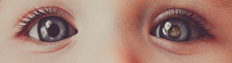
Leukocoria means ‘white pupil’ or ‘cat’s eye pupil.’ Leukocoria is an abnormal pupillary reflex more clearly seen after mydriasis or photography. It is often the first sign of a range of serious intraocular disorders, including congenital cataracts, Coats disease, retinoblastoma, retinopathy of prematurity, toxocariasis, Norrie disease, and retrolental fibroplasia. Immediate diagnosis and management are mandatory as most conditions are vision-threatening, and some are life-threatening. The normal red reflex of the human eye is due to the retro-illumination of normal choroidal vasculature reflecting through the retina, vitreous, lens, pupil, and cornea. Any interference in these structures would give an altered red reflex or leukocoria. This activity highlights the importance of the evaluation and management of leukocoria and the role of interprofessional teamwork in managing patients with underlying conditions.
Objectives:
- Identify the etiology and epidemiology of leukocoria.
- Describe the evaluation of leukocoria.
- Summarize the management options available for leukocoria.
- Highlight the role of interprofessional collaboration in improving outcomes in patients with leukocoria.
Introduction
Leukocoria means ‘white pupil.’ Leukocoria is an abnormal pupillary reflex usually more prominent after pupillary dilation. It is often the first sign of a range of serious intraocular disorders. Leukocoria is commonly seen in congenital cataracts, Coats disease, retinoblastoma, retinopathy of prematurity, toxocariasis, Norrie disease, retrolental fibroplasia, and other disorders. Immediate diagnosis and management are mandatory as most conditions are vision-threatening, and retinoblastoma may be life-threatening. Immediate referral to an ophthalmologist, an interdisciplinary liaison with a pediatric ophthalmologist, retina specialist, and ocular oncologist will help in the management of eyes with leukocoria in an appropriate manner.
Etiology
The normal red reflex of the human eye is due to the retro-illumination of normal choroidal vasculature reflecting through the retina, vitreous, lens, pupil, and cornea. Any interference in these structures would give an altered red reflex or leukocoria. Some of the common causes of leukocoria include:
- Retinoblastoma
- Coats disease
- Retinopathy of prematurity (ROP)
- Persistent fetal vasculature (PFV)
- Ocular toxocariasis and toxoplasmosis
- Astrocytic hamartoma
- Familial exudative vitreoretinopathy (FEVR)
- Vitreous hemorrhage
- Retinochoroidal coloboma,
- Endogenous endophthalmitis
- Incontinentia pigmenti (Bloch-Sulzberger syndrome)
- Retinal detachment
- Pediatric uveitis
- Medullated retinal nerve fibers (MRNF)
- Medulloepithelioma
- TORCH (Toxoplasmosis, Other agents, Rubella, Cytomegalovirus, and Herpes simplex syndrome)
Epidemiology
In a retrospective study spanning ten years, the mean age of children presenting with leukocoria was 42.5 months.[1] Bilateral involvement was seen in 54% with a male-female sex ratio of 1.5. 76% of children were under six years of age. Cataracts caused seventy-five percent of the cases, and 21% had retinoblastoma. Other causes of leukocoria included retinal detachment (1%), retinopathy of prematurity (1%), persistent pupillary membrane (1%), persistent hyperplastic primary vitreous (0.6%), endophthalmitis (0.64%), optic nerve coloboma (0.3%), iris heterochromia (0.3%), and ametropia (0.3%).[1]
The Third National Cancer survey data indicates an annual incidence of 11 new cases of retinoblastoma per million children under the age of 5 years.[2] Retinoblastoma is the most common intraocular malignancy in children and accounts for 2% of all childhood cancers. The estimated incidence ranges from 3 to 42 cases per million live births. In the United States, the incidence is 12 cases per million live births in children less than five years of age, with a prevalence of 6 % of all cancers in this age group.[3] Retinoblastoma affects both sexes equally. Retinoblastoma shows the highest incidence in children less than four years of age.[4] Coats disease has an incidence of 0.09 per 100,000 population, with 85% of cases affecting males.[5]
The global prevalence of childhood cataracts ranges from 0.3 to 23 per 10,000, with a median of 1 per 10,000 and 0.6% to 9.8% per 10,000 for congenital cataracts, with a median of 1.7 per 10,000 based on many studies that reported congenital cataracts.[6] In a study involving 602 newborns, the incidence of retinopathy of prematurity (ROP) at any stage was 34%, and Type 1 pre-threshold ROP was 5% with a mean gestational age of 31 weeks.[7] The incidence of neonatal endophthalmitis in the United States is 4.2 cases/1000000 live births.[8]
Pathophysiology
Leukocoria is a whitish or altered pupillary reflex that appears due to obstruction of the normal retinochoroidal vasculature, which reflects through the pupil. The various causes of leukocoria, their etiopathogenesis, and clinical features are as follows:
- Retinoblastoma: Retinoblastoma (RB) results from the malignant transformation of the primitive retinal cells, most likely a multipotent retinoblast. Forty-eight percent of cases show autosomal dominant transmission with 90% penetrance at birth and maps to chromosome 13q14.[9] Ninety-five percent of cases are sporadic, while only 5% have a family history of RB. RB is caused by a mutation in the RB1 tumor suppressor gene located on sub-band 13q14.2. Seventy-five percent of RB is caused by a mutation in a single retinal cell by inactivation of both alleles of the RB1 gene. These are unilateral and unifocal—60% of cases present with leukocoria within two years of age. Pathological microscopy shows Flexner-Wintersteiner rosettes (ring of nuclei surrounding a clear central lumen, also seen in Medulloepithelioma), Homer-Wright rosettes (neural filaments with no precise, distinct lumen, also seen in adrenal neuroblastoma and medulloblastoma), pseudo-rosettes, and fleurettes (bouquet of pink bulbous neoplastic photoreceptor inner segments; highly specific for RB). Intratumoral calcification (90%) is common. Retinoblastoma presents as leukocoria (60%), squint (20%), and decreased vision (5%). Clinical features are yellowish-white retinal mass, rubeosis iridis, pseudohypopyon, hyphema, angle-closure glaucoma, and uveitis. Two types are seen: the endophytic type, which starts at the inner retina and grows into vitreous simulating endophthalmitis and causes pseudohypopyon, and the exophytic type, which starts from the outer retina and grows towards the choroid extending into the sclera simulating Coats disease or traumatic retinal detachment. Intratumoral calcification is seen along with tumor necrosis when it outgrows its blood supply—trilateral retinoblastoma (3%) in the presence of pineoblastoma or parasellar neuroblastoma along with bilateral RB. Retinocytoma or retinoma is a benign tumor composed of fleurettes.
- Coats disease: Coats disease is a retinal telangiectatic condition that commonly affects one eye of young males.[10] Though it is mostly a sporadic condition, multiple genetic changes have been described. Chromosomes involved are 3 and 13, with the involvement of NDP (Norrie disease protein), CRB1, and PANK2 genes. Clinical features are mid-peripheral or peripheral telangiectasia, retinal exudation, exudative retinal detachment, neovascular glaucoma, and bulb-like microaneurysms (Leber’s military aneurysms). The choroid and sclera remain unaffected. The most devastating complication is neovascular glaucoma, which is seen in 10% of cases and might often require enucleation. The timely intervention of retinal detachment can prevent this.
- Persistent fetal vasculature: Persistent fetal vasculature (PFV) or Persistent hyperplastic primary vitreous (PHPV)is a congenital condition due to failure of regression of the intraocular fetal vasculature. It’s the second most common cause of infantile leukocoria.[11] It is non-heritable and commonly causes unilateral involvement. Leukocoria results from a complete pupillary membrane, cataract, fibrovascular sheath attached to the posterior lens capsule, strabismus, and a rarely cloudy cornea due to glaucoma. Anterior PFV presents with microphthalmia, cataract, elongated ciliary process, shallow anterior chamber, retrolental fibrovascular membranes, dilated iris vessels, glaucoma, and ectropion uveae. Posterior PFV presents with microphthalmia, tractional retinal detachment, hypoplastic optic nerve and macula, vitreous membranes and stalk, clear lens, and strabismus.
- Pediatric cataract: Over 60% of children presenting with leukocoria have congenital cataracts [unilateral (18%) and bilateral (42%)]. Unilateral cataracts are usually sporadic, while bilateral cataracts are often associated with other systemic diseases like intrauterine infections (rubella, toxoplasmosis, cytomegalovirus, and herpes simplex virus infections), metabolic disorders (galactosemia), chromosomal anomalies (trisomy 21, Stickler syndrome and myotonic dystrophy).
- Pediatric uveitis: Pars-planitis with its ‘snowball’ or ‘snow-banking’ in the vitreous commonly presents as leukocoria. Most children have bilateral symmetry. Other causes of uveitis in children are juvenile idiopathic arthritis (JIA), tubulointerstitial nephritis, uveitis (TINU), leukemia, retinoblastoma, juvenile xanthogranuloma, and Blau syndrome.
- Toxocariasis: Toxocariasis is caused by a nematode, Toxocara canis, which causes both systemic and ocular toxocariasis. Definitive hosts are dogs and cats, so close contact with domestic pet dogs and cats is an important risk factor for children. Ocular toxocariasis is mostly unilateral and presents with blurred vision, floaters, leukocoria, and strabismus. Toxocariasis clinically presents as retinal granuloma (localized at the posterior pole or peripheral), endophthalmitis, vitreous abscess, temporal dragging of the macula, and tractional retinal detachment.
- Astrocytic hamartoma: Retinal astrocytic hamartoma is a benign retinal tumor composed of glial cells, especially astrocytes, and is frequently associated with tuberous sclerosis. Other features are hamartomas of the iris and ciliary body with neovascular glaucoma and giant retinal astrocytoma with exudative retinal detachment.[12] Tuberous sclerosis transmits autosomal dominantly and results from mutation of either of the two genes, TSC1 (encoding hamartin) and TSC2 (encoding tuberin).[13]
- Retinopathy of prematurity (ROP): This is also known as Retrolental Fibroplasia. ROP results from abnormal proliferation of retinal vessels in premature infants weighing less than 1250 gm or under 32 weeks of gestation. These children usually receive oxygen therapy and present signs of ROP between 35 and 45 weeks of post-conceptional age. Children under 1000 gm pose the highest risk for blindness, while most cases regress spontaneously over 3 to 4 months. ROP shows four stages, as mentioned below. Leukocoria might present from stage 3 onwards.
- Endogenous endophthalmitis: Endogenous endophthalmitis is rare and occurs commonly in premature, very low birth weight neonates. Klebsiella pneumoniae, Methicillin-resistant Staphylococcus aureus, Pseudomonas aeruginosa, and Candida albicans are the common causative organisms.[14] The pathogen most commonly reaches the eye through the posterior circulation in the form of a septic embolus. The infection then extends from the Retinochoroidal circulation into the vitreous cavity and then to the anterior chamber of the eye.
- Retinochoroidal coloboma: Coloboma is a congenital defect in the iris, ciliary body, choroid, retina, and optic nerve due to a failed or incomplete closure of the embryonic fissure. It usually involves the inferonasal quadrant of the eye. Retinochoroidal coloboma usually presents as leukocoria. Rhegmatogenous Retinal detachment is the most common complication.
- Familial exudative vitreoretinopathy (FEVR): Familial exudative vitreoretinopathy (FEVR) is a hereditary retinal vascular disorder characterized by an avascular peripheral retina, especially temporal quadrant with a ‘V-shaped demarcation, temporal dragging of retinal vessels and macula, retinal falciform folds, retinal ischemia with neovascularization, subretinal exudates, and retinal detachment. Inheritance is Autosomal Dominance with 100% penetrance. FEVR develops as a consequence of a genetic abnormality (Wnt signaling) that affects the growth and development of retinal blood vessels. An important differential is ROP, but cases with ROP have a history of preterm birth and low birth weight. FEVR cases have a family history of a similar disease, and clinical examination or fundus fluorescein angiogram (FFA) may reveal peripheral avascular retina in the family members.
- Retinal detachment: Pediatric retinal detachments account for 3.2% to 6.6% of all cases of retinal detachment.[15] Pediatric retinal detachments are commonly secondary to ocular trauma, ROP, Myopia, Stickler’s syndrome, X-linked retinoschisis, Persistent hyperplastic primary vitreous, FEVR, retinochoroidal coloboma, Marfan’s syndrome, Coats disease, uveitis, and following intraocular surgery.
- Medulloepithelioma: Medulloepithelioma (diktyoma) is a non-hereditary tumor of the non-pigmented ciliary epithelium. These slow-growing tumors present with visual decline, raised intraocular pressure, localized angle closure, leukocoria, cataracts, and red-eye. It appears in the ciliary body region as a whitish-pink mass and is associated with the DICER-1 gene. 5% of cases are associated with pleuropulmonary blastoma, family tumor, and dysplasia Syndrome (PPB-FTDS). Ultrasound biomicroscopy (UBM) and anterior segment optical coherence tomography (OCT) would help assess the size and extent of the tumor.
- TORCH syndrome: TORCH syndrome includes toxoplasmosis, other agents (Treponema pallidum, Varicella-zoster virus, Parvovirus B19), rubella, cytomegalovirus, and herpes simplex. It’s a neonatal infection that spreads transplacentally from an infected mother and presents with leukocoria.
- Medullated retinal nerve fibers: Normal retinal nerve fibers usually are devoid of myelin sheath anterior to lamina cribrosa. Medullated retinal nerve fibers are those fibers that still possess the myelin sheath anterior to lamina cribrosa. They are asymptomatic except for causing leukocoria and are associated with myopia, amblyopia, and strabismus. They appear as a white or yellowish striated patch with feathered borders in the peripapillary area or around the macula.
- Incontinentia pigmenti (IP): Incontinentia Pigmenti (Bloch-Sulzberger syndrome) is a rare X-linked dominant disorder caused by mutation of the NEMO/IKK gene located at the Xq28 loci. It affects mostly females, and the disease is lethal for males in-utero. IP is characterized by skin, ocular, neurological, and dental abnormalities. Ocular findings are retinal non-perfusion areas, tractional retinal detachment, vitreoretinal fibrosis, and retinal pigment epithelial defects. IP may clinically resemble retinoblastoma and presents as leukocoria due to vitreoretinal fibrosis. Skin lesions initially present as erythematous vesicular rashes, which are later replaced with verrucous lesions, swirling macular hyperpigmentation, and linear hypopigmentation.[16][17][18][19]
History and Physical
A thorough medical history of the present illness, past ocular history, medical history, and family history should be elicited. A family photograph would help know if the leukocoria was present from birth or later in childhood. Persistent fetal vasculature is most often present at birth. A history of prematurity would lead us to investigate in line with retinopathy of prematurity, which presents as leukocoria due to retinal detachment and retrolental fibroplasia. Birth trauma can lead to hyphema or vitreous hemorrhage, which presents as an altered pupillary reflex. Family history is essential in retinoblastoma, FEVR, and coloboma.
The color of the pupillary reflex also gives a hint of the diagnosis. Retinoblastoma presents as a whitish pupillary reflex, while congenital cataracts show a blue-grey reflex. Coats disease and retinal detachment show a yellowish reflex. In 20% of cases, retinoblastoma presents as strabismus. Retinal detachments, vitreous hemorrhage, Coats disease, and retinoblastoma (60%) usually present as unilateral disease, while FEVR, endogenous endophthalmitis, astrocytic hamartomas, and 40% of retinoblastomas are bilateral.[20]
Evaluation
Any child presenting with leukocoria should be evaluated for vision, pupillary reflexes, slit-lamp examination of both anterior segment and fundus, indirect ophthalmoscopy, FFA, OCT of the retina, and B scan ultrasonography. Neuroimaging, blood workup, genetic testing, and fine-needle aspiration cytology are other ancillary tests done to confirm the diagnosis.
- Pupillary reflex: In a dark room, looking through the direct ophthalmoscope set at ‘0’ from a distance of 18 inches away from the eye, when light is projected onto the eyes, a normal red reflex should emanate from both eyes and should be symmetrical in color and contrast. A markedly diminished reflex, a white reflex, dark spots in the reflex, or an asymmetry (Bruckner reflex) are all considered abnormal. When in doubt, dilate the pupil with dilating drops to examine the reflex in detail. For infants under nine months of age, a combination of cyclopentolate 0.2% with phenylephrine 1% eye drops are used 45 minutes prior to the examination.[21] Atropine drops may be avoided due to their anticholinergic side effects. For infants of more than nine months of age, either tropicamide 1% with phenylephrine 2.5% combination or cyclopentolate 0.2% with phenylepherine1% combinations drops can be used 45 minutes prior to examination.
- Bruckner reflex: When viewed through a direct ophthalmoscope, when strabismus is present, the fixating eye has a darker reflex than the deviating eye. This test would help differentiate leukocoria from altered reflexes produced in small-angle deviations and amblyopia (anisometropia).
- Ultrasonography: This is recommended in all patients presenting with leukocoria. It is highly specific for detecting intraocular calcifications (90%) in retinoblastoma. Retinoblastoma is seen as a solid retinal mass with high reflectivity due to calcification. Coats disease is seen as a retinal detachment, and the point echoes in the subretinal fluid with no retinal mass lesion. Toxocariasis shows an elevated granuloma with no calcification. Astrocytic hamartomas present as glistening yellow calcification in contrast to the dull, chalky white calcification of retinoblastoma. Ultrasonography helps in differentiating an isolated retinal detachment from one associated with a retinal mass lesion. Medulloepithelioma shows a pattern of heterogeneous areas of high internal reflectivity. Clear cysts are visible within the mass.
- Fundus fluorescein angiography (FFA): Retcam photography with FFA is very useful in evaluating leukocoria in infants. Coats disease shows retinal telangiectasia as a ‘lightbulb’ appearance with late leakage and peripheral non-perfusion areas. Retinoblastoma shows a rapid homogenous hyperfluorescence. Toxocariasis granulomas show reticular hyperfluorescence with retinal traction. ROP and FEVR demonstrate a peripheral avascular zone.
- Optical coherence tomography (OCT): It is a noninvasive test to assess fluid, edema, or fibrosis in the posterior pole lesions. Anterior segment OCT (AS-OCT) would help delineate the size and extent of Medulloepithelioma.
- Computerized tomography (CT): CT is avoided in children due to high exposure to radiation but can be useful if intraocular calcification is doubtful.
- Magnetic resonance imaging (MRI): It is a diagnostic modality of choice to detect the intracranial spread of retinoblastoma and other secondary tumors like pinealoblastoma or parasellar neuroblastoma. Retinoblastoma is seen as an isointense to a hyperintense lesion on T1-weighted images and hypointense on T2-weighted images.
- Blood investigations: These are helpful in toxocariasis, TORCH infections, and endogenous endophthalmitis.
- Fine needle aspiration cytology (FNAC): Vitreous tap helps in endogenous endophthalmitis and chronic endophthalmitis secondary to toxocariasis. It is contraindicated in retinoblastoma as it leads to seeding and metastasis. Anterior chamber tap shows eosinophils in toxocariasis. The ratio of aqueous to plasma lactate dehydrogenase (LDH) is more than 1.0 in retinoblastoma. It is usually less than 1.0 in ocular diseases other than RB. Aqueous LDH serves as a histologic tumor marker and correlates with the severity of retinoblastoma (types 4 and 5). Isoenzymes LDH4 and LDH5 are characteristically elevated.
- Genetic testing: This helps in the diagnosis and genetic counseling of retinoblastoma, FEVR, astrocytic hamartoma, and Coats disease (NDP gene). Analysis of the retinoblastoma gene is done by direct and indirect methods. The direct method aims to detect the initial mutation that formed the tumor and subsequently find out whether the mutation is in the germline of the patient. Indirect methods are used if the initial mutation cannot be located. DNA can be evaluated from the tumor cells or from leukocytes. The deletion of the RB gene is detected by karyotyping and the Southern blot technique. Point mutations are detected by ribonuclease protection, gel electrophoresis, and direct DNA sequencing.
Treatment / Management
Leukocoria is a sign of an underlying ocular disease. The management of common ocular conditions causing leukocoria is as follows:
Retinoblastoma: Chemoreduction followed by adjuvant consolidative treatment (laser photocoagulation, thermotherapy, cryotherapy, chemotherapy) has replaced external beam radiotherapy as the primary mode of treatment for intraocular retinoblastoma.[22] The following are the various methods of treatment for retinoblastoma:
- Enucleation: Treatment of choice for unilateral group E and D intraocular retinoblastoma and in all blind and painful eyes. This involves minimal manipulation with the ‘no-touch’ technique and excision of at least 10 mm of the optic nerve to prevent the intracranial spread.
- Chemoreduction: Six monthly cycles of vincristine (0.05mg/kg), etoposide (5mg/kg), and carboplatin (18.6mg/kg) (VEC) are used to reduce the tumor size.
- Thermotherapy: Heating up to 42 to 60 degrees C is indicated in tumors with a basal diameter of less than 3 mm without any vitreous seeding. It has a synergistic effect with chemotherapy.
- External beam radiation therapy (EBRT) is indicated in Large tumors with vitreous seeding, salvageable eyes, multifocal tumors, and eyes that failed coagulation therapy. RB is a very radiosensitive tumor. A dose of 4000 cGy is used with 200 cGy fractions.
- Episcleral Plaque brachytherapy: Episcleral application of ruthenium-106 or iodine-125 plaque is made. This is indicated in single medium-sized tumors not involving the optic nerve or macula.
- Photocoagulation/Cryotherapy is used for small posteriorly located tumors, not involving the optic nerve or macula.
- Cryotherapy is used for small anteriorly located tumors.
- Intra-arterial chemotherapy using agents including melphalan, carboplatin, and topotecan has been used successfully in some RB cases.[23]
Coats disease: Mild cases may be observed. Other management options include laser treatment of telangiectatic vessels, microaneurysms, capillary nonperfusion areas, cryotherapy for exudation with subretinal fluid, intravitreal anti-vascular endothelial growth factor (anti-VEGF) agents, and retinal surgery for retinal detachment are other available therapy.
PFV: The anterior PFV is treated with observation, cataract, and glaucoma management. Treatment aims to prevent amblyopia and correct strabismus by treating cataracts and glaucoma. Severe intraoperative bleeding should be anticipated from persistent tunica vasculosa lentis. Retinal lesions of posterior PFV are managed by lensectomy, membranectomy, and vitrectomy through either anterior limbal or pars-plana approach.
Pediatric cataract: Cataracts operated between 2 months to 6 months yield good results. Emmetropization of an eye generally is achieved by nine years of age.[24] Unilateral cataracts should be operated on before six weeks, and bilateral cataracts before eight weeks of age.[25] Surgery is advised for visually significant cataracts (more than 3 mm of opacity).
Pediatric uveitis: Corticosteroids are the mainstay of treatment of noninfectious uveitis in children. Periocular or sub-tenon corticosteroid injections are used in pars planitis, especially in unilateral cases. Steroid-induced glaucoma and cataract are potential complications of corticosteroid therapy and should always be looked for. Methotrexate is the most commonly used first-line immunomodulatory drug in children with uveitis. Second-line agents are azathioprine, cyclosporine, and mycophenolate. Anti-TNF (tumor necrosis factor) agents like infliximab and adalimumab are successfully used to treat resistant pediatric uveitis.[26]
Toxocariasis: Topical or systemic corticosteroids are the mainstay of treatment in controlling inflammation. The role of anti-helminthic therapy in ocular toxocariasis is controversial and should always accompany steroids.[27] Pars plana Vitrectomy is the most common surgical treatment for ocular toxocariasis.
Astrocytic hamartoma should be observed. Glaucoma should be treated accordingly. Tuberous sclerosis should be ruled out.
Retinopathy of prematurity: Observation, retinal laser, cryotherapy, and retinal surgery are the different treatment modalities for ROP. Immature retinal vessels alone can be observed, and screening repeated. The cryo-ROP study showed positive results of cryotherapy for the entire avascular retina in the threshold ROP. Early treatment of the ROP study (ETROP) recommended the ablation of the peripheral avascular retina for type 1 ROP.
Endogenous endophthalmitis: Management depends on the causative organism and the severity of the infection. Empirical treatment includes intravitreal injection of vancomycin (1 mg/0.1 ml) plus either ceftazidime (2.25 mg/0.1 ml) or amikacin (0.4 mg/0.1 ml). Vancomycin covers gram-positive organisms, while ceftazidime and amikacin cover gram-negative organisms.[28][29][30] For fungal vitritis, vitrectomy with intravitreal injection of amphotericin (5-10 mcg/0.1 ml) or voriconazole (100 to 200 mcg/0.1 ml) and systemic antifungal therapy is indicated. Pars plana vitrectomy is indicated for all severe sight-threatening endophthalmitis. Systemic antifungal or antibacterial therapy should be started depending on the blood and vitreous culture reports.
Retinochoroidal coloboma: Retinal detachment and cataracts are the most common associations of retinochoroidal coloboma. Poor vision, amblyopia, and strabismus may be noted. Prophylactic barrage laser along the coloboma margin creates a strong chorioretinal adhesion and reinforces the margins, thereby may reduce the occurrence of extra-colobomatous retinal detachment.[31] Pars-plana vitrectomy with or without scleral buckling, drainage of subretinal fluid, and endotamponade with long-acting gas or silicone oil is the treatment of choice for retinal detachment. The exact localization of the retinal breaks that cause the retinal detachment may be difficult to localize because of the thin atrophic retina, transparent intercalary membrane, nystagmus, small cornea, and cataract.
Familial exudative vitreoretinopathy: Stage 1 FEVR without exudation can be observed. Stage 2 with exudation is treated with a laser to the avascular retina. Surgery is indicated for stages 3 to 5 and is aimed at relieving traction by excision or incising the scar tissue, thereby encouraging reattachment of the retina.
Retinal detachment: Two surgical options for pediatric rhegmatogenous retinal detachments are a scleral buckle with or without an encircling band and the vitrectomy. This is the preferred surgery, even in Grade C proliferative vitreoretinopathy (PVR). Vitrectomy with or without encirclage is done in retinal detachment with more than PVR Grade C. In tractional retinal detachment, the scleral buckle may be sufficient if the break is in the periphery.
Medulloepithelioma: Enucleation is the standard treatment for advanced ciliary body tumors. Exenteration of the orbit is required when the orbit is involved. Local excision can be done for small tumors involving 3 to 4 clock hours.
Incontinentia pigmenti: Laser ablation of retinal non-perfusion areas and cryotherapy may be helpful.
Differential Diagnosis
Leukocoria is an altered pupillary reflex presenting as a white/greying reflex compared to the bright red/orange reflex of the fellow eye. The following conditions mimic leukocoria, but they have not actually altered pupillary reflexes; instead, a differently looking eye which the parents bring to the doctor's attention:
- Pediatric glaucoma
- Strabismus and Bruckner reflex
- Hypopyon
- Hyphema
- Corneal opacities
- Foreign body
Causes of Intraocular calcification:
- Calcified cataract
- Asteroid hyalosis[32][33]
- Retinoblastoma
- Calcified drusen
- Astrocytic hamartomas
- Retinocytoma
- Astrocytomas
- Optic nerve meningioma
- Optic nerve head drusen
- Phthisis
Staging
Tartarella et al. have classified leukocoria as below:
- Pre-lenticular leukocoria
- Lenticular leukocoria
- Retrolenticular leukocoria
- Mixed presentation leukocoria
International Classification of Retinoblastoma (ICRB)[34]
- Group A: Small intraretinal tumors (less than 3 mm) away from the foveola and the disc.
- Group B: Tumors more than 3 mm, macular or juxtapapillary in location, or with subretinal fluid.
- Group C: Tumor with focal subretinal or vitreous seeding more than 3 mm from the tumor.
- Group D: Tumor with diffuse subretinal or vitreous seeding more than 3 mm from the tumor.
- Group E: Extensive retinoblastoma occupying more than 50 percent of the eye-globe volume with or without neovascular glaucoma, hemorrhage, tumor extension to the anterior chamber, or optic nerve.
International Classification of Retinopathy of Prematurity[35]
Zones
- Zone 1: a small circle of the retina around the disc with a radius twice the distance from the macula to the center of the disc.
- Zone 2: circle surrounding zone 1, which extends to the nasal ora Serrata
- Zone 3: crescent-shaped area of the Temporal retina
- Extent: involved areas measured in clock hours
- Stages:
- Stage 1: Demarcation ‘line’ between the vascular and avascular retina
- Stage 2: Capillary growth over the demarcation line forms a ‘Ridge’
- Stage 3: Tufts of extraretinal blood vessels grow over the ridge
- Stage 4: Subtotal retinal detachment (4A-extrafoveal,4B-foveal)
- Stage 5: Total retinal detachment.
‘Plus’ disease: Dilated tortuous retinal vessels surrounding the optic disc, vitreous haze, and iris vascular congestion.
Aggressive posterior ROP(AP-ROP): ‘Plus’ disease in zone 1 or posterior zone 2 with vascular loops. Such aggressive disease may worsen rapidly and not follow the typical stages of ROP.
Staging of Persistent hyperplastic primary vitreous (FEVR)[36][37]
- Stage 1: Avascular retinal periphery
- Stage 2: Retinal neovascularization (2A-without exudate,2B-with exudate)
- Stage 3: Extramacular retinal detachment (3A-without exudate,3B-with exudate)
- Stage 4: Subtotal retinal detachment involving the macula (4A-without exudate,4B-with exudate)
- Stage 5: Total retinal detachment.
Classification of retinochoroidal coloboma[38]
- Type 1: Coloboma extending above the disc
- Type 2: Coloboma extending up to the superior border of the disc
- Type 3: coloboma extending below the inferior border of the disc
- Type 4: Coloboma involving the disc
- Type5: Coloboma present inferior to the disc with normal retina above and below the coloboma
- Type 6: pigmentation present in the periphery
- Type 7: coloboma involving only the periphery
Prognosis
Retinoblastoma (RB) has a survival rate of 90% to 95% with a good prognosis, although the affected eye might lose vision. Poor prognostic signs of retinoblastoma are optic nerve/extra-scleral/uveal invasion, multifocal tumors, delayed diagnosis, and degree of differentiation. Bilateral involvement doesn’t worsen the prognosis, while the degree of necrosis and calcification does not influence the prognosis. The prognosis of RB depends on the status of the tumor in the worst eye. High-risk features for recurrence are optic nerve invasion, massive choroidal involvement, orbital spread, and involvement of anterior segment and uveal tissue.
Congenital cataract has a good prognosis if diagnosed early and operated on before six weeks. Developmental cataract has a better prognosis than congenital cataract. Bilateral cataracts have a better prognosis than unilateral cataracts. Even a mild opacity can cause significant amblyopia. Unilateral cataracts also pose a higher risk of anisometropia.
Endogenous endophthalmitis doesn’t have a favorable prognosis. Most cases result in retinal detachment and uncontrolled infection, eventually requiring enucleation or evisceration.
FEVR is a lifelong disease and can reactivate at any time. Following treatment, patients with FEVR should be followed up every six months with fundus examination and fluorescein angiography. The prognosis of retinal detachment surgery is very poor because children often present with poor visual acuity with a higher percentage of macular involvement. Besides these, the child possesses a lifelong risk of cataracts, glaucoma, and recurrence in retinal detachment.[39]
Medulloepithelioma, which is confined to the globe, has an excellent prognosis with a 5-year survival rate of 90-95% after enucleation.[40]
The prognosis for retinochoroidal coloboma is reasonably good if amblyopia is managed in time and if the prophylactic laser is done to prevent retinal detachment. Foveal involvement in coloboma causes severe loss of vision.
Complications
Complications of Various Disorders causing Leukocoria include:
Retinoblastoma
- Choroidal and scleral invasion
- Metastasis
- Vitreous seeding
- Tumor invasion into the anterior chamber
- Rubeosis iridis and glaucoma
- Recurrence of the tumor and subsequent neoplasms
- Elevated intracranial pressure
- Phthisis[41]
- Death
Pediatric Uveitis
- Amblyopia
- Cataracts
- Band-shaped keratopathy
- Glaucoma
- Cystoid macular edema
Retinochoroidal Coloboma
- Cataract
- Retinal detachment
- Amblyopia
- Zonular weakness
Cataract and Cataract Surgery
- Amblyopia
- Anisometropia
- Secondary capsular opacification
- Secondary glaucoma
- Postoperative uveitis and endophthalmitis
- Retinal detachment
Coats Disease
- Neovascular glaucoma
- Orbital cellulitis
- Anterior chamber cholesterolosis
- Vitreous hemorrhage
- Neovascularization of disc and retina
- Intraretinal microcysts
- Secondary vasoproliferative tumor
Deterrence and Patient Education
Leukocoria is a pediatric manifestation. So parents of the child should be properly educated about investigating the child fully to know the cause of the leukocoria. It could be as benign as an altered Bruckner reflex to the most sinister retinoblastoma, which could be sight or life-threatening. The family doctor or the pediatrician, or the primary care physician is the first to diagnose leukocoria, and thus they play an essential role in the outcome of the eye condition. Any complaint of altered pupil reflex should be taken seriously and thoroughly investigated.
Parents should be reassured and counseled appropriately about the cumbersome process of the full investigation, including neuroimaging and also screening siblings for a hereditary disorder. Genetic counseling for retinoblastoma is complex and very much essential. Only 5 percent of retinoblastoma patients have a family history of RB. A bilateral retinoblastoma survivor parent has a 45 percent chance of having an affected child. A unilateral retinoblastoma survivor parent has a 7 percent chance of having an affected child. Normal parents of a child with bilateral RB have less than a 5 percent risk of having another child with RB. If two or more siblings are affected, the chance of another child having RB is 45 percent.
Enhancing Healthcare Team Outcomes
Primary care physicians, pediatricians, and optometrists are the first to assess leukocoria, but diagnosis and management require the efforts of an interprofessional team. Early diagnosis and referral to an ophthalmologist are mandatory for a successful outcome. Inter-sub-specialty liaison between a general ophthalmologist, pediatric ophthalmologist, ocular oncologist, and a retinal surgeon is essential in a faster diagnosis and managing of the child as conditions like retinoblastoma are life-threatening in addition to being a vision-threatening disease. Siblings and parents should all be involved in care management as some of the conditions causing leukocoria are hereditary. Involving a geneticist early in care management helps in the genetic counseling of the parents. American Academy of Pediatrics has formulated recommendations for children with leukocoria.[42] These include:
- Pediatricians or other primary care clinicians should check the red reflex of all neonates, infants, and children before discharging them from their care.
- The result of the red reflex examination should be rated as normal if color, intensity, and clarity are the same in both eyes when checked simultaneously.
- All infants or children with altered Bruckner reflex or absent red reflex should be referred to a pediatric ophthalmologist immediately.
- The referring practitioner must communicate directly with the ophthalmologist and chart a plan for proper follow-up.
- Relatives of children with high-risk disorders like retinoblastoma, cataracts, glaucoma, and retinal dysplasia should be examined for red reflex and must be referred to a pediatric ophthalmologist.
- The pediatric ophthalmologist should examine for subtle retinal lesions when parents bring the children with a complaint of leukocoria.
Nurses will also play a crucial role in helping coordinate activities between clinicians and are valuable counseling resources for patients and their families. Pharmacists will contribute to the medical management of the condition, performing medication reconciliation, verifying appropriate dosing, and counseling parents on the proper administration of the drugs. Interprofessional communication and care coordination also include accurate and updated record-keeping of all interactions and interventions with the patient so that everyone on the care team has access to the same patient data. This approach to patient care will yield optimal outcomes with the fewest adverse events. [Level 5]