Continuing Education Activity
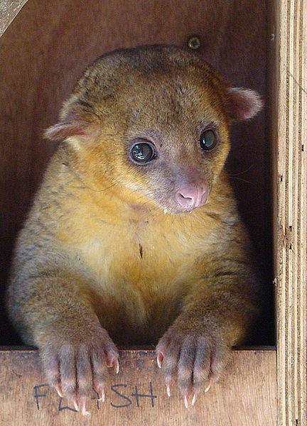
Kinkajou (Potos flavus) is an omnivorous rainforest mammal that lives in the tropical forests of Central and South America. It is considered related to raccoons despite its resemblance to primates. The current medical literature lacks specific guidelines for the medical management of Kinkajou bites and any subsequent soft tissue infections. The goal of this activity is to outline basic guidelines for primary healthcare providers in their approach toward managing patients with kinkajou bites that have either a high risk of soft tissue infection or present with soft tissue infections at the bite site. Animal bites, in general, account for 1% of the total emergency department (ED) visits, which are approximately 2 to 5 million ED patients annually. Ten percent of those patients presenting in the ED requires suturing, and only 1% to 2% need hospitalization for treatment with intravenous (IV) antibiotics or observation. Any animal bite, including kinkajou bites, warrants proper inspection to evaluate their capacity to become infectious. This activity reviews the evaluation and treatment of kinkajou bites and highlights the role of the interprofessional team in managing these injuries.
Objectives:
- Identify the most common locations of the kinkajou.
- Describe the evaluation of an animal bite wound.
- Outline the treatment of a kinkajou bite.
- Review the evaluation and treatment of kinkajou bites and highlight the role of the interprofessional team in managing these injuries.
Introduction
Kinkajou (Potos flavus) is an omnivorous rainforest mammal that lives in the tropical forests of Central and South America. It is considered related to raccoons despite its resemblance to primates. The current medical literature lacks specific guidelines for the medical management of Kinkajou bites and any subsequent soft tissue infections. The goal of this article is to outline basic guidelines for primary healthcare providers in their approach toward managing patients with kinkajou bites that have either a high risk of soft tissue infection or present with soft tissue infections at the bite site.[1]
Animal bites, in general, account for 1% of the total emergency department (ED) visits, which are approximately 2 to 5 million ED patients annually. Ten percent of those patients presenting in the ED requires suturing, and only 1% to 2% need hospitalization for treatment with intravenous (IV) antibiotics or observation. Any animal bite, including kinkajou bites, warrants proper inspection to evaluate their capacity to become infectious.[2]
Etiology
Animal bites account for significant morbidity and mortality worldwide. Most patients presenting to the ED with animal bites usually have been bitten by dogs and cats. Kinkajou bites, although rare, must undergo an evaluation with similar urgency. Medical literature on kinkajou bites is scarce, and the only reported bacterial isolates from kinkajou bites are that of alpha-hemolytic streptococci, mixed anaerobic bacteria, and Kingella potus which forms the bulk of the strains.[3][4]
Reported isolates were from a female zookeeper who had to undergo exploratory surgery of her hand due to suspected infection of her flexor tendons after a kinkajou bite. She was treated successfully with clarithromycin, ciprofloxacin, and metronidazole for 14 days.[1]
The only other case of a kinkajou bite in literature reports zoonotic transmission of Blastomyces dermatitidis to a man from his pet kinkajou after a bite. This reported patient had a recalcitrant infection after being on antibiotics. Physical exam findings suggested chronic ascending lymphadenitis, and he fully recovered after being treated with itraconazole for six months.[5]
Epidemiology
Most animal bites presenting to the emergency department are due to dogs and cats; kinkajou bites are rare. There are no reported incidence rates of kinkajou bites in the United States. Based on case reports in the literature, caretakers of these kinkajous have the most bite wounds.[6]
Pathophysiology
Kinkajou bite wounds both in literature and including the author's one reported case had been small puncture wounds with minimal tissue tear. There is no reported blunt force trauma to the bones. Following the bite injury, there are reports of soft tissue infections treated successfully with antibiotics. The wound discussed in this case was narrow and deep, with the mechanism of the bite described by the patient as biting and holding down.[7]
History and Physical
Clinicians should take a detailed history and physical exam of every patient with a kinkajou bite. These should include information about the circumstances surrounding the bite incident, for example, the bite location and how long it took the patient to seek medical care after the bite. The patient might need local anesthesia during the exploration of the bite site.[7]
Evaluation
A patient's bite site should have a thorough evaluation to assess for underlying neurovascular compromise or foreign body presence. The patient must be provided with appropriate pain management for associated swelling at the bite site. An active and passive range of motion testing should be performed to assess for underlying tendon involvement. The patient further is evaluated for the need for any imaging at the bite site such as X-ray, ultrasound, MRI, or CT. If need be, a patient should be taken to the operating room for irrigation and exploration under local anesthesia.
Treatment / Management
Given the lack of literature and guidelines on how to manage kinkajou bites and subsequent development of cellulitis, the following is a proposed protocol based on the reported case and current animal bite treatment and management guidelines.
- Wound management as per current wound-care guidelines for animal bites including irrigation, exploration, and debridement of the wounds
- Proper imaging (X-ray, ultrasound, CT, or MRI) are necessary imaging modalities for the assessment of deeper structure involvement.
- Assess for any need for surgical exploration at the wound site.
- Based on the type of wound, if the risk of infection is high at the kinkajou bite site patient should be admitted to the hospital.
- Obtain wound and blood cultures before the start of any antibiotics.
- The patient should be started on broad-spectrum antibiotics to cover both gram-positive and gram-negative infections.
- Administer tetanus toxoid, DTaP, or Td as indicated.
- Kinkajou bite patients should get rabies vaccine series, which consists of a regimen of one dose of immune globulin at patient presentation and four doses of the rabies vaccine over 14 days.
Differential Diagnosis
The following should merit consideration in the differential diagnosis:
- Cellulitis
- Abscess
- Sepsis
Prognosis
With prompt evaluation by a qualified health care provider and interprofessional team, prognosis is good.
Complications
If the bite wound is not cleaned and adequately and inspected, complications such as cellulitis or abscess formation may occur.
Consultations
Consider consulting infectious disease organizations for antibiotic management. Surgical consultation may be warranted if there is concern about the involvement of underlying structures or foreign body removal.
Pearls and Other Issues
A kinkajou is considered an exotic pet, and pet ownership seems to be a growing trend in the United States. The reported patient was at a gas station where she tried to be friendly with a truck driver's pet kinkajou when it bit her. The truck driver, with his pet, escaped when the women decided to get help.
Enhancing Healthcare Team Outcomes
Kinkajou bites are very rare in the U.S. and only seen in zookeepers and pet owners. This wild animal from South America has razor-sharp teeth, but its bites are not deep. In general, the majority of kinkajou bites are manageable in the emergency department physician, nurse practitioner, and the primary care provider. Kinkajou bites require the usual tetanus prophylaxis, rabies vaccine, and wound cleaning. In most cases, antibiotics are prescribed to prevent the development of cellulitis or osteomyelitis. The majority of patients have a good outcome after treatment. An interprofessional team approach to treating these injuries should yield favorable results without any long-term sequelae.