Continuing Education Activity
The Seidel test assesses for the presence of aqueous humor leakage from the anterior chamber. This leakage is from a defect in the cornea or sclera from multiple causes, including trauma, post-surgical leak, corneal perforation, and corneal degeneration. The test was first described in 1921 by Dr. Erich Seidel (1882-1948), a German ophthalmologist, for which the test is named. He used the test to evaluate leakage in the postoperative patient but later expanded its use to other causes or anterior chamber leakage. This activity reviews the role of the Seidel test, its indications, and highlights the interprofessional team's role in the evaluation of orbital trauma.
Objectives:
Describe the technique of performing the Seidel test.
Outline the indications for the Seidel test.
Summarize the clinical relevance of the Seidel test.
Review the importance of improving care coordination among interprofessional team members to improve outcomes for patients with orbital trauma.
Introduction
Ophthalmologic visits account for about 3% of emergency department visits annually. About 38 to 52% of these visits are for ocular trauma. These injuries range from simple abrasions to catastrophic globe rupture. More than 1 million people worldwide have vision loss bilaterally secondary to trauma. Further, there is an incidence of 500000 cases of unilateral vision loss secondary to trauma, placing it among the leading causes of vision loss. A thorough evaluation of ocular injuries is critical in identifying injuries in an attempt to preserve vision.[1][2] One test that helps evaluate ocular trauma is the Seidel test. The Seidel test assesses for the presence of aqueous humor leakage from the anterior chamber. This leakage is from a defect in the cornea or sclera from multiple causes, including trauma, post-surgical leak, corneal perforation, and corneal degeneration. The test was first described in 1921 by Dr. Erich Seidel (1882-1948), a German ophthalmologist, for which the test is named. He used the test to evaluate leakage in the postoperative patient but later expanded its use to other causes or anterior chamber leakage.[3]
Anatomy and Physiology
The eye is an incredibly complex organ; multiple components and intricate mechanisms must collaborate for the eye to function correctly. The Seidel test assesses for disruption of the cornea, sclera, or a combination of both. The sclera is a fibrous, opaque, “white of the eye,” the structure that provides support and protection to the deep structures of the eye. Anteriorly at the limbus, the sclera is continuous with the cornea.[4] The cornea, the clear outermost part of the eye, sits anterior to the pupil, iris, and lens. Light enters the eye through this construct and accounts for a large portion of the focusing power of the eye. The cornea is composed of five layers that include from superficial to deep; the corneal epithelium, Bowman’s layer, corneal stroma, Descemet’s membrane, and corneal endothelium.
All five layers combined are approximately 550 microns or just over half a millimeter thick. The epithelium is about 5 to 7 cells thick that provides the eye with a smooth surface for the tears to form a film. This film spreads across and keeps the eye moist, healthy, and allows for clear vision. The epithelium has a high turnover rate and is replaced entirely over about 7 days. The Bowman layer is the next layer; it is a dense fibrous sheet that protects the deeper layers. Once a scratch passes Bowman’s layer, the probability of scaring increases significantly. The following layer is the stromal layer that is about 90% of the cornea and is composed of a connective tissue called collagen fibrils. They are uniform in size and are stacked parallel to one another in bundles called lamellae. Their arrangement makes it so they are a transparent layer. The next layer is Descemet’s membrane, which is another extremely thin layer that separated the stroma from the endothelial layer. The final layer is the endothelium that is also one cell layer think and is in direct communication with the aqueous humor of the anterior chamber. The anterior chamber is located behind the cornea and in front of the Iris and pupil. Behind the iris and pupil lies the posterior chamber, which includes multiple structures out of the scope of this discussion.[5]
Indications
The Seidel test is indicated anytime one suspects orbital trauma with concern for an ocular leak. Conditions that should raise suspicion for potential trauma and ocular leak including but are not limited to:
- Pupillary defect
- Laceration through eyelid
- Shallow anterior chamber
- Blood in the anterior chamber
- Bullous subconjunctival hemorrhage
- Post-surgical with concern for ocular leak
- Evaluation of corneal laceration to evaluate if it sealed or not
- Corneal perforation secondary to degeneration
Contraindications
Contraindications to the Seidel test include several conditions, such as:
- Obvious globe rupture
- Full-thickness eye laceration
- Obvious corneal perforation
- Hypersensitivity to fluorescein dye
Equipment
The Seidel test does not require significant resources, but specific components are required to obtain an accurate analysis include:
- Fluorescein strip
- Topical ophthalmic anesthetic
- Slit-lamp with cobalt blue light
Personnel
The Seidel test can be performed by any medical provider that can instill the dye and interpret the results. Usually performed by physicians and physician extenders, and does not require additional support personnel.
Preparation
Prepare the room for evaluation and obtain all necessary equipment and medications. The cornea is very sensitive, and any lesion to it can cause severe photophobia limiting the exam. Dim the lights in the room as much as possible to ensure patient comfort and improving the evaluation.
Technique or Treatment
The following steps are generally required to complete the Seidel test[6]:
- Prepare the room and obtain all equipment.
- Prepare the slit lamp.
- Explain the procedure to the patient.
- Apply topical anesthetic.
- Moisten fluorescein dye strip with normal saline.
- Apply fluorescein above lesion or the superior conjunctival fornix.
- Ask the patient to blink to help spread the stain.
- Visualize the injured site under cobalt blue light.
Interpretation
Fluorescein, when concentrated, is an orange to red color. When it becomes diluted, it turns green under cobalt blue light. When instilled into the eye, the dye is taken up by defects in the cornea, such as abrasions or lacerations. Seidel test is positive when the fluorescein dilutes in the aqueous humor and causes it to fluoresce bright green and stream down the eye with gravity. The streaming is sometimes described as a waterfall by some with more brisk leaks. There may be just a focal area or dilution if the leak is not brisk. The fluorescent green color will be located above the lesion and along the sides of the aqueous that has leaked. The center of the waterfall will not have fluorescein present, as it will be just aqueous humor. A positive test indicates a full-thickness corneal or scleral injury.[3]
Possible false negatives:
- A small defect that has self-sealed
- A large laceration that has plugged
- Retrobulbar rupture
If there is a strong suspicion for a globe rupture and the Seidel test is negative, the next set in evaluation is to obtain an orbital CT scan, which can evaluate for a flat anterior chamber and may demonstrate an intraocular foreign body.[7]
Complications
The patient must remove contact lenses before staining the eye as the fluorescein will permanently stain them. The eye should be flushed with saline, and contacts should be left out about 1 hour after staining if no injury is identified.
Staining of the skin around the eye will fade over a few hours.
Another possible complication occurs as a missed ruptured globe due to the laceration or perforation being already sealed or in a location unable to be tested by Seidel test (posterior globe rupture).[7]
Clinical Significance
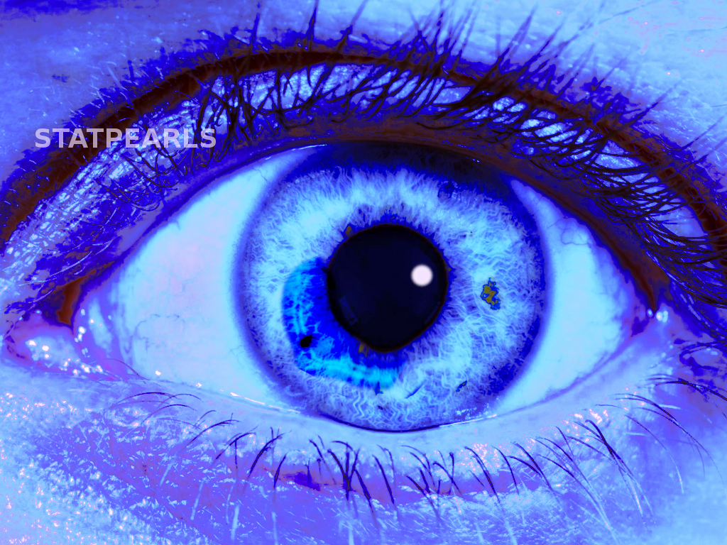
A positive test indicates leakage of aqueous humor for the anterior chamber, which is an ocular emergency (see Image. Positive Seidel Test).
- Management of positive Seidel test
- Consult ophthalmology immediately for surgical repair
- Prevent further Injury
- Do not manipulate the eye
- Do not check intraocular pressure or perform an ocular ultrasound
- Cover the eye with a metal shield (Fox Shield) or a cover that does not touch or apply pressure to the globe
- Do not place a patch over the eye
- Minimize elevation of intraocular pressure
- Bed rest; no Valsalva maneuvers, bending, or lifting
- Consider anti-emetic and Foley catheter
- Pain control
- Tetanus prophylaxis
- IV antibiotics to prevent
- No intra-ocular foreign body
- 1line: Fluoroquinolone, OR
- 2line: Vancomycin and ceftazidime
- Intra-ocular foreign body present
- Ceftazidime and vancomycin
- Penicillin allergy: Cipro and vancomycin
- No intravitreal antibiotics[8]
Enhancing Healthcare Team Outcomes
Ocular injuries with a positive Seidel test require multiple healthcare workers and specialties in an interprofessional team approach. Most ocular traumas present to the emergency department, where they will likely first come into contact with nursing staff that will initially evaluate the patient. They may notice the injury and begin to protect the eye by covering it. They may also obtain medications and equipment needed for further patient evaluation. Usually, during triage, they also obtain a visual acuity that is one of the best prognostic indicators once there has been a definitive repair to the defect.[9] [Level 4]
Once triage is complete, the patient will be evaluated and treated by a provider and possibly multiple providers, depending on if they have any other injuries. The entire staff coordinates care to assure the patients get a fast, accurate exam. Once found to have a positive Seidel test, an ophthalmologist will be contacted immediately for definitive repair and continue to follow the patient on an outpatient basis once repaired. A pharmacist will also be involved in care not only during the acute setting but also on an outpatient. Ocular injuries are real emergencies, and it takes a team to ensure the patient receives the best care possible. [Level 5]