Continuing Education Activity
Kohler disease is a disease only seen in pediatric patients. Although the etiology is not fully understood, it is thought to be caused by the compression of the navicular bone prior to ossification. This leads to blood flow abnormalities resulting in avascular necrosis. Kohler disease is most commonly seen in males ages 4 to 7 years old. Kohler disease is typically unilateral, although one report in the literature found that 25% of Kohler disease is bilateral. Patients typically present with medial-sided foot pain, swelling of the medial foot, and/or a limp. On plain films, the navicular will have standard characteristics of avascular necrosis, including sclerosis, fragmentation, and flattening. Kohler disease is a self-limiting condition with an excellent prognosis. There have been no reported cases of Kohler disease developing long-term clinical or radiologic abnormalities. This activity reviews the evaluation and treatment of Kohler disease and the role of the interprofessional team in managing patients with this condition.
Objectives:
- Describe the most likely cause of Kohler disease.
- Describe the consequences of Kohler disease.
- Outline the radiographic findings expected in Kohler disease.
- Summarize the evaluation and treatment of Kohler disease and the role of the interprofessional team in managing patients with this condition.
Introduction
Kohler disease was first described by Alban Kohler in 1908 and referred to avascular necrosis of the navicular bone of the foot. Kohler disease is a disease only seen in pediatric patients. Although the etiology is not fully understood, it is thought to be caused by the compression of the navicular bone prior to ossification. This leads to blood flow abnormalities resulting in avascular necrosis. Kohler disease is most commonly seen in males ages 4 to 7 years old. Kohler disease is typically unilateral, although one report in the literature found that 25% of Kohler disease is bilateral. Patients typically present with medial-sided foot pain, swelling of the medial foot, and/or a limp. On plain films, the navicular will have standard characteristics of avascular necrosis (AVN), including sclerosis, fragmentation, and flattening. Kohler disease is a self-limiting condition with an excellent prognosis. There have been no reported cases of Kohler disease developing long-term clinical or radiologic abnormalities.[1][2][3]
Etiology
The navicular bone possesses a dual blood supply. A branch of dorsalis pedis artery supplies the dorsal aspect of the bone, while the plantar blood supply arises from the medial plantar branch of the posterior tibial artery. Both the dorsal and plantar blood supplies enter the navicular and branch to supply the medial and lateral thirds of the bone. This creates an avascular zone in the central one-third of the bone. Vascular foramina help to supply this avascular area and are found on the dorsal, plantar, medial, and lateral surfaces of the navicular bone. However, in a study of 100 cadaveric navicular bones, 97% of vascular foramina were smaller than 1 millimeter in adults. In theory, any compression of these small vascular foramina could result in decreased blood flow and put the navicular bone at risk for avascular necrosis.
Kohler disease is thought to be due to an abnormal strain on the navicular. In children, the navicular bone is the last of the tarsal bones to ossify. In girls, it ossifies between 18 to 24 months and in boys 30 to 36 months old. This theory points out that given the navicular bone’s slow ossification, it is weaker than the other tarsal bones. As the child grows and becomes heavier, the navicular can be compressed between the already ossified talus and cuneiform bones. The compression of the non-ossified navicular results in the squeezing of the perforating vessels in the central spongy bone, which could lead to ischemia and later avascular necrosis.[4][5][6][7]
Epidemiology
The incidence of Kohler disease is not well known, considering that not all patients with Kohler disease are symptomatic. One publication estimated Kohler disease to be present in 2% of all children. Kohler disease is five times more likely to affect males than females, and it is most commonly seen in children 4 to 7 years old.
Histopathology
A diagnosis of Kohler disease does not require a bone biopsy, and a biopsy is not recommended for the diagnosis unless there is a need to rule out infection or malignancy. Kohler disease looks similar to other forms of avascular necrosis histologically. The primary histologic feature of avascular necrosis is dead trabeculae with empty lacunae that will stain deeper than healthy bone. The lacunae will be enlarged and with cystic changes. Bone marrow is noted to be a more sensitive indicator for AVN than the bone itself and will show fat necrosis and calcium deposits.
History and Physical
Kohler disease can be asymptomatic. However, patients often present to their pediatrician with concerns for dorsomedial midfoot pain. On physical exam, the patient may have point tenderness over the navicular with or without redness, warmth, and swelling. When asked to walk, the patient may show an antalgic limp in which they walk on the lateral side of their foot.
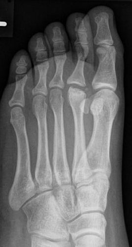
Evaluation
Plain radiographic images are the imaging modality of choice in the diagnosis of Kohler disease. The navicular will appear wafer-thin with bony collapse. The bone will appear fragmented with a loss of a trabecular pattern. There will be patchy sclerosis of the bone and increased radiodensity. Soft tissue swelling around the affected navicular bone can also be seen on plain radiographs. Advanced imaging such as CT and MRI are not required for diagnosis, although they may become necessary if the patient's symptoms do not improve with treatment. While navicular sclerosis may be consistent with a normal variant in asymptomatic patients, it is important to correlate radiographic findings with clinical suspicions.
Basic labs, such as a complete blood count (CBC), C-reactive protein (CRP), and erythrocyte sedimentation rate (ESR) are needed in cases in which an infection is suspected. If any of these are elevated, further diagnostic testing is warranted.
Treatment / Management
If Kohler disease is suspected, patients should be referred to a pediatric orthopedic surgeon for further evaluation.[8]
The treatment of Kohler disease is conservative. NSAIDs can be used to decrease symptoms but have not been shown to shorten the duration of disease symptoms. Immobilization via a short leg walking cast for 4 to 6 weeks can be used in patients to shorten the duration of symptoms. The effect of weight-bearing casts compared to non-weight bearing casts is unclear and often surgeon dependent. Some reports of using offloading orthotics for symptomatic relief has been reported; however, it does not appear orthotics shorten the duration of symptoms.
There is no indication for surgery in Kohler disease. If symptoms do not improve, physicians should consider an alternative diagnosis. Both symptoms and radiographs should start to show improvement in around six months.
Differential Diagnosis
Kohler disease is often misdiagnosed as osteomyelitis in children. However, basic labs (WBC, CRP, ESR) will help to differentiate between the two diagnoses. If a child has an elevated ESR or CRP, there should be a high index of suspicion for infection. Kohler disease will not have elevated inflammatory markers, and a pediatric patient should not have elevated inflammatory markers. If an infection is suspected, bone aspiration, bone biopsy, or blood cultures may be warranted.
Prognosis
Kohler disease has an excellent prognosis, and to date, there have been no reports of long-term symptoms or disability in children with Kohler disease. Radiographs will improve around 6 to 48 months from the onset of symptoms. Without casting, symptoms typically resolve in 6 to 9 months. In a review of case reports, patients treated in plaster casts (non-weight bearing) were pain-free at an average of 3 months. Arch support orthopedics were shown to decreased local pain but found that symptoms lasted an average of 7 months.
Consultations
If a clinician suspects Kohler disease, a consultation should always be made with a pediatric orthopedic surgeon.
Enhancing Healthcare Team Outcomes
The management of Kohler disease is best by an interprofessional team. While the primary care provider may initially see the disorder, the definitive management is by an orthopedic surgeon. The team may include a radiologist, physiatrist, and an orthopedic nurse. In most cases, the disorder is treated conservatively with close follow up. The outlook for patients with Kohler disease is excellent with no long term radiological or clinical manifestations reported in the literature. [Level 5]