Continuing Education Activity
The cottonmouth or water moccasin, Agkistrodon piscivorus, is a semi-aquatic pit viper found throughout the southeastern United States and into east Texas. Cottonmouth snakes are part of the Crotalinae family of pit vipers which includes rattlesnakes and copperheads. Like the other North American pit viper species, identifying features include elliptical pupils, triangular-shaped heads, heat-sensing pits, and either a rattle or single row of ventral scales distal to the anal plate. Pit viper venom is used to facilitate the capture and digestion of prey and can cause significant toxicity in humans. Water moccasins typically feed on fish, turtles, and small mammals but bite humans when provoked or disturbed. There is very little data on the specific evaluation and treatment of cottonmouth envenomation. This activity, therefore, will discuss cottonmouth envenomation in the context of other pit piper envenomation, as well as how the interprofessional health team can manage such incidents.
Objectives:
Identify the most common symptoms seen after a cottonmouth bite/envenomation.
Assess the pathophysiology of the venom from cottonmouth snakes and its effects on local tissue.
Identify the physical characteristics of pit vipers that distinguish them from nonvenomous snakes.
Collaborate with an interprofessional team to address cottonmouth snake envenomation.
Introduction
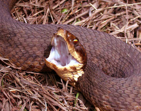
The water moccasin or cottonmouth, Agkistrodon piscivorus, is a semi-aquatic pit viper found throughout the Southeastern United States and into West Texas (see Image. Cottonmouth Snake).[1] Their 2 common names derive from the white-colored membranes in its mouth and living near water sources. Cottonmouth snakes are part of the Crotalinae family of pit vipers which includes rattlesnakes and copperheads. Like the other North American pit viper species, identifying features include elliptical pupils, triangular-shaped heads, heat-sensing pits, and either a rattle or single row of ventral scales distal to the anal plate.[2][3] Pit viper venom is used to facilitate the capture and digestion of prey and can cause significant toxicity in humans. Water moccasins typically feed on fish, turtles, and small mammals but bite humans when provoked or disturbed. There is not much data specific to the evaluation and treatment of cottonmouth envenomation. This activity, therefore, will discuss cottonmouth envenomation in the context of other pit piper envenomation.
Etiology
Pit vipers are generally not aggressive and typically strike in a defensive manner or when provoked. Pit viper envenomation is almost exclusively unintentional. Causative factors resulting in envenomation are either while handling snakes or during outdoor activities, resulting in unintentional exposure to the snake. In 1 registry of north American snakebites, about half of all bites were on the lower extremity. Of these, 27% of patients were not wearing shoes.[4] There is an increased risk of severe envenomation for patients arriving at medical care greater than 6 hours after the bite, patients younger than 12, and envenomation by larger snakes. Bites from cottonmouth and copperhead snakes have a decreased likelihood of severe envenomation.[5][6]
Epidemiology
In the United States, most venomous snake bites occur from snakes in the Crotalinae family. Data specific to cottonmouth envenomation is limited. The North American Snakebite Registry (NASBR) and Toxicology Investigator's Consortium (ToxIC) between January 2013 and December 2017 reported 31 cottonmouth envenomations. Most of these bites occurred on the lower extremities, and swelling was the most common symptom reported. Nineteen percent of patients developed gastrointestinal symptoms and 19% developed coagulopathy. Eighty-four percent of envenomations received antivenom, and 61% of patients required admission for greater than 24 hours.[4] Data from the National Poison Data System 2017 revealed 4071 pit viper envenomations. Of these, there were 2035 copperheads, 753 rattlesnakes, 255 cottonmouths, and 1,028 unknown crotalid bites.[7] There were 2 deaths, 1 from a rattlesnake and 1 from an unknown crotalid envenomation. Of the cottonmouth envenomations, 242 were seen at healthcare facilities, 122 were reported to have moderate outcomes, 10 had significant outcomes, and no deaths.[7]
Pathophysiology
The venom of cottonmouth snakes contains enzymes that cause local tissue necrosis and potentially coagulopathy. The local tissue swelling and ecchymosis enzymes include metalloproteinases, hyaluronidase, and phospholipases A2. The venom of the A. piscivorous is cytotoxic and causes local tissue destruction. Like the other pit vipers, their venom contains phospholipase A2 (PLA2), an enzyme that hydrolyzes phospholipids. Phospholipids are the primary component in the membranes that surround cells, and when broken down by PLA2, they cause tissue damage.[3]
Metalloproteinases, among other venom constituents, contribute to hematologic and soft-tissue toxicity through various actions. Hematologic effects include activating the coagulation cascade, having fibrinolytic and prothrombin activation, and inhibiting platelet aggregation.[8] Metalloproteinases, including fibrolase, a fibrinolytic enzyme, have been found in Agkistrodon species.[9] Soft tissue toxicity from the proteases can cause myonecrosis, skin injury, and robust inflammatory response.[10] The breakdown of capillaries by these enzymes results in reduced blood flow, which further contributes to tissue breakdown, worsening the edema and ecchymosis around the bite.[5] The indirect effects of reduced blood flow from damaged blood vessels can cause local ischemia.[7]
Toxicokinetics
Specific data on cottonmouth venom toxicokinetics in humans is extremely limited. Pit vipers have curved hollow fangs that inject venom subcutaneously. In rats who had an intramuscular injection of Agkistrodon halys ussuriensis, the absorption half-life was 2.5 hours, distribution half-life of 4.8 hours, and elimination half-life of 125 hours with an apparent volume of distribution of 19 L/kg.[11]
History and Physical
After initial assessment for acute life-threatening conditions, a thorough history and physical examination are necessary. The clinician should determine when the bite occurred, obtain a description of the snake if possible, and evaluate the envenomation site. Common physical exam findings from a pit viper envenomation include 1 or more fang marks, pain, erythema, ecchymosis, and progressive edema. Nausea, vomiting, coagulopathy, and systemic signs of envenomation can also occur. Patients will also occasionally bring in the snake, which can help to identify venomous versus non-venomous species. In general, it is not advised to catch the snake to bring it to the hospital as this may risk further bites. Even dead snakes can still cause envenomation if not handled carefully.[12][13]
Evaluation
Prehospital considerations include immobilizing the extremity and transportation to a healthcare facility. There is no role for constrictive tourniquets in pit viper envenomation. Additionally, there is no role for any venom extraction device.[14] After arrival at the emergency department, airway, breathing, and circulation should undergo assessment. Acute, life-threatening signs or symptoms would be extremely uncommon but can occur from either an anaphylactic or anaphylactoid reaction to venom or if the envenomation injection was into a vein.[15][16] Unstable patients should undergo customary resuscitation to secure the airway, ensure adequate oxygenation and ventilation, and support hemodynamics.
Hematologic laboratory abnormalities are common after pit viper envenomation, especially with rattlesnakes. Significant abnormalities are less frequent and less severe with cottonmouth or copperhead envenomations.[17] Hematologic abnormalities of moderate to severe pit viper envenomation may include thrombocytopenia, elevated prothrombin and activated partial thromboplastin time, depressed fibrinogen concentration, and elevated fibrin degradation products and d-dimer. This constellation of laboratory abnormalities can mimic disseminated intravascular coagulopathy (DIC). Pit viper envenomation, however, unlike DIC, rarely causes clinically significant bleeding or thrombosis.[18] There is very little data evaluating thromboelastography in the setting of pit viper envenomation, and it requires additional study.[19] Hematologic abnormalities can reoccur several days after treatment. Specific data on cottonmouths is limited. However, reports exist of recurrence of coagulopathy up to two weeks post-envenomation despite initial treatment with antivenom.[20]
Most patients with a confirmed or presumed envenomation require observation for 8 hours. Suppose there are no physical or laboratory signs of envenomation during this observation period. In that case, the envenomation is presumed to be a "dry bite," meaning that a bite occurred with little or no venom injected. In these cases, patients can be discharged from the emergency department with customary return precautions. If there are signs of progressive soft tissue injury during the observation, then hospital observation would be reasonable. All patients receiving antivenom require admission. Reasonable laboratory studies for a cottonmouth envenomation can include a complete blood count, basic metabolic panel, fibrinogen, and prothrombin time.[8] The clinician should perform a serial examination of the envenomated extremity or site. There is no consensus on the exact evaluation method, but a reasonable approach is the neurovascular status of the limb, the overall degree of limb edema, and determining the proximal line of envenomation; this is the proximal edge where the patient reports pain. Measuring limb circumferences at the bite site and several proximal sites to compare to the contralateral side has some appeal due to its simplicity and objectivity. However, there is no clear indication of how much circumference asymmetry would prompt antivenom. Furthermore, there may be inaccuracies with repeated measures, especially from different operators.[21]
Compartment syndrome is a theoretical concern given many similarities between envenomation and compartment syndrome. Most pit viper envenomations occur in the subcutaneous space, and muscle involvement is very uncommon. If compartment syndrome does occur, it could be in the setting of a severe rattlesnake envenomation with myonecrosis.[15]
Treatment / Management
After a snakebite, patients should immobilize the limb and seek medical attention. Many folklore treatments for venomous snakebites include venom extraction, electric current, tourniquets, and applying ice packs. These therapies, including mechanical venom extraction devices, are not useful.[14] Local wound care and tetanus should be updated if needed. Elevation of the limb may be useful to reduce distal edema and pain. Patients presenting after a cottonmouth bite should undergo observation for 8 hours postenvenomation. If there are no physical or hematologic signs within 8 hours, the patient can be discharged home.[22] If the patient starts showing signs and symptoms of envenomation, they should be admitted for observation. Patients who exhibit progressive proximal edema, hematologic toxicity, or systemic effects should receive antivenom.[22][15] There are two commercially available antivenom products available in the United States. Crotalidae polyvalent immune Fab (Fab) is processed from sheep immunoglobulins inoculated from 4 North American pit vipers: eastern and western diamondback (C. adamanteus and atrox), Mojave rattlesnake (C. scutulatus), and cottonmouth (Agkistrodon piscivorus).[15] Crotalidae Immune F(ab’)2 is a newer antivenom processed from horse immunoglobulin inoculated from the South American rattlesnake (C. durissus) and fer-de-lance (Bothrops asper) snakes.[15] It is worth noting that F(ab)2 antivenom has not been derived from an Agkistrodon species, and its efficacy for cottonmouth envenomation is unknown.
The initial dose of Crotalidae polyvalent immune Fab antivenom is 4 to 6 vials given intravenously. Antivenom will not reverse the soft tissue effects of pit viper envenomation but should halt progression. Control of the progression of soft tissue injury or hematologic toxicity is the goal of antivenom therapy. If initial control is not achieved, another loading dose of 4 to 6 vials should be given. More than 6 vials can be initially administered if there are signs of shock or severe bleeding.[22] Once progression control has occurred, maintenance dosing includes giving 2 vials every 6 hours for 3 doses (6 additional vials).[9]
The dose of Crotalidae Immune F(ab’)2 is 10 vials intravenously given once; this can be repeated in 1 hour if failing to achieve initial control. Since F(ab’)2 is a larger fragment with slower elimination kinetics, there is no maintenance dosing. Treatment of venom-induced hematologic toxicity is an additional antivenom. If clinically significant bleeding is occurring, treatment with blood products in addition to antivenom may be necessary.[15]
Despite being rare, if compartment syndrome develops, it should be treated with additional doses of antivenom. There is a very limited role for fasciotomy for North American pit viper envenomation, and it is a consideration for persistent concerns despite adequate antivenom, or for decompression of digits.[23][15]
Differential Diagnosis
Often, patients see the snake, which leaves very few differential diagnoses to consider except for the identification of the snake species. Patients occasionally do not see the actual snake when bitten, so there may be questions about whether the wound is from a snake or due to another process. Two puncture wounds next to one another are suggestive of a pit viper envenomation. However, not all patients have 2 puncture wounds. Punctures from other sharp objects or thorns could be considerations. Punctures from non-snake sources could result in cellulitis that may potentially mimic a pit viper envenomation; however, the temporal course and appearance are different. As such, the differential diagnosis can include the following:
- Arthropod envenomation
- Cellulitis
- Vascular trauma
- Wasp sting
- Deep vein thrombosis
Prognosis
Specific data on prognosis from cottonmouth envenomation are very limited. These envenomations are considered to be less serious on average than rattlesnake bites. Cottonmouth envenomation has a lower likelihood of being a severe systemic envenomation.[5] In 2017, there were 255 cottonmouth envenomations reported to the US poison center, but only ten serious outcomes and no deaths.[7] In a retrospective study of snakebites, cottonmouth envenomation, regardless of whether they received antivenom, did not require surgical intervention and only needed pain control and wound care.[10] Patients bitten by a cottonmouth snake will require observation, local wound care, the elevation of the affected limb, and potentially antivenom. Patients who have any signs of coagulopathy or worsening pain, edema, or ecchymosis should receive antivenom and require admission. Prospective data from copperhead bites, a pit viper in the same family as cottonmouths, reported resolution of envenomation typically between 7 to 13 days.[11] It is essential to inform patients that they may experience symptoms for up to 4 weeks before fully resolving limb dysfunction.
Complications
Serious complications from cottonmouth envenomation are uncommon. Data are limited for cottonmouth snakes. The copperhead (Agkistrodon contortrix) is another North American pit viper of the same species with some clinical data available. Hematologic abnormalities occur in about 14% of copperhead envenomations; however, clinical bleeding is rare.[17] True coagulopathy and serious bleeding are rare, but reports do exist.[24] Less serious complications from cottonmouth envenomation include limb dysfunction, pain, and edema for up to 30 days post-envenomation.[25] Antivenom can cause immediate hypersensitivity reactions and require prompt treatment. A meta-analysis from 1997 to 2010 reported that when Crotalidae polyvalent immune Fab antivenom was used, immediate hypersensitivity was 8%, and serum sickness was 13%.[12]
Consultations
- Medical toxicology consultation can be useful but may not be accessible at all hospitals
- Any US regional poison control center is available by calling (800) 222-1222. Medical toxicologists are available to discuss treatment 24 hours per day.
Deterrence and Patient Education
The following are summary recommendations in the management of pit viper envenomation:
- Patients should avoid mechanical venom extraction
- Do not apply heat or ice to the wound
- Avoid tight and constrictive bandages
- Do not use a tourniquet
- Elevate the affected limb to help with pain and edema
- Patients should be evaluated in an emergency department and observed for 8 hours
Pearls and Other Issues
Key facts to keep in mind about water moccasin snake toxicity are as follows:
- Cottonmouth snakes are venomous pit vipers that range throughout the Southeast United States.
- Their venom contains enzymes that cause local destruction of tissue through the metabolism of cellular membranes and cause an inflammatory response.
- Systemic effects and coagulopathy from cottonmouth envenomation are uncommon. The most common symptoms are pain, ecchymosis, and edema.
- Crotalidae polyvalent immune Fab antivenom should be administered for patients exhibiting progressive signs of proximal edema, significant hematologic abnormalities, or severe systemic symptoms.
- Patients with early signs of envenomation require admission for observation. Those who do not have symptoms after 8 hours can be discharged.
Enhancing Healthcare Team Outcomes
When a patient presents with a cottonmouth envenomation, it requires the prompt evaluation and management of an interprofessional healthcare team. This team includes nursing, an emergency provider, a toxicologist, and the regional poison center. The collaborative effort from these teams can properly triage patients and appropriately manage patients with snake bite envenomation. Patients in the emergency department should have the following:
- Vital signs are recorded and monitored by the nurse with reporting of changes to the clinician
- Assess the wound and provide local wound care
- Record the patient's last tetanus shot and update if needed
- Elevate the affected limb to help with pain and swelling
- Avoid tourniquets, ice packs, or manipulating the wound
- Observe all patients with suspected envenomation for at least 8 hours
- Administer antivenom if indicated
While most patients improve with local wound care, the patient requires monitoring for the development of compartment syndrome; this is where nursing can fill a vital role, informing the physician or contacting toxicology if they have any concerns. If there is any concern about blood flow compromise, the surgeon should be asked to perform a fasciotomy. If antivenom is necessary, it will be obtained from the pharmacy, and the pharmacists should be sure to familiarize themselves with the interaction and adverse event profile since it is an infrequently encountered agent; this will enable them to be a better asset to the care team. Today, with prompt treatment, the outcomes following cottonmouth envenomation are good, but only if the interprofessional healthcare team, including physicians, toxicologists, pharmacists, and nurses, work collaboratively to ensure patients receive the care they need in a timely fashion.