Continuing Education Activity
Harlequin ichthyosis (HI) is an extremely rare, autosomal recessive congenital disorder of the epidermal skin layer. Neonates with HI have a high mortality rate due to fulminant sepsis and acute respiratory failure in the newborn period. This activity reviews the evaluation and treatment of harlequin ichthyosis and highlights the role of the interprofessional team in the care of patients with this condition.
Objectives:
- Describe the etiology of harlequin ichthyosis.
- Outline the typical findings of harlequin ichthyosis during physical exam and evaluation.
- Review the approach in the management of a newborn with harlequin ichthyosis.
- Explain the importance of improving care coordination amongst the interprofessional team to enhance the delivery of care for patients with harlequin ichthyosis.
Introduction
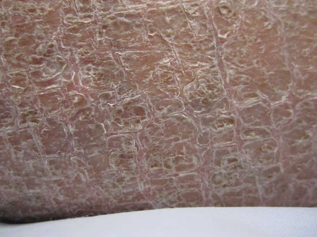
Harlequin ichthyosis (HI), also known as 'ichthyosis fetalis', is an extremely rare autosomal recessive congenital ichthyosis (ARCI) affecting the ABCA12 gene. It is the most severe subtype of ichthyosis. Classified as an autosomal-recessive disease, mutations of the ABCA12 gene in HI causes thickening of the keratin layer in the skin (hyperkeratosis) in the stratum corneum. Moreover, HI is characterized by thickened, dry "armor-like" plaques that give an appearance of 'scales' that are separated by deep fissures covering the entire surface of the body. Infants affected by this disease are at higher risk of infection due to compromise of the skin's protective barrier.[1][2]
Etiology
ABCA12 functions as a keratinocyte transmembrane lipid transporter protein. It is a component of the adenosine triphosphate (ATP)-binding cassette transporter that bind and hydrolyze ATP to mediate transport of lipid molecules across extracellular membranes. ABCA12 functions to transport across extracellular membranes via lamellar granules forming the extracellular lipid layers of the epidermis, specifically the stratum corneum. Mutations in the ABCA12 transport system cause defective transportation of lipids to the stratum corneum. Consequently, malformation of the lipid barrier leads to over-accumulation of lipids in the epidermal keratinocytes known as hyperkeratosis. Intracellular accumulation of lipids in the epidermal keratinocytes gives rise to the characteristic phenotypes that are seen in HI.[3][4]
Epidemiology
The incidence of harlequin ichthyosis has been reported to be 1 in 300,000 births. No evidence to support susceptibility in either males or females. No evidence of frequency seen in the disease has been identified in racial groups or sex distribution.[5]
Histopathology
Under light microscopy, a thickened stratum corneum will be seen from samples of any skin region. The remaining layers (granular, spinous, basal), as well as the dermis, are not affected. Characteristic features of HI seen in the epithelial keratinocytes include absent lamellar granules, multivesicular bodies, and autolysosomes. Between the first cornified cell layer and the granular layer, extracellular lamellar structures are absent. Also, abnormal lipid droplets and vacuoles, which contain dense granules or laminated structures are found in immature keratinized cells.[6]
History and Physical
Neonates affected by HI are typically born prematurely and encased in markedly thickened, hardened stratum corneum. This casing cracks and results in yellow plaque-like “armor” plates separated by deep longitudinal and transverse erythematous fissures encompassing the entirety of the body. The hyperkeratotic plates restrict movement of the chest wall, upper and lower limbs. Enlarged scales on the chest wall restrict expansion leading to respiratory distress, hypoventilation, and respiratory failure. Moreover, pseudo-contractures of the limbs from the tension of hyperkeratotic plates restrict movement and impair perfusion, leading to digital pallor and ischemic necrosis to distal extremities.
Edematous hands and feet are often enveloped in a mitten-like casing from hardened scales. Furthermore, severe bilateral ectropion (out turning of the eyelids) involving upper and lower eyelids exposes the conjunctiva leaving the cornea at risk for abrasions and desiccation. The nose may appear flattened, and nares may be obstructed by overlying skin. Also, flattened ears are observed with external auditory canal obstructed by the skin. Severe traction on the lips causes eclabium (eversion of the lips) with an appearance of a wide gaping mouth. Additionally, the loss of skin barrier leads to impairment of thermoregulation, dysfunction of sweat glands, hyperthermia, and dehydration from excess water loss. Generalized poor hair growth and nail deformities are also commonly seen.[5][7][8][9]
Evaluation
The diagnosis of HI is usually made by clinical examination of the newborn because of the severe characteristic clinical features at birth. Due to the severity of the disease, fatal outcomes in the prenatal period are frequent. In families with a known history of HI, prenatal genetic testing via fetal DNA analysis may be offered. Amniocentesis or chorionic villus sampling for fetal DNA analysis is less invasive and replaced the old and more invasive techniques of fetal skin biopsy. Another modality of testing for HI in patients with a known family history of HI is with the use of ultrasound. Prenatal 3-D ultrasonography show features suggestive of HI, including ectropion with bulging eyes, eclabium with a large immobile mouth, flattened nose, rudimentary ears, flexion contractures with abnormal limbs.
Other common features on ultrasound also include echogenic dense floating particles in amniotic fluid described as ("snowflake sign"), polyhydramnios, and growth restrictions. Infants face complications secondary to the compromise of the skin barrier. Monitoring for systemic infections is warranted and includes comprehensive blood work that should be obtained routinely, including white blood cell count and blood cultures. Wound cultures can be obtained if a purulent discharge is observed in the fissures, and infection is suspected. Moreover, close observation to minimize the risk of dehydration includes measurements in urinary output, weight, and electrolyte that may be abnormal secondary to impaired barrier function.[5][8][9][10]
Treatment / Management
Newborns require admission to a Level III neonatal intensive care unit and an interprofessional team approach to tackle the obstacles faced with caring for neonates affected by HI. Along with the ICU clinicians and nurses, the team should include specialists in a variety of fields such as dermatology, neonatology, plastic surgery, genetics, ophthalmology, otolaryngology, physical therapy, occupational therapy, nutritionist, and social work. ICU management is mainly supportive. Neonates should be placed in an isolated room in an incubator with added humidity to counter thermoregulatory issues. Neonates are at high risk of bacterial infection and sepsis. Patients require close monitoring of vitals, blood cultures if hemodynamically unstable or lethargy noted, and routine serum electrolytes with depletion.
Loss of skin barrier leads to transepidermal water loss and electrolyte imbalance. To avoid dehydration, clinicians need to monitor daily weight changes, fluid intake, and urinary output. Deep fissures may pose a risk for infection if purulent drainage is identified, then acquire would cultures. Topical antibiotics can be applied to fissures. Extensive fissuring can cause pain and discomfort. Adequate pain control should be provided with acetaminophen, NSAIDs, or narcotics. Furthermore, neonates with HI are at risk for complications related to impaired flexibility such as restrictions to the anterior chest-wall expansion that may lead to restriction of lung movement, increased risk of pneumonia, difficulty feeding, and risk for respiratory failure. Neonates that display signs of acute respiratory distress require intubation to secure a patent airway.
Other impairments in flexibility include contractures in distal extremities in the hands and feet. These require plaque removal and a liberal application of emollients to avoid ischemic digit necrosis and compartment syndrome from epithelial constrictions. Moreover, ectropion increases the risk of exposure keratitis and will require ophthalmology consult. Adequate lubrication of the eyes with hourly artificial tears or other ocular lubricants are necessary.
Eclabium caused by tension from the plaques may lead to immobile mouth and inadequate feeding. This will require a nasogastric tube for sufficient caloric intake. Neonates will also require once to twice daily bathing with only water and routine application of bland emollients, such as petroleum-based products, to promote shedding of stratum corneum. Avoid potent topical emollients due to the risk of systemic absorption. The use of self-adherent wraps or gauze to secure lines or tubing is recommended instead of tape due to the fragility of the skin. In addition to supportive therapy and the liberal use of bland emollients, the use of systemic retinoids is the standard of care in HI. Acitretin is a systemic retinoid with a short half-life and reduced side effect profile, which promotes the shedding of hyperkeratotic plates and reduces scaling.
There is enhanced mortality and survivability with early use of systemic retinoids. Early use improves digital and thoracic constrictions, thus improving breathing and functional movement, and decreases the risk of digital necrosis. In order to implement systemic retinoid therapy, a pre-therapeutic assessment is required, followed by serial labs including complete blood count, comprehensive metabolic panel, cholesterol, triglyceride levels, and urinalysis. Acitretin is dosed between 0.5 and 1 mg/kg per day. Acitretin should be titrated to the lowest dose based on physical exam assessments and side effects. Systemic retinoids can be discontinued at 6 months of age. In case oral therapy cannot be tolerated, topical retinoids with 0.1% tazarotene cream is an effective alternative with limb contractures and ectropion management. Beyond the neonatal period, children with HI require long-term care with frequent follow-up care with physical and occupational therapy to optimize range of motion, also speech and language therapy for children that display cognitive and social dysfunction.[5][8][11]
Differential Diagnosis
Ichthyoses represent a group of cutaneous disorders with a common finding of abnormal epidermal differentiation mostly inherited in an autosomal recessive manner. Collodion baby is a common presentation of the autosomal recessive congenital ichthyoses (ARCI), which includes congenital ichthyosiform erythroderma (CIE), lamellar ichthyosis (LI), and self-healing collodion baby (shed the initial membrane with no further skin pathology). ARCI phenotypes range from extremely fatal HI to less severe CIE and LI. Other disorders that present with "collodion baby" include Sjogren-Larsson Syndrome, trichothiodystrophy, and neutral lipid storage disease.[8][12]
Prognosis
The mortality in HI is extremely high. A review of 45 cases of HI by Rajpopat et al. showed of the 45 cases, there were 25 survivors and 20 deaths with a mortality rate of 44%. The ages of survivors varied from 10 months to 25 years of age. The most common cause of death in the first 3 months was attributed to fulminant sepsis 75%, respiratory failure 25%, and or a combination of both. Early introduction of oral retinoids may improve survivability, 83% that were treated survived when compared to 76% who did not receive retinoids died.[9]
Complications
The complications that are commonly seen in the neonatal period include fulminant sepsis, respiratory failure, ectropion, eclabium, transepidermal water loss (TEWL), electrolyte imbalances, contractures, constricting skin bands leading to limb and or digital ischemic necrosis. Rajpopat et al. reported recurrent skin infections in early childhood and plantar changes leading to delay in walking due to palmoplantar keratoderma. Both heat and cold intolerance have been reported. Pruritis affected almost half of the survivors.
Generalized poor hair growth and nail deformities were commonly reported. Ophthalmologic problems included persistent ectropion, recurrent conjunctivitis, and keratitis. Difficulty maintaining weight despite high-caloric diets has been reported. Developmental delays in reaching age-appropriate developmental milestones were common. Height and weight were below average. The abnormal keratotic epithelium at birth transitions to severe ichthyosiform erythroderma in 4 to 6 weeks. Children with HI require long-term interprofessional care.[5][6][9]
Deterrence and Patient Education
Newborns with HI pose a great challenge to families, including possible loss of the child and long-term issues that require continuous care to provide adequate quality of life. Families should be educated about HI thoroughly. They should be made to understand that the initial presentation of the neonate with HI is transient, pain can be controlled, and the underlying skin disordered can be managed. Clinicians have a duty to any expected parents with a known family history of HI to offer genetic counseling and genetic testing.[5]
Enhancing Healthcare Team Outcomes
The care for a neonate affected by harlequin ichthyosis requires an interprofessional team approach involving specialty-trained clinicians who need to provide the proper education to families to work together to provide adequate treatment. [Level V]