Continuing Education Activity
Subconjunctival hemorrhage is a common ocular complaint that most clinicians will see in their practice. This article describes its causes and the context in which SCH can be present. The article will summarize the evaluation and further management to help clinicians treat and guide patients who present with this condition.
Objectives:
- Review the etiologies of subconjunctival hemorrhage.
- Identify pertinent aspects of the medical history regarding the context surrounding a subconjunctival hemorrhage.
- Outline the treatment and management options available for subconjunctival hemorrhage.
- Describe interprofessional team strategies for improving care coordination and communication to advance subconjunctival hemorrhage and improve outcomes.
Introduction
The red eye is a common complaint in emergency departments and outpatient clinics. One frequent cause is a subconjunctival hemorrhage. Subconjunctival Hemorrhage (SCH) is a disorder that can occur for the most part from benign situations. However, there are certain times when subconjunctival hemorrhages can occur as a manifestation of a more dangerous underlying diagnosis, especially if persistent or recurrent. Subconjunctival Hemorrhage is generally painless but can appear as diffusely hyperemic. Therefore physicians, advanced practice providers, and ophthalmologists can encounter SCH many times throughout their clinical practice. The conjunctiva is divided into two sections. The bulbar conjunctiva covers the sclera and the tarsal conjunctiva covers the inside of the eyelids. The blood from an SCH comes from small blood vessels on the surface of the eye over the sclera and not from the inside of the eye. Blood leaks under Tenon's capsule and the condition becomes more apparent when blood leaks into the externally exposed part of the bulbar conjunctiva. Elderly patients, especially those with underlying vascular disorders such as hypertension and diabetes, are most at risk. Younger patients tend to have more spontaneous or traumatic causes. Nevertheless, SCH usually does not require any specific treatment and should resolve in 1-2 weeks.[1][2][3]
Etiology
SCH can be differentiated into two categories: traumatic vs spontaneous.
Traumatic incidences of SCH have risen secondary to the increased use of contact lenses as well as the number of people undergoing ocular surgeries. Contact lens wearers have a higher tendency to have conjunctivochalasis, pinguecula, and superficial punctate keratitis. These conjunctival diseases can cause increased inflammation through dryness and friction between the lenses and conjunctiva itself as well as a possible disruption of tear flow. Material defects and surface deposits in hard lenses, as well as defects at the rims with prolonged use of disposable contact lenses, can promote SCH. [4][5]
Ocular surgeries, especially in patients on anticoagulation, increase the risk for SCH. Cataract surgery, refractive surgery, local anesthesia such as sub-Tenon's injections can potentiate SCH.
Often times local minor trauma such as eye rubbing or foreign body can cause SCH. For this reason, the patient may not recall any minor trauma. In cases with extensive trauma, SCH may be present in the scope of a more devastating injury such as an open globe. SCH may develop after orbital fractures. Basilar skull fractures can be identified if there is SCH coming from the fornix when globe trauma is not present. [6]
Nonaccidental trauma should be considered in infants who present with bilateral isolated subconjunctival hemorrhages especially if they are associated with facial petechiae. Traumatic asphyxia syndrome which is caused by prolonged compression of a child's upper abdomen and chest can cause sudden severe venous congestion. [7] Conversely, SCH in newborns can be normal after a vaginal delivery with the estimated incidence of 1-2%. The mechanism is the same as above however uterine contractions provide the compression. [8]
The biggest risk factor for spontaneous SCH is hypertension and other vascular disorders like diabetes and hyperlipidemia. These diseases can cause blood vessels to become fragile and spontaneously rupture. Hypertension has been shown to be the major risk factor for SCH regardless of whether the blood pressure is controlled by medication. Spontaneous SCH has also been shown to be a predictor of hypertension if shown to be high on initial presentation subsequently at a 1 and 4 week follow-up. [9][10]
People who have vascular disorders also may be placed on anticoagulation such as warfarin or heparin. NSAIDs such as aspirin and P2Y12 inhibitors such as clopidogrel can also increase the risk for SCH. A risk of SCH is still present even if INR is in the therapeutic range. [11] Other spontaneous causes include elevated venous pressures such as coughing, vomiting, strenuous exercise/lifting, Valsalva maneuvers. Acute hemorrhagic conjunctivitis caused most commonly by enterovirus 70 can cause widely extensive SCH, however, the prevalence of this disease is declining. Menstruation can cause SCH likely secondary to an underlying blood dyscrasia and/or venous pressure. There are many other diseases whose initial presentation has been associated with SCH including Steven-Johnson syndrome, hemochromatosis, and dermatologic vasculature diseases such as Kaposi's sarcoma, pyogenic granuloma, telangiectasias, and hemangiomas. Still, almost half of spontaneous cases of SCH are idiopathic in etiology.
Epidemiology
Subconjunctival hemorrhages, in general, do not have any gender discrepancy. However traumatic SCH was shown to be more prevalent in young males most likely related to performing heavy work and tendency to do more aggressive activities. The rate of spontaneous vs. traumatic varies depending on the population characteristics themselves. One study showed the incidence rate of non-traumatic SCH to be higher in women with a men to women ratio of 0.8. It is with a wide consensus that spontaneous SCH increases with age, especially after the age of 50. This is due to the higher probability of comorbidities such as hypertension, hyperlipidemia, and diabetes mellitus. There is also an increased incidence of SCH in the summer according to one study however this may be secondary to children presenting more often during summer vacation months. [3][12][13]
Pathophysiology
Subconjunctival hemorrhage results from bleeding of the conjunctival or episcleral blood vessels and subsequently leaks into the subconjunctival space. Blood vessels can wear and tear over time. The elastic and connective tissues become fragile with age and underlying comorbidities which can result in the ease of spread of the hemorrhage in the elderly. Traumatic SCH is more localized to the site of impact compared to spontaneous. There is a predilection for SCH to develop on the temporal aspect of the eye since the bulbar conjunctiva of the temporal aspect is larger than the nasal aspect. Other reasons include increased incidence of conjunctivochalasis, protection of the nose on the nasal aspect, and more difficulty detecting projectiles on the temporal aspect. The inferior aspect was noted to have an increased incidence of SCH compared to superior likely to blood gravitating downwards after insult. [14][2][4]
Histopathology
Histopathologically, the hemorrhage itself occurs between the conjunctiva and the episclera. Specifically, the blood elements are found in the substantia propria. The eye may turn blue and yellow as the hemoglobin and other blood elements break down similar to a bruise.
History and Physical
A careful history and physical examination are key to determine whether an SCH is benign or a sign of something more malignant. Sometimes a patient may be unaware of a problem until he or she looks in a mirror or is told by someone else. A clinician should determine what type of ocular trauma if any occurred. A subconjunctival hemorrhage in the setting of blunt trauma is worrisome and should be evaluated for possible ruptured globe or retrobulbar hematoma. Be sure to obtain past medical history especially vascular disorders like hypertension, hyperlipidemia, and diabetes. Be sure to note any anticoagulant therapy, underlying coagulopathy, or blood dyscrasia. Note any non-compliance with medications, use of contact lenses, and any prior ocular surgeries. The clinician should also determine any viral-like illnesses as well as any coughing, vomiting, or constipation. A history of any visual loss, discharge, photophobia, foreign body sensation, headache should prompt the clinician to investigate other etiologies.
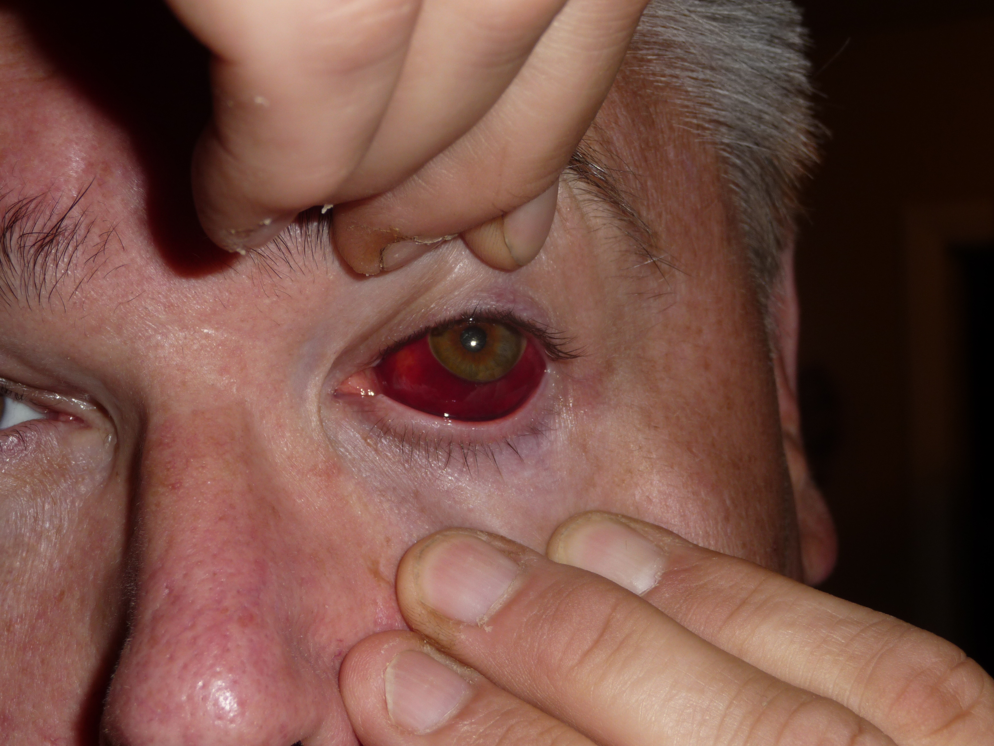
On the physical exam, SCH is a painless, acute, sharply demarcated area of extravasated blood just beneath the surface of the eye. SCH is generally unilateral. There is no reduction in visual acuity. A traumatic SCH should be more localized and if spontaneous and elderly, the SCH could be more diffuse. The inferotemporal aspect of the conjunctiva is the most common site. A simple SCH should not have any chemosis, proptosis, purulent discharge, ophthalmoplegia. In instances of scleral rupture, intraocular blood can leak through a defect and collect in the subconjunctival space which can create an elevated, bullous appearing hemorrhage. [2]
A key aspect of the physical exam is to distinguish between conjunctival versus ciliary injection. Conjunctival hemorrhage is caused by dilation of the posterior and more superficial conjunctival vessels. This can cause the eye to appear more dramatically red in a continuous pattern over the sclera. In contrast, ciliary injection involves dilation of the anterior ciliary arteries which could imply intraocular inflammation to the iris, cornea, or ciliary body. Ciliary injection can also be known as circumcorneal flush and appears as a halo of redness. The distinction is important since ciliary injection is associated with potentially more dangerous diagnoses such as iritis, acute glaucoma, episcleritis, and scleritis. [15]
SCH can also be confused for viral or bacterial conjunctivitis. However, there is usually some degree of pain associated with these diagnoses. Additionally, on physical exam, the redness is more diffuse and not a discreet, confluent area of hemorrhagic change as seen in SCH. Viral conjunctivitis is bilateral and in most cases SCH is unilateral. [15]
Evaluation
The initial evaluation and determination of SCH is clinical and based on appearance itself. However, a careful slit-lamp with fluorescein examination is important to determine any ocular trauma or possible underlying local ocular condition, which can lead to an SCH. All patients presenting with SCH should have blood pressure checked routinely. An INR should be checked if a patient is taking warfarin. If the SCH becomes persistent or recurrent then a workup regarding bleeding disorders and other hypocoagulable states should be done. However, it should be noted that extensive hemostatic testing is not warranted in the absence of other bleeding symptoms and solely SCH. Fundoscopy is generally not indicated. [2][9][16]]
Treatment / Management
Generally, there is no treatment indicated for SCH unless associated with a certain serious condition. The blood is typically resorbed over 1-2 weeks depending on the amount of extravasated blood. Recovery may take up to 3 weeks if patients are on anticoagulation. Ice packs and artificial tears can be utilized to minimize tissue swelling and provide some relief of discomfort respectively. Emergent ophthalmology consultation is warranted if SCH occurred via trauma and intraocular or additional retinal trauma is suspected. Dilute brimonidine and oxymetazoline have been indicated to improve patient comfort and decrease the incidence of SCH after intravitreal injections. [2][17][18]
Differential Diagnosis
If orbital trauma is suspected, globe rupture and retrobulbar hematoma is a diagnosis that must be ruled out as this is sight-threatening and requires emergent ophthalmology consult. In the setting of trauma, one must also consider corneal abrasion, conjunctival laceration, ocular foreign body, traumatic iritis, traumatic hyphema. Differential for non-traumatic cases includes conjunctivitis, episcleritis, inflamed pterygium or pinguecula, corneal erosions, keratitis, anterior uveitis. One must also consider and rule out acute angle-closure glaucoma, corneal ulcer, endophthalmitis, and scleritis as these are ophthalmologic emergencies.
Most of the time a more dangerous etiology can be found just by simple observation. A patient in the setting of a globe rupture or retrobulbar hematoma could have some degree of proptosis, chemosis, decrease in visual acuity, or a tear-drop shaped pupil. The physical exam can also help to distinguish other eye disorders such as an afferent pupillary defect in the setting an optic neuropathy or consensual photophobia in the setting of iritis. The slit lamp exam with fluorescein staining can be a very useful adjunct in the exam to better investigate possible erosions, ulcers, dendrites. Many of these patients could have symptoms of grittiness or a foreign body sensation all of which should not be present in the case with a simple SCH.
Prognosis
SCH offers a good visual prognosis after resolution. Vision is generally not impaired. The recurrence rate for spontaneous SCH is about 10% without identifiable risk factors and higher if patients are taking anticoagulant or antiplatelet therapy. [16]
Complications
There are no complications surrounding subconjunctival hemorrhage as most resolve around 2 weeks. Subconjunctival hemorrhage itself may be a sign of a more underlying dangerous disorder such as coagulopathy, severe asthma exacerbation, non-accidental trauma, or severe orbital trauma. [19]
Deterrence and Patient Education
Subconjunctival hemorrhages generally subside within two weeks. If patients notice recurrence or persistence of SCH and/or bruising on other parts of the body, especially if taking any anticoagulation or antiplatelet therapy, then their general practitioner or cardiologist may organize further tests. Artificial tears may help if the eye feels gritty or full. Patients should contact their primary care doctor or seek a specialist if there is vision loss ophthalmoplegia, or increasing pain and swelling.
Enhancing Healthcare Team Outcomes
Subconjunctival hemorrhage is a frequently encountered complaint in medical practice. Many patients do not experience any symptoms at all except for the physical appearance of the hemorrhage itself. The cause of a subconjunctival hemorrhage can be usually from no identifiable etiology. For the most part, they are of benign origin including increased strain, Valsalva, contact lens usage. But in some instances, there can be a systemic predisposing risk factor and further work-up is needed to prevent further morbidity and mortality.
There are many different types of medical professionals that can encounter subconjunctival hemorrhages. The primary care provider, emergency physicians, and ocular specialists can all be involved in the care of patients with subconjunctival hemorrhage. It can be important to consult with your interprofessional team to discuss the patient's diagnosis and collaborate further care. Nurses and pharmacists are vital members of the interprofessional team as they will monitor the patient's vital signs as well as potentially adjust medications respectively. The ophthalmologist is an important component of the interprofessional team since patients are hopefully able to follow up with these specialists and further coordinate care. In addition, some patients may have subconjunctival hemorrhages caused by anticoagulation. Therefore, it is important to consult with one's cardiologist/vascular surgeon or whoever is monitoring a patient's anticoagulation. Subconjunctival hemorrhages can also be present in newborns and children, therefore, neonatalogists, pediatricians, pediatric emergency physicians could be involved. It is important to note that subconjunctival hemorrhages can be present in the context of non-accidental trauma so the clinician must be vigilant for signs of child abuse.