Continuing Education Activity
Elbow dislocations are a common large joint dislocation experienced in children and adults. They can occur with associated fractures or damage to surrounding supporting structures. This activity outlines the evaluation and management of elbow dislocations and reviews the role of the interprofessional healthcare team in improving care for patients with this condition.
Objectives:
- Identify the typical patterns and common patient presentations with elbow dislocations.
- Summarize the treatment considerations for patients with elbow dislocations.
- Describe and review common complications of patients with elbow dislocations.
- Review the importance of improving care coordination among the interprofessional team to enhance care coordination for patients affected by elbow dislocations.
Introduction
The elbow is among the most common large joints to dislocate. Dislocations may be isolated, involve damage to static supportive structures of the elbow, and may even cause fractures about the elbow. Because of this, it is important to recognize elbow dislocations and know the appropriate management to avoid any complications.
Simply put, the elbow is the articulation between the distal humerus with the proximal ulna and radius.[1] The distal humerus contains the trochlea medially and capitulum laterally. Three separate articulations comprise the elbow. The medial aspect of the elbow includes the ulnotrochlear joint, which is primarily responsible for flexion and extension. The lateral radiocapitellar joint and the proximal radioulnar joint are mainly responsible for pronation and supination.[2] This anatomy creates a combination of a hinge joint and a pivot joint.[1]
The anatomy of the ulna allows two articulations and serves as an area of attachment for many structures. The greater sigmoid notch of the ulna creates the ulnotrochlear joint, and the lesser sigmoid notch creates the proximal radioulnar joint. The proximal anterior surface of the ulna also contains the coronoid process. The anteromedial facet (called the sublime tubercle) creates the insertion point for the medial collateral ligament, specifically, the anterior bundle.[3] The anterior band of the medial collateral ligament is a basic structure for valgus stability of the elbow.[4]
The elbow is generally stable due to the congruity articular surfaces. It is further supported by static supporting structures, including the collateral ligaments on the medial and lateral side of the elbow and the joint capsule. Dynamic stabilizers of the joints are composed of the surrounding musculature.[1]
The vascular anatomy of the elbow is composed of a few structures. The brachial artery is a central component of the anterior elbow and eventually divides into the radial and ulnar artery in the proximal forearm. The radial and ulnar artery, along with the brachial and the deep brachial artery, comprise an intricate anastomosis of vessels around the elbow, including the radial and ulnar collateral arteries and the radial and ulnar recurrent arteries.[5]
There are many nerves that exist around the elbow, and whose function can be compromised by an elbow dislocation. The radial nerve runs in the posterior compartment of the arm in the radial groove of the humerus and wraps laterally to its position near the elbow, where it is anterior to the lateral epicondyle.[6] It then divides into a superficial and deep branch, the deep branch giving rise to the posterior interosseus nerve after it has passed through the heads of the supinator muscle.[7] The median nerve runs in the medial bicipital groove of the arm along with the brachial artery, passing over the brachial artery and under the bicipital aponeurosis near the elbow, staying medial to the brachial artery.[8] The anterior interosseus nerve is a terminal branch of the median nerve after it passes through the heads of the pronator teres, giving rise to the motor function a majority of the deep flexor compartment of the forearm.[9] The ulnar nerve lies in the medial arm, and near the elbow, passes just posterior to the medial epicondyle of the humerus.[10]
Etiology
Falling onto an outstretched hand is the most common mechanism leading to elbow dislocations.[11] For posterior elbow dislocations, the overall forces involved in creating the dislocation include axial compression, valgus stress, and supination.[1] As the hand takes the force from the ground, it places an axial load onto the elbow. The forearm also experiences a supination moment as the body externally rotates about the hand on the ground.[12] This motion leads to a valgus moment about the elbow.[13] For anterior elbow dislocations, the mechanism usually involves a fall on a flexed elbow, with an anterior-directed force on the proximal ulna.[14]
Importantly, soft-tissue structures can also undergo compromise during an elbow dislocation. Disruption of supportive structures occurs, from lateral to medial, called the “Horii circle.”[12] Therefore, the order of disruption is as follows: the lateral collateral ligament, the anterior capsule, posterior capsule, and the medial collateral ligament.[1]
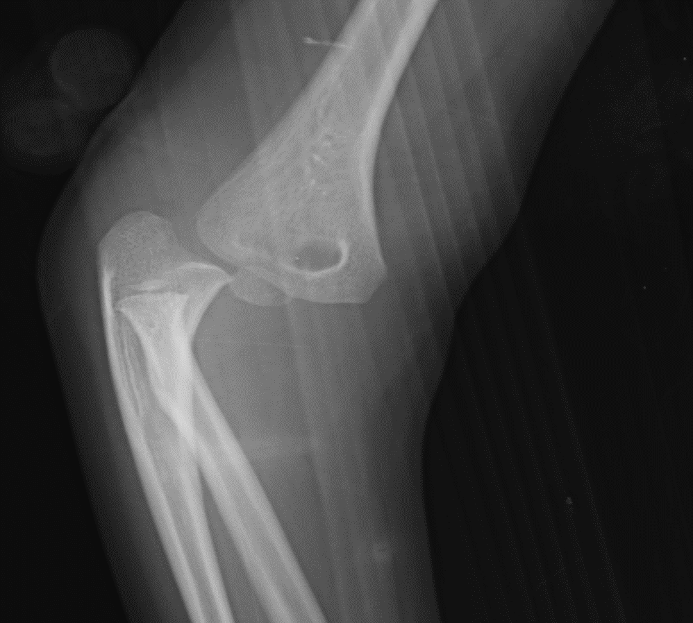
Elbow dislocations differentiate by the direction of the olecranon relative to the humerus. Posterolateral, posteromedial, posterior, anterior, medial, and lateral are different possibilities. Posterolateral is the most common pattern of dislocation.[13] In fact, 90% of dislocations are either posterior or posterolateral.[15]
Dislocations can also be complete or subluxated (also referred to as perched). A perched dislocation shows the coronoid resting on the trochlea instead of complete dissociation, as seen in a complete dislocation.[13]
Finally, dislocations can be termed simple or complex. A simple dislocation involves injury to only capsular or ligamentous structures. A complex dislocation involves fractures of the surrounding bony structure.[3] These commonly include the radial head, coronoid process, olecranon, distal humerus, and medial or lateral epicondyles of the humerus.[15] There is one fracture-dislocation that is specifically termed “the terrible triad” due to historically poor outcomes. The terrible triad is an elbow dislocation with both a radial head fracture and a coronoid process fracture of the ulna.[3]
Epidemiology
Elbow dislocations represent the second most common large joint dislocation.[13] In children, it is the most common large joint dislocation.[16] The incidence of elbow dislocations is 5.21 per 100000 person-years. Elbow dislocations are also more common in males than females, 53% versus 47%, respectively. Of note, males have nearly double the incidence of dislocations in the second decade (10 to 19) of life.
The second decade age group also accounts for nearly half (43.5%) of the elbow dislocations. Age is inversely proportional to elbow dislocation rates: as age increases, elbow dislocation rates decrease. Over half of dislocation events occur at home, followed by at work and at school. The most popular sports with elbow dislocations from highest to lowest rates are football, roller-blading or skateboarding, and wrestling.[2]
History and Physical
Objective evaluation should begin with a visual inspection of the elbow, looking for any obvious deformity or malpositioning of the extremity. The elbow should have an assessment for any palpable effusion, and skin also inspected for ecchymosis. Next, the patient's elbow should be palpated to evaluate for any tenderness.[3] The wrist must also be palpated and examined for instability to rule out the involvement of the distal radioulnar joint, which would indicate interosseous membrane disruption.[15] Also, a cautious range of motion of the elbow can allow the examiner to evaluate which articulations are involved in the dislocation, as well as to feel any potential crepitus indicating a possible intra-articular fracture.[3]
Subsequently, the neurovascular status of the extremity requires documentation. It is rare for arterial or neurologic injury to occur with elbow dislocations. However, it is crucial to document due to potential changes in neurovascular status after a reduction attempt.[15]
Therefore, the function of the motor nerves is an essential part of the physical exam and requires testing in patients who have sustained an elbow dislocation. The median nerve is evaluated by checking the function of the flexor digitorum superficialis, or by asking the patient to make a fist.[8] Specifically, testing can involve holding the fingers flat to an exam table and asking the patient to flex an isolated finger. The anterior interosseus nerve is tested by evaluating the function of the flexor pollicis longus, or by asking the patient to make an “OK” sign, evaluating for flexion of the thumb at the interphalangeal joint.[9] The radial nerve is tested by the motor function of the extensor digitorum muscles and by evaluating the patient’s ability to extend their wrist; if they cannot, the term for this condition is “wrist drop.”[6] The posterior interosseus nerve innervates the extensor pollicis longus and the patient is asked to make a “thumbs up” gesture.[7] Lastly, the ulnar nerve innervates the palmar and dorsal interossei muscles, and the patient is asked to abduct and adduct their fingers.[10]
Evaluation
The examiner must understand the direction of the dislocation before attempting reduction.[15] Plain radiographs of the elbow are necessary, which generally include anteroposterior and lateral views.[13] Oblique views of the elbow may help with the identification of the direction of the dislocation and any surrounding fractures.[15]
Post-reduction films are ordered to evaluate concentric reduction of the joint and to evaluate for any associated fractures.[13] In pediatrics, it is crucial to look for entrapment of the medial epicondyle within the joint.[4] Computed tomography of the elbow is useful to help identify associated injuries or fractures such as a terrible triad, which may be difficult to see on plain films. If associated fractures are small and nondisplaced, they can be treated non-operatively as a simple elbow dislocation.[3]
Treatment / Management
Attempting closed reduction of the elbow is the initial management of choice.[3] If an elbow dislocation occurs at a sporting event, an experienced physician may be able to perform the reduction immediately on the field, even before obtaining X-rays.[13] The reduction can decrease the pain and swelling of the injury.[3]
Generally, reduction requires appropriate intravenous sedation and relaxation of the patient, often administered by a team in the emergency department. If there is displacement of the elbow in a medial or lateral direction, the displacement first gets translated before the application of longitudinal traction of the forearm. As posterolateral dislocations are most common, the elbow is often extended.[13] Pressure on the posterior aspect of the olecranon is applied to guide the olecranon distal and anterior to the trochlea on the humerus, allowing reduction of the elbow. Often, a palpable or audible clunk can occur during reduction, which may be an indicator of likely stability.[13][15]
After reduction, elbow stability requires assessment by performing a gentle range of motion in flexion/extension, valgus/varus, as well as pronation/supination.[3] Often, instability is present after reduction with an attempted extension of the elbow, which can point to an unstable joint.[13] The patient requires splinting after reduction, often with a posterior long arm splint that prevents extension of the elbow. Elbow splinting is generally in 90 degrees of flexion.
A repeat neurovascular assessment is then performed to assure no arterial or nerve injuries due to reduction. A brachial artery injury or median nerve entrapment may occur, which can lead to operative management with exploration.[3]
After adequate reduction with a stable joint, patients can proceed with non-operative management with posterior long-arm splinting, usually in 90 degrees of flexion. Follow-up in the outpatient clinic allows for further radiographs to evaluate the maintenance of the reduction and that it remains stable. After two weeks, if the joint is stable, the splint can be removed, and physical therapy is initiated utilizing range of motion exercises to help avoid loss of terminal extension. In a less stable joint, the splint will remain, and the use of extension blocking splints can allow early flexion.[15] It is often helpful to begin range of motion under supervision and to avoid prolonged immobilization.[13]
Surgical management is required only in a few situations. If the dislocation is unable to be reduced, closed reduction under anesthesia in the operating room is a possible option, as muscular relaxation is difficult to reproduce with sedation in the emergency department.
Another indication for surgery is if the elbow is unstable with extension during range of motion, exhibited by dislocation once the elbow reaches 50 to 60 degrees of extension.[15] Traditionally, the goal of surgery is to restore a stable, functional arc of motion to include 30 to 130 degrees in flexion and extension, and to allow 50 degrees of pronation and supination. However, this dogma is changing and is potentially patient-specific, depending on the occupation.[17] Instability of the elbow may also result from damage to the collateral ligaments, which may need to be repaired to restore stability and proper function.[15]
Surgery is also necessary if there are any associated unstable fractures with a complex elbow dislocation.[15] This includes incarcerated fragments within the joint, or if the medial epicondyle becomes entrapped in the joint, as can be seen in pediatric elbow dislocations.[4] The surgical approach chosen for the dislocation depends on the fracture, pattern of instability, and laterality of soft-tissue injury.[3]
Differential Diagnosis
Elbow dislocations are often a clinical presentation confirmed by radiographs. The patient's elbow, as well as surrounding bony structures, should be palpated to evaluate for any tenderness.[3] Tenderness overlying the elbow may be due to other causes, such as distal humerus fractures, medial or lateral condyle fractures, or proximal radial or ulnar shaft fractures. It is also possible that there may isolated injuries to ligaments about the elbow, such as the medial collateral ligament, which leads to valgus instability of the elbow.[4]
Chronic elbow dislocation is another differential. Chronic elbow dislocations are elbows that have remained dislocated for more than two weeks. It is essential to determine if there is a history of instability, previous attempts at reduction, or multiple episodes of dislocation. Ulnar neuropraxia in chronic dislocations occurs in 15% of patients. Due to this, fibrosis of the surrounding structures is common, and reduction is often difficult. Surgical intervention with open reduction is generally necessary due to extensive fibrosis and granulation tissue that can be present within the joint. Surgery often involves anterior and posterior capsule release and release of any surrounding fibrotic areas. Patients may also require triceps lengthening procedures or transposition of the ulnar nerve.[16]
Prognosis
It is not uncommon for patients to lose 10 to 15 degrees of terminal extension. Limiting the time of immobilization to less than three weeks can help to avoid contracture. Dynamic elbow splinting along with therapy can be used if the patient is not improving at an expected rate. It may take up to 5 months for patients to regain extension. If these fail, surgical intervention for contracture release may be required.[15]
In children, the prognosis after elbow dislocation is usually more optimistic. Children generally have lower rates of contracture and loss of motion and have a higher tendency to regain normal elbow function.[16]
Complications
One of the most common complications with elbow injuries is loss of range of motion. In particular, extension loss is the most common and is usually related to the time of immobilization in a static splint. Greater than 25 days of immobilization can lead to a loss of 30 degrees in extension and nearly 20 degrees in flexion. Extrinsic causes may be due to flexion contracture of the anterior capsule, or contracture of the collateral ligaments and the surrounding musculature.[17] Contracture of the anterior capsule is the most common cause of extension block due to fibrosis of the capsule.[15] Intrinsic causes, such as articular incongruency or fragments that cause a mechanical block, could also cause loss of motion.[17]
Ectopic bone formation is common, but is usually periarticular and does not block motion.[15] Patients with myositis ossificans have calcium deposition in the soft tissue structures and musculature surrounding the joint, which is why it rarely prevents movement.[17] Heterotopic ossification that blocks motion occurs in less than 5% of patients with elbow dislocations. There is an association between ectopic bone formation and closed head injury.[15] Other risk factors include males older than 60, burn patients, and genetic disorders.[17] Prevention for high-risk patients consists of the use of nonsteroidal anti-inflammatory medications, also known as NSAIDs, and low dose local radiation.[15] Indomethacin is the most studied and commonly used NSAID for the prevention of heterotopic ossification, usually dosed three times daily at 25 milligrams or 75 milligrams twice daily. Radiation prophylaxis is possible in many ways, but most commonly as a single dose of 700 cGy to the local area within 72 hours. If these fail, surgical excision may be necessary, and it is recommended to wait until the ossification has reached maturity and has been radiographically stable.[17]
Elbow instability is another common complication. Due to the mechanism of injury, the laterality and involvement of ligaments can change and requires appropriate management. Lateral collateral ligament insufficiency is common, leading to valgus instability, and is treatable with the patient in a pronated position during splinting to allow soft tissue to heal.[15] If surgical treatment is necessary for elbow dislocation or associated fractures, these structures may require repair.
Neurovascular injury from acute elbow dislocations is uncommon; however, ulnar nerve neuropraxia is the most commonly reported. Patients typically undergo observation, as these injuries generally resolve following closed reduction. Other neuropraxia such as median nerve and anterior interosseous nerve occur less frequently. Median nerve entrapment can occur, especially in the pediatric population, and is commonly associated with entrapment in the fracture site or posterior to a displaced medial epicondyle, which may be visible on radiographs as a widening of a medial condyle due to the median nerve displacing it, or neuropraxia may be apparent after closed reduction. Median nerve involvement after closed reduction requires open exploration. In patients that have an observed neuropraxia, electromyography is ordered only after three months of persistent symptoms. Lastly, brachial artery injury can occur. Initial assessment of pulse and perfusion is important, and if absent requires emergent reduction. Pulses often return following closed reduction. In patients without return of pulses or perfusion, the clinician should have a high suspicion of vascular injury.[17]
Deterrence and Patient Education
Certain activities are more prone to elbow dislocations due to the nature of the forces encountered. The most common sports being football, roller-blading or skateboarding, and wrestling.[2] These patients should be counseled, wear protective equipment, and, if possible, try to avoid excessive stresses to these areas.
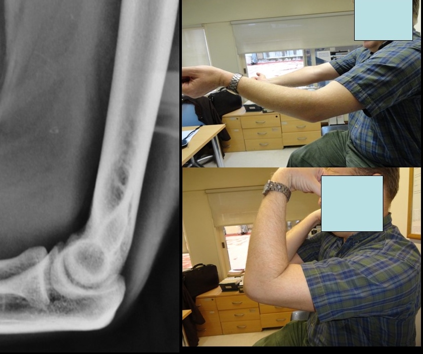
Patients quite commonly lose 10-15 degrees of terminal extension after elbow dislocations.[15] Patients should be counseled about this early and should work towards increasing the range of motion as soon as allowed.
Lastly, elbow dislocations are the most common large joint dislocation in children.[16] These patients tend to regain more range of motion and elbow function than adults. However, parents need to understand the possibility of growth disturbances.[16]
Pearls and Other Issues
Pediatric elbow dislocations have many similarities to adult elbow dislocations. However, in children, medial epicondyle fractures may occur. Up to 60% of medial epicondyle fractures are associated with elbow dislocations in children. Importantly, the medial epicondyle is the last ossification center in the distal humerus to fuse, anywhere from 15 to 20 years old. The medial epicondyle serves as the common flexor origin and the origin of the inferior aspect of the medial collateral ligament, leaving it vulnerable to avulsion injuries. It also lays anterior to the ulnar nerve.[4] Due to the higher prevalence of associated injury, it is crucial to assess post-reduction radiographs, as the medial epicondyle can become incarcerated in the joint after the reduction.[13]
Enhancing Healthcare Team Outcomes
Interprofessional care is fundamental to the treatment of patients with elbow dislocation. If an elbow dislocation occurs on the field of a sporting event, a member of the healthcare team may be able to reduce it immediately.[13] Education of these healthcare providers, such as physical therapists or athletic trainers, may allow prompt treatment if a physician is not available.
Plain radiographs of the elbow are needed to understand the direction of the dislocation, including anteroposterior, lateral, and oblique views.[13][15] Coordination with radiology technicians is needed to produce accurate and reproducible images of the elbows.
Closed reduction is often the initial management, and likely to occur in the emergency department (ED).[3] For adequate relaxation for reduction, the emergency department often sedates the patients. Length of stay in the ED can improve through interprofessional communication strategies and has been shown to improve patient satisfaction. Follow-up care should involve a nurse with orthopedic specialty training to ensure therapeutic progress, answer questions, and evaluate compliance. This care should be coordinated with the clinical team.
A quality improvement study from Boston Children's Hospital looked at ways to reduce this, including meetings involving clinical assistants, emergency department physicians, orthopedic physicians, and nurses. They initiated new strategies, roles, and checklists to attempt to improve efficiency. They found a 5.8% decrease in length of stay in patients requiring sedation for long bone fracture reduction; this also allowed the engagement of interprofessional personnel to help increase efficiency together.[18] [Level V]