Sebaceous Gland Carcinoma

Earn CME/CE in your profession:


Continuing Education Activity

Sebaceous carcinomas are rare malignant tumors of the sebaceous glands. They most commonly develop on the eyelid, but may arise from anywhere sebaceous glands are present. These tumors may arise spontaneously or in association with Muir-Torre syndrome. This activity reviews the diagnosis, evaluation, and treatment of sebaceous carcinomas and highlights the role of the interprofessional team in evaluating and treating patients with this condition.

Objectives:

  • Identify the etiology of sebaceous carcinomas.

  • Review the process for the evaluation of sebaceous carcinomas.

  • Outline the management options available for sebaceous carcinomas.

  • Describe interprofessional team strategies for improving care coordination and communication to advance sebaceous carcinoma treatment and improve outcomes.

Introduction

Sebaceous carcinoma (SC) is a rare and aggressive malignancy that develops from the sebaceous glands. Sebaceous carcinoma is most common in the periocular area, head, and neck, but can occur anywhere in the body where sebaceous glands are present.[1] 

Sebaceous carcinomas may behave as primary neoplasms or may occur in association with Muir-Torre syndrome (a form of Lynch syndrome characterized by sebaceous tumors and other internal cancers, being the most common those affecting the gastrointestinal tract). SC behaves aggressively both locally and distantly; locally, it correlates with a multifocal origin, and causes destruction and sometimes a pattern of pagetoid spread, which is challenging to diagnose. Moreover, this tumor easily causes distant metastasis. For those reasons, the SC is considered an aggressive disease, which causes significant morbidity and mortality. 

Etiology

As mentioned above, SC has its origin in sebaceous cells. When occurring in the periocular region, the most frequent, SC usually arise from Meibomian glands, which are modified sebaceous glands, but also from the sebaceous glands of Zeiss, and the sebaceous glands in the ocular caruncle.[2] 

The majority of sebaceous carcinomas arise from de novo mutations. They may also develop from benign sebaceous neoplasms, although this appears to be relatively uncommon. Sebaceous carcinomas associated with Muir-Torre syndrome have a loss of mismatch repair gene expression and exhibit microsatellite instability.[3] Sebaceous carcinomas that occur sporadically do not exhibit loss of mismatch repair or microsatellite instability.[4]

Epidemiology

SC is more frequent in older women. It is a rare malignancy in White race individuals, whereas in the Asian population represents up to 28% of all lid malignancies. Overall, SC has an estimated incidence of approximately 1 to 2 cases per 1,000,000 person-years.[5][6] More than 98% of SC occurs in patients over the age of 40, and the peak incidence occurs in the seventh and eighth decades of life.[7] There have only been reports of it appearing in a handful of childhood cases.[8] Sebaceous carcinoma is approximately three times as common in whites as in non-White patients. Regarding the anatomy, these tumors are more frequent in the upper eyelid, as there are more meibomian glands there.[9][7] 

Risk factors for the development of sebaceous carcinoma include prior radiation exposure, immunosuppression, and Muir-Torre syndrome.

Histopathology

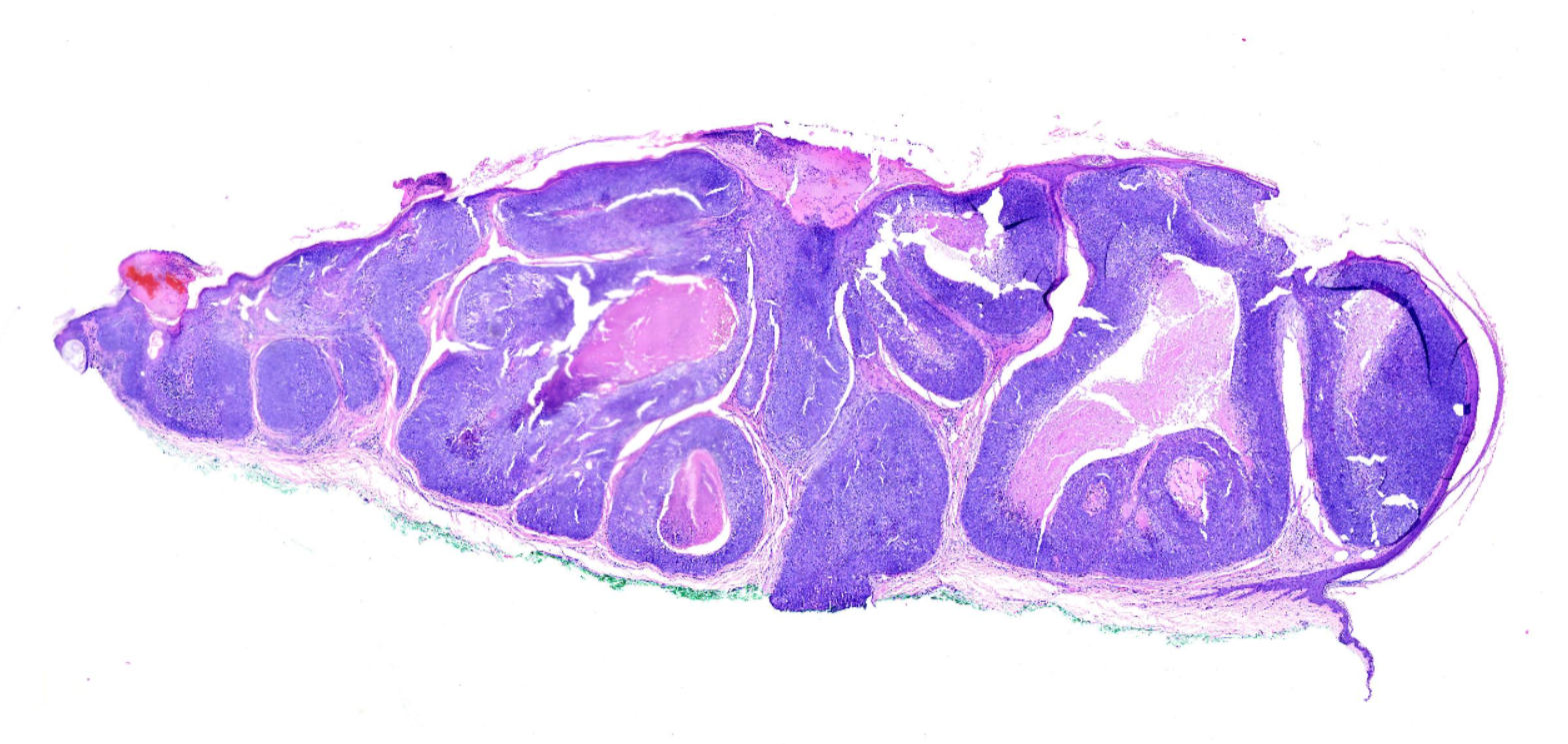
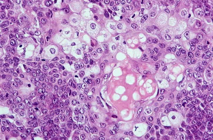
Histopathology typically shows irregular, asymmetric sebaceous lobules within the dermis. Malignant cells show significant amounts of pleomorphism, hyperchromatism, mitotic activity, and nuclear atypia. Lesions may be classified as poorly, moderately, or well-differentiated (see Images. Sebaceous Carcinoma, Low-Power Field; Sebaceous Carcinoma, High-Power Field). Well-differentiated lesions may demonstrate foamy cytoplasm, comedo-necrosis, and pagetoid spread.[10] Sebaceous carcinomas fall into four histopathologic categories: papillary, lobular, comedocarcinoma, and mixed.[11] Immunohistochemical stains for sebaceous carcinoma include adipophilin, EMA, androgen receptor, and nuclear factor XIIIa.[12][13] Adipophilin in a vesicular instead of granular staining pattern and nuclear factor XIIIa are the most specific and sensitive among the stains.[13][14][15] Negative staining for S100 and HMB-45 may help differentiate sebaceous carcinoma from balloon cell or amelanotic melanoma.[10]

History and Physical

Approximately 40% of all sebaceous carcinomas present on the eyelids, with 80% occurring on the head or neck.[1] On the eyelid, sebaceous carcinomas typically develop as a firm, round, enlarging nodule. They are two to three times more frequent in the upper lid due to the more number of sebaceous glands there. SC may also present with inflammation and thickening of the eyelid, typically mimicking other conditions, especially chalazion and blepharitis, and even conjunctivitis, and causing a delayed diagnosis. Local spread is frequent and may lead to involve wide areas of the conjunctiva and even the cornea. Advanced infiltration may cause invasion of the orbit and adjacent periorbital structures.

SC of extra-ocular locations generally presents as a yellowish nodule, often with ulceration. The head and neck are the most common locations; however, the trunk and extremities may also have involvement. There are also rare reports of cases of SC development at extracutaneous sites, such as the parotid gland, breast, ovary, and prostate.[16]

Evaluation

SC must always be in mind when treating a patient with unilateral resistant blepharoconjunctivitis, or a chalazion or stye that does not resolve or recur after treatment. An incisional biopsy might be necessary to confirm the diagnosis. Excisional biopsy with wide margins is considered the gold standard treatment, which is also useful to confirm the diagnosis. Routine histologic staining with hematoxylin and eosin will generally provide a diagnosis and is useful for staging of the lesion. Immunohistochemistry may also be useful to confirm the diagnosis and to differentiate from other non-melanoma skin cancers. Sebaceous carcinomas can exhibit nearly 100% EMA, ADP, and AR positivity.[12][14][17] 

While the majority of sebaceous carcinomas occur sporadically, some experts recommend screening all patients with this diagnosis for Muir-Torre syndrome.

A comprehensive physical exam to assess the globe and the periocular region for disease, as well as palpation of the preauricular and cervical areas for enlarged lymph nodes, should be performed. If the eyelid lesion appears to be diffuse or if there is any evidence of lymphadenopathy, then head and neck or even systemic imaging are warranted to evaluate the degree of disease spread. Fine-needle aspiration biopsy of suspicious lymph nodes can help diagnose sebaceous carcinoma.

Treatment / Management

The primary goal of treatment is complete surgical excision of the tumor. Wide local excision with the use of frozen sections, or Mohs micrographic surgery, are first-line treatments for sebaceous carcinoma of the head or neck.[18][1] If conjunctival epithelial involvement is identified, treatment may involve cryotherapy, surgical resection of the affected bulbar epithelium, or use of topical mitomycin C.[19] In cases where surgical resection is not possible, mainly in cases of extensive conjunctival spread or orbital extension, orbital exenteration is often required. 

 The role of radiation therapy for primary sebaceous carcinomas is uncertain. Some small studies have reported good outcomes from the use of radiation as the primary treatment of ocular sebaceous carcinoma, but surgical resection remains the primary treatment modality.[20][21]

Adjuvant therapies include cryotherapy, and topical mitomycin C. The use of these therapies is primarily reserved for pagetoid spread to the conjunctiva. 

Differential Diagnosis

Several benign and malignant conditions merit consideration in the differential diagnosis for SC. These tumors may often mimic a chalazion, especially if they become painful or inflamed, resistant to the usual treatment, or if they recur after surgery. An extensive list of eyelid masses may be part of the differential diagnosis of SC, including apart from the most frequent chalazion, other tumors such us inclusion cyst, papilloma, keratosis, keratoacanthoma, or dermoid cyst, among others.[22] 

Benign sebaceous neoplasms such as sebaceous adenomas may present similarly to SC, but these benign lesions are uncommon in the periocular region.[23] Basal cell carcinomas, squamous cell carcinomas, and Merkel cell carcinomas may all have clinical presentations similar to SC; the histologic examination is essential for differentiating these. 

Staging

Evaluation for evidence of local tumor spread should be performed immediately after diagnosis. Regional lymph nodes should be palpated for evidence of metastasis, with a biopsy performed on concerning lymph nodes. Ophthalmologists usually diagnose patients diagnosed with SC of the eyelids; a comprehensive ophthalmic examination is mandatory to assess for the clinical tumor extension.

There is no clear consensus and guidelines concerning the systemic evaluation of these patients; however, patients with large tumors, or those where there has been delayed diagnosis, should undergo imaging with CT or MRI to evaluate for a distant extension. Patients with Muir-Torre may also receive benefits from a PET scan for early detection of visceral malignancies.[24]

Prognosis

Sebaceous carcinomas tend to be aggressive with significant potential for nodal and distant metastasis. Ocular sebaceous carcinomas have demonstrated a recurrence rate of 11 to 30% after excision and a rate of metastasis of 25%.[25] For extraocular sebaceous carcinoma, rates of recurrence and distant metastases are 29% and 21%, respectively.[1] 

Regional lymph nodes are the most common sites of spread; lungs, brain, liver, small intestine, and urinary tract metastasis have also been reported. Overall mortality rates are between 9 and 50%.[1] Five-year survival rates are more favorable for the ocular disease at 75.2%, compared to the extraocular disease at 68%.[26] Negative prognostic factors include size greater than 1 cm, poor differentiation on histopathology, lymphovascular invasion, and lesions involving both the upper and lower eyelid.

Complications

Recurrence, local invasion, and distant metastasis by lymphatic or hematogenous spread are all relatively common complications of sebaceous carcinoma. 

Deterrence and Patient Education

No known modifiable risk factors for the development of sebaceous carcinoma exist. Patients with a diagnosis of Muir-Torre should receive education regarding the risk of sebaceous carcinoma, and to seek medical care if a concerning skin lesion develops. Also, patients with chronic inflammatory processes of the eye and eyelids should attend an ophthalmologist for an examination.

Enhancing Healthcare Team Outcomes

Sebaceous gland carcinoma is a rare tumor that masquerades as benign ocular conditions and hence requires the clinicians to work in a multidisciplinary team to identify the tumor, make a correct pathological diagnosis, and perform an early surgical therapy trying to cure the disease. Delayed management of these cases might be fatal due to the aggressive nature of the tumor. Whenever sebaceous tumors are identified, clinicians should consider internal malignancies.

Early recognition aids in the prompt treatment of patients and screening of family members for early intervention. Many health professionals may be involved in the care of patients affected with SC, such as physicians and surgeons of different specialties (depending on the location of the tumor), radiologists, pathologists, and nurses.

Disclosure: The views expressed in this article are those of the author and do not reflect the official policy of the Departments of Navy/ Army/ or Air Force, Department of Defense, or US Government.


(Click Image to Enlarge)
<p>Sebaceous Carcinoma, Low-Power Field</p>

Sebaceous Carcinoma, Low-Power Field


Contributed by Dipti Anand, MD


(Click Image to Enlarge)
<p>Sebaceous Carcinoma, High-Power Field</p>

Sebaceous Carcinoma, High-Power Field


Contributed by Dipti Anand, MD

Details

Author

Ryan Gall

Updated:

8/14/2023 10:42:41 PM

References


[1]

Dasgupta T, Wilson LD, Yu JB. A retrospective review of 1349 cases of sebaceous carcinoma. Cancer. 2009 Jan 1:115(1):158-65. doi: 10.1002/cncr.23952. Epub     [PubMed PMID: 18988294]

Level 2 (mid-level) evidence

[2]

Salazar-Villegas Á, Matas J, Palma-Carvajal F, González-Valdivia H, Alos L, Ortiz-Pérez S. Sebaceous carcinoma of the caruncle. Journal francais d'ophtalmologie. 2019 Oct:42(8):925-927. doi: 10.1016/j.jfo.2019.04.014. Epub 2019 Jun 6     [PubMed PMID: 31178073]


[3]

Kraft S, Granter SR. Molecular pathology of skin neoplasms of the head and neck. Archives of pathology & laboratory medicine. 2014 Jun:138(6):759-87. doi: 10.5858/arpa.2013-0157-RA. Epub     [PubMed PMID: 24878016]


[4]

Rajan Kd A, Burris C, Iliff N, Grant M, Eshleman JR, Eberhart CG. DNA mismatch repair defects and microsatellite instability status in periocular sebaceous carcinoma. American journal of ophthalmology. 2014 Mar:157(3):640-7.e1-2. doi: 10.1016/j.ajo.2013.12.002. Epub 2013 Dec 7     [PubMed PMID: 24321472]


[5]

Kuzel P, Metelitsa AI, Dover DC, Salopek TG. Epidemiology of sebaceous carcinoma in Alberta, Canada, from 1988 to 2007. Journal of cutaneous medicine and surgery. 2012 Nov-Dec:16(6):417-23     [PubMed PMID: 23149197]


[6]

Dores GM, Curtis RE, Toro JR, Devesa SS, Fraumeni JF Jr. Incidence of cutaneous sebaceous carcinoma and risk of associated neoplasms: insight into Muir-Torre syndrome. Cancer. 2008 Dec 15:113(12):3372-81. doi: 10.1002/cncr.23963. Epub     [PubMed PMID: 18932259]


[7]

Tripathi R, Chen Z, Li L, Bordeaux JS. Incidence and survival of sebaceous carcinoma in the United States. Journal of the American Academy of Dermatology. 2016 Dec:75(6):1210-1215. doi: 10.1016/j.jaad.2016.07.046. Epub 2016 Oct 6     [PubMed PMID: 27720512]


[8]

Omura NE, Collison DW, Perry AE, Myers LM. Sebaceous carcinoma in children. Journal of the American Academy of Dermatology. 2002 Dec:47(6):950-3     [PubMed PMID: 12451386]


[9]

Rao NA, Hidayat AA, McLean IW, Zimmerman LE. Sebaceous carcinomas of the ocular adnexa: A clinicopathologic study of 104 cases, with five-year follow-up data. Human pathology. 1982 Feb:13(2):113-22     [PubMed PMID: 7076199]

Level 3 (low-level) evidence

[10]

Nelson BR, Hamlet KR, Gillard M, Railan D, Johnson TM. Sebaceous carcinoma. Journal of the American Academy of Dermatology. 1995 Jul:33(1):1-15; quiz 16-8     [PubMed PMID: 7601925]


[11]

Jakobiec FA, Mendoza PR. Eyelid sebaceous carcinoma: clinicopathologic and multiparametric immunohistochemical analysis that includes adipophilin. American journal of ophthalmology. 2014 Jan:157(1):186-208.e2. doi: 10.1016/j.ajo.2013.08.015. Epub 2013 Oct 7     [PubMed PMID: 24112633]


[12]

Mulay K, White VA, Shah SJ, Honavar SG. Sebaceous carcinoma: clinicopathologic features and diagnostic role of immunohistochemistry (including androgen receptor). Canadian journal of ophthalmology. Journal canadien d'ophtalmologie. 2014 Aug:49(4):326-32. doi: 10.1016/j.jcjo.2014.04.004. Epub 2014 Jul 19     [PubMed PMID: 25103648]


[13]

Uhlenhake EE, Clark LN, Smoller BR, Shalin SC, Gardner JM. Nuclear factor XIIIa staining (clone AC-1A1 mouse monoclonal) is a sensitive and specific marker to discriminate sebaceous proliferations from other cutaneous clear cell neoplasms. Journal of cutaneous pathology. 2016 Aug:43(8):649-56. doi: 10.1111/cup.12726. Epub 2016 Jun 10     [PubMed PMID: 27153339]


[14]

Ansai S, Takeichi H, Arase S, Kawana S, Kimura T. Sebaceous carcinoma: an immunohistochemical reappraisal. The American Journal of dermatopathology. 2011 Aug:33(6):579-87. doi: 10.1097/DAD.0b013e31820a2027. Epub     [PubMed PMID: 21778832]


[15]

Ostler DA, Prieto VG, Reed JA, Deavers MT, Lazar AJ, Ivan D. Adipophilin expression in sebaceous tumors and other cutaneous lesions with clear cell histology: an immunohistochemical study of 117 cases. Modern pathology : an official journal of the United States and Canadian Academy of Pathology, Inc. 2010 Apr:23(4):567-73. doi: 10.1038/modpathol.2010.1. Epub 2010 Jan 29     [PubMed PMID: 20118912]

Level 3 (low-level) evidence

[16]

Gnepp DR. My journey into the world of salivary gland sebaceous neoplasms. Head and neck pathology. 2012 Mar:6(1):101-10. doi: 10.1007/s12105-012-0343-x. Epub 2012 Mar 20     [PubMed PMID: 22430772]


[17]

Plaza JA, Mackinnon A, Carrillo L, Prieto VG, Sangueza M, Suster S. Role of immunohistochemistry in the diagnosis of sebaceous carcinoma: a clinicopathologic and immunohistochemical study. The American Journal of dermatopathology. 2015 Nov:37(11):809-21. doi: 10.1097/DAD.0000000000000255. Epub     [PubMed PMID: 26485238]


[18]

Hou JL, Killian JM, Baum CL, Otley CC, Roenigk RK, Arpey CJ, Weaver AL, Brewer JD. Characteristics of sebaceous carcinoma and early outcomes of treatment using Mohs micrographic surgery versus wide local excision: an update of the Mayo Clinic experience over the past 2 decades. Dermatologic surgery : official publication for American Society for Dermatologic Surgery [et al.]. 2014 Mar:40(3):241-6. doi: 10.1111/dsu.12433. Epub 2014 Jan 25     [PubMed PMID: 24460730]


[19]

Shields JA, Demirci H, Marr BP, Eagle RC Jr, Stefanyszyn M, Shields CL. Conjunctival epithelial involvement by eyelid sebaceous carcinoma. The 2003 J. Howard Stokes lecture. Ophthalmic plastic and reconstructive surgery. 2005 Mar:21(2):92-6     [PubMed PMID: 15778660]


[20]

Azad S, Choudhary V. Treatment results of high dose rate interstitial brachytherapy in carcinoma of eye lid. Journal of cancer research and therapeutics. 2011 Apr-Jun:7(2):157-61. doi: 10.4103/0973-1482.82922. Epub     [PubMed PMID: 21768703]


[21]

Hata M, Koike I, Maegawa J, Kaneko A, Odagiri K, Kasuya T, Minagawa Y, Kaizu H, Mukai Y, Inoue T. Radiation therapy for primary carcinoma of the eyelid: tumor control and visual function. Strahlentherapie und Onkologie : Organ der Deutschen Rontgengesellschaft ... [et al]. 2012 Dec:188(12):1102-7. doi: 10.1007/s00066-012-0145-9. Epub 2012 Oct 28     [PubMed PMID: 23104519]


[22]

Wali UK, Al-Mujaini A. Sebaceous gland carcinoma of the eyelid. Oman journal of ophthalmology. 2010 Sep:3(3):117-21. doi: 10.4103/0974-620X.71885. Epub     [PubMed PMID: 21120046]


[23]

Shields JA, Demirci H, Marr BP, Eagle RC Jr, Shields CL. Sebaceous carcinoma of the eyelids: personal experience with 60 cases. Ophthalmology. 2004 Dec:111(12):2151-7     [PubMed PMID: 15582067]

Level 3 (low-level) evidence

[24]

Ishiguro Y, Homma S, Yoshida T, Ohno Y, Ichikawa N, Kawamura H, Hata H, Kase S, Ishida S, Okada-Kanno H, Hatanaka KC, Taketomi A. Usefulness of PET/CT for early detection of internal malignancies in patients with Muir-Torre syndrome: report of two cases. Surgical case reports. 2017 Dec:3(1):71. doi: 10.1186/s40792-017-0346-7. Epub 2017 May 23     [PubMed PMID: 28537014]

Level 3 (low-level) evidence

[25]

Watanabe A, Sun MT, Pirbhai A, Ueda K, Katori N, Selva D. Sebaceous carcinoma in Japanese patients: clinical presentation, staging and outcomes. The British journal of ophthalmology. 2013 Nov:97(11):1459-63. doi: 10.1136/bjophthalmol-2013-303758. Epub 2013 Sep 13     [PubMed PMID: 24037608]


[26]

Snow SN, Larson PO, Lucarelli MJ, Lemke BN, Madjar DD. Sebaceous carcinoma of the eyelids treated by mohs micrographic surgery: report of nine cases with review of the literature. Dermatologic surgery : official publication for American Society for Dermatologic Surgery [et al.]. 2002 Jul:28(7):623-31     [PubMed PMID: 12135523]