Neuroanatomy, Superior Colliculus


Introduction

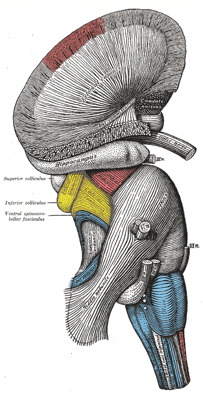
The superior colliculi are paired rostral midbrain structures involved in processing optical stimuli, orienting attention, and coordinating eye and head movements (see Image. Hindbrain, Superficial Dissection). The counterpart of the superior colliculi in other vertebrates is the optic tectum, which possesses a topographic map of the contralateral visual field and inputs from somatosensory and auditory pathways.[1] Similarly, the superior colliculi receive input from the eyes and higher brain regions involved in vision and other sensory modalities. Thus, the superior colliculi are the primary brain regions that integrate visual and nonvisual information.

The superior colliculi drive young children to follow faces and react to emotional stimuli, which are actions impaired in autism. Neurodegenerative conditions such as Parkinson disease may disrupt superior colliculus function, manifesting with abnormal fixation and saccadic eye movements. Understanding the role of this structure in these disorders is important for developing therapeutic interventions.

Structure and Function

The superior colliculus is on the posterior midbrain rostral to the inferior colliculus and caudal to the pineal gland. This structure has 7 internal cell layers, divided into superficial, intermediate, and deep layers.

The superficial layers consist of the stratum zonale, stratum griseum superficiale, and stratum opticum. These purely sensory layers respond to bilateral retinal inputs from contralateral visual stimuli.

The intermediate layers consist of the stratum griseum intermedium and stratum album intermedium. The deep layers are composed of the stratum griseum profundum and stratum album profundum. The intermediate and deep layers receive information from visual, auditory, and somatosensory pathways.

The superior colliculus contributes to motor functions that orient the head and eyes toward or away from a stimulus.[2] When head movement is restricted, the superior colliculi are involved in saccadic eye motion. Thus, the superior colliculi are associated with skeletomotor and oculomotor pathways responsible for producing neck and saccadic eye movements.

The inferior colliculus coordinates with the superior colliculus in orienting the gaze toward or away from visual and auditory stimuli. Other nonmotor functions of the superior colliculus are less studied and include multimodal sensory processing and attention.[3][4] The superior colliculi represent a subcortical node in a general sensorimotor network.

Visual looming is when a stimulus is perceived as a threat and evokes a defensive response. For example, when a snake approaches an individual, information processing in the superior colliculi identifies details orienting the person away from the snake, which is perceived as a threat. This internal orienting can occur even without overt eye movements.

The superior colliculi's deeper layers encode the visual looming process and response. This information is relayed to subcortical structures like the periaqueductal grey, ventral tegmental area, and thalamus, leading to defensive behavior development.[5]

The functions of the superior colliculi can be summarized as follows:

  • Visual detection
  • Orientation movements of the eyes, head, and arms
  • Saccade generation and target selection
  • Detecting and responding to looming motion
  • Reaching movements of the forelimbs, target selection, and decision-making
  • Detecting faces [6][7]

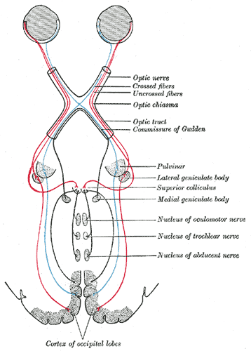
The superior colliculi contribute to the ability to interact with the surrounding environment and respond to relevant stimuli in a coordinated manner (see Image. Central Connections of the Optic Nerves and Tracts).

Embryology

The mesencephalon serves as the midbrain's embryonic precursor. The superior colliculus arises during the primary and secondary vesicle stages of brain development at weeks 4 and 5. Expression of the genes Pax7 and engrailed correlate with the formation and specialization of the superior colliculus. Pax7 expression is essential in initiating superior colliculus formation and all tectal development stages. The expression of the engrailed gene is necessary for regulating cell migration during tectal lamina formation.[8][9]

Blood Supply and Lymphatics

The superior colliculus receives blood from the collicular and posteromedial choroidal arteries.[10] These blood vessels are proximal posterior cerebral artery branches. The superior cerebellar artery, which sometimes branches from the basilar artery, may also supply the superior colliculus. Lymphatics of the brain were recently discovered. One study reported the presence of brain lymphatic endothelial cells over the zebrafish's optic tectum that selectively take up specific macromolecules.[11][12]

Nerves

The super colliculi's neural connections are complex and still not entirely understood. These structures receive visual information mainly from the retina via the optic nerve and tract and less from the visual cortex. The superior colliculi also receive auditory input from the inferior colliculi to coordinate movement responses. Additional somatosensory projections to the deep laminae allow the superior colliculi to respond to tactile stimuli.

The superior colliculi also receive prefrontal cortex inputs involved in attention and distractibility regulation. These efferent fibers leave the internal capsule, follow a pedunculotegmental route to the lower midbrain, and rise to the caudal superior colliculi. Outward tracts from the superior colliculi have been shown to project to the central gray matter overlying the rostral oculomotor nucleus and areas adjacent to the contralateral trochlear nucleus and contralateral abducens nucleus.[13][14]

Muscles

During gaze shifts, oculomotor and neck muscles receive stimuli from the superior colliculus. As stated above, the oculomotor, trochlear, and abducens nuclei all receive projections from the superior colliculus. Thus, the 6 extraocular muscles innervated by these nerves ultimately connect to the superior colliculus. The contralateral deep neck muscles are likewise stimulated by the superior colliculus during a gaze shift. These muscles are ipsilateral to the visual field perceiving the stimulus. The effect is to turn the head and eyes toward the stimulus.

Surgical Considerations

Surgery of the superior colliculi should be approached with caution as the midbrain is an eloquent area. Surgical manipulation of these structures poses significant risks.

Surgery in this region may be indicated for tectal gliomas. However, potential complications include visual defects, Parinaud syndrome, mutism, and reduction to a vegetative state followed by death. Thus, surgical resection is carefully considered based on the tumor's location, size, progression, and operability.

Tectal glioma management is still controversial and up for debate. However, this once inaccessible area is now approachable with increased safety due to novel microneurosurgical techniques.[15][16]

Clinical Significance

The effects of direct superior colliculus injury have been observed in rhesus monkeys and rats. Researchers found that the monkeys with damage to this region have visual deficits and gaze shift impairments.[17][18]

Rats with collicular damage displayed a lack of orienting reflex and were not distracted when presented with visual or auditory stimuli compared with normal rats.[19] In humans, damage to the connection between the superior colliculus and prefrontal cortex has been shown to affect attention. Lesions interrupting inhibitory dorsolateral prefrontal cortex signals to the superior colliculus increased distractibility in one patient.

Tectal glioma, a rare low-grade tumor, is a condition of particular concern. This tumor affects the superior and inferior colliculi and cerebral aqueduct, resulting in increased intracranial pressure and long-term sequelae. Tectal glioma primarily affects the pediatric population. Symptoms include chronic headaches, visual deficits, and neurological impairment. Treatment traditionally involves cerebrospinal fluid shunting with radiation therapy, chemotherapy, or possible resection if the tumor continues to enlarge.[14][15]


(Click Image to Enlarge)
<p>Hindbrain, Superficial Dissection

Hindbrain, Superficial Dissection. This lateral-view illustration shows the relationships between the hindbrain (rhombencephalon) structures. The structures included are the corona radiata, external capsule, anterior commissure, hippocampus, cerebral peduncle, superior and inferior colliculus, brachium of the inferior colliculus, lateral lemniscus, ventral spinocerebellar fasciculus, pons, superior, middle, and inferior peduncles, optic, oculomotor, trigeminal, abducens, facial, and vestibulocochlear nerves, olive, and medulla oblongata.


Henry Vandyke Carter, Public Domain, via Wikimedia Commons


(Click Image to Enlarge)
<p>Central Connections of the Optic Nerves and Tracts

Central Connections of the Optic Nerves and Tracts. This circuit shows the crossed fibers, uncrossed fibers, optic chiasma, optic tract, c commissure of Gudden, pulvinar, lateral geniculate body, superior colliculus, medial geniculate body, nucleus of oculomotor nerve, nucleus of trochlear nerve, and nucleus of the abducens nerve.


Henry Vandyke Carter, Public Domain, via Wikimedia Commons

Details

Editor:

Joe M. Das

Updated:

1/30/2024 2:44:27 AM

References


[1]

Lane RH, Allman JM, Kaas JH, Miezin FM. The visuotopic organization of the superior colliculus of the owl monkey (Aotus trivirgatus) and the bush baby (Galago senegalensis). Brain research. 1973 Oct 12:60(2):335-49     [PubMed PMID: 4202853]


[2]

Gandhi NJ, Katnani HA. Motor functions of the superior colliculus. Annual review of neuroscience. 2011:34():205-31. doi: 10.1146/annurev-neuro-061010-113728. Epub     [PubMed PMID: 21456962]


[3]

King AJ. The superior colliculus. Current biology : CB. 2004 May 4:14(9):R335-8     [PubMed PMID: 15120083]


[4]

Sparks DL, Gandhi NJ. Single cell signals: an oculomotor perspective. Progress in brain research. 2003:142():35-53     [PubMed PMID: 12693253]

Level 3 (low-level) evidence

[5]

Thieu MK, Ayzenberg V, Lourenco SF, Kragel PA. Visual looming is a primitive for human emotion. bioRxiv : the preprint server for biology. 2024 Jan 3:():. pii: 2023.08.29.555380. doi: 10.1101/2023.08.29.555380. Epub 2024 Jan 3     [PubMed PMID: 37693448]


[6]

Prabhu NG, Himmelbach M. Activity in the human superior colliculus associated with reaching for tactile targets. NeuroImage. 2023 Oct 15:280():120322. doi: 10.1016/j.neuroimage.2023.120322. Epub 2023 Aug 15     [PubMed PMID: 37586443]


[7]

Yu G, Katz LN, Quaia C, Messinger A, Krauzlis RJ. Short-latency preference for faces in the primate superior colliculus. bioRxiv : the preprint server for biology. 2023 Sep 9:():. pii: 2023.09.06.556401. doi: 10.1101/2023.09.06.556401. Epub 2023 Sep 9     [PubMed PMID: 37886488]


[8]

Thompson J, Lovicu F, Ziman M. The role of Pax7 in determining the cytoarchitecture of the superior colliculus. Development, growth & differentiation. 2004 Jun:46(3):213-8     [PubMed PMID: 15206952]


[9]

Omi M, Nakamura H. Engrailed and tectum development. Development, growth & differentiation. 2015 Feb:57(2):135-45. doi: 10.1111/dgd.12197. Epub 2015 Feb 25     [PubMed PMID: 25716935]


[10]

Tatu L, Moulin T, Bogousslavsky J, Duvernoy H. Arterial territories of human brain: brainstem and cerebellum. Neurology. 1996 Nov:47(5):1125-35     [PubMed PMID: 8909417]


[11]

Ruchalski K, Hathout GM. A medley of midbrain maladies: a brief review of midbrain anatomy and syndromology for radiologists. Radiology research and practice. 2012:2012():258524. doi: 10.1155/2012/258524. Epub 2012 May 22     [PubMed PMID: 22693668]


[12]

van Lessen M, Shibata-Germanos S, van Impel A, Hawkins TA, Rihel J, Schulte-Merker S. Intracellular uptake of macromolecules by brain lymphatic endothelial cells during zebrafish embryonic development. eLife. 2017 May 12:6():. pii: e25932. doi: 10.7554/eLife.25932. Epub 2017 May 12     [PubMed PMID: 28498105]


[13]

Edwards SB, Henkel CK. Superior colliculus connections with the extraocular motor nuclei in the cat. The Journal of comparative neurology. 1978 May 15:179(2):451-67     [PubMed PMID: 641226]

Level 2 (mid-level) evidence

[14]

Gaymard B, François C, Ploner CJ, Condy C, Rivaud-Péchoux S. A direct prefrontotectal tract against distractibility in the human brain. Annals of neurology. 2003 Apr:53(4):542-5     [PubMed PMID: 12666125]


[15]

Liu APY, Harreld JH, Jacola LM, Gero M, Acharya S, Ghazwani Y, Wu S, Li X, Klimo P Jr, Gajjar A, Chiang J, Qaddoumi I. Tectal glioma as a distinct diagnostic entity: a comprehensive clinical, imaging, histologic and molecular analysis. Acta neuropathologica communications. 2018 Sep 25:6(1):101. doi: 10.1186/s40478-018-0602-5. Epub 2018 Sep 25     [PubMed PMID: 30253793]


[16]

Lapras C, Bognar L, Turjman F, Villanyi E, Mottolese C, Fischer C, Jouvet A, Guyotat J. Tectal plate gliomas. Part I: Microsurgery of the tectal plate gliomas. Acta neurochirurgica. 1994:126(2-4):76-83     [PubMed PMID: 8042559]


[17]

Kurtz D, Butter CM. Impairments in visual discrimination performance and gaze shifts in monkeys with superior colliculus lesions. Brain research. 1980 Aug 25:196(1):109-24     [PubMed PMID: 6772274]


[18]

MacKinnon DA, Gross CG, Bender DB. A visual deficit after superior colliculus lesions in monkeys. Acta neurobiologiae experimentalis. 1976:36(1-2):169-80     [PubMed PMID: 823800]


[19]

Goodale MA, Murison RC. The effects of lesions of the superior colliculus on locomotor orientation and the orienting reflex in the rat. Brain research. 1975 May 2:88(2):243-61     [PubMed PMID: 1148825]