Posttraumatic Syringomyelia

Earn CME/CE in your profession:


Continuing Education Activity

Posttraumatic syringomyelia refers to the development of a cerebrospinal fluid-filled cavity within the spinal cord several months or years after a known trauma. It is distinct from the type of syringomyelia that is often associated with congenital malformations such as the Chiari type 1 malformation, where obstruction of the normal cerebrospinal fluid flow can result in a syrinx occurring as a secondary phenomenon. This activity reviews the presentation and management of posttraumatic syringomyelia and highlights the role of the interprofessional team in the recognition and management of this disease.

Objectives:

  • Describe the etiology of posttraumatic syringomyelia.
  • Describe the epidemiology of posttraumatic syringomyelia.
  • Describe the evaluation of posttraumatic syringomyelia.
  • Incorporate a structured interprofessional team approach to provide effective care and appropriate surveillance of patients with posttraumatic syringomyelia.

Introduction

Posttraumatic syringomyelia (PTS) refers to the development of a cerebrospinal fluid (CSF) filled cavity within the substance of the spinal cord. As the name suggests, this particular type of syringomyelia occurs after a previous trauma with or without clinical spinal cord injury. It is distinct from syringomyelia occurring due to congenital malformations, where obstruction of normal CSF flow can result in a syrinx occurring as a secondary phenomenon. Other terms that are found in literature in traumatic cases include cystic myelomalacia, or spinal cord cysts. As the name suggests, it is a cyst or cavity within the spinal cord. Management of PTS is complicated as it can result in slow, progressive, and potentially devastating loss of sensory and/or motor function in many cases. Most patients will have chronic pain which can affect their daily living function and emotional health.

Etiology

Obstruction of normal spinal CSF flow dynamics is felt to be important in the generation of a spinal cord syrinx. This can occur due to vertebral fractures, scar tissue, arachnoid adhesions, penetrating injury, posttraumatic kyphotic deformity, and arachnoid scarring without obvious recognized trauma. The presence of spinal canal stenosis and bony deformity such as kyphosis is known to increase the likelihood of syrinx formation. Blood products may be a contributing factor.

Epidemiology

There is no known racial or ethnic predisposition for PTS. It is more common in males as they are involved more frequently in motor vehicle accidents and extreme activities that give origin to the spinal trauma. Around 1-7% of persons with spinal cord injury have been reported to develop clinically symptomatic syringomyelia.[1][2] With the advent and increased use of advanced imaging technology, a higher percentage of persons may be found to have a syrinx. The incidence is higher in series with longer follow-up, and because nowadays patients survive longer with spinal cord injuries. A syrinx can develop at any time following a spinal cord injury. It can show early after as a hematoma cavity resolves. The median duration of post-injury to diagnosis is 9-15 years, but onset has been reported as early as 1 month and as late as 45 years after injury.[3] It is more common in thoracic than in cervical trauma. Older patients and those with a complete spinal cord injury have a higher incidence.[4]

Pathophysiology

The precise pathophysiology is not completely understood, but several theories have been proposed. All relate to abnormalities of CSF flow dynamics.[5] CSF may be forced into the spinal cord due to obstruction of flow by dural adhesion or scarring, and unable to exit because of a one-way valve phenomenon. During coughing and sneezing, changes in CSF pressure which are normally dissipated cannot due to blockage of normal CSF flow. High versus lower pulse pressure regions may play a role as well. Others have proposed a "Venturi effect" whereby more rapid CSF flow in a region of dural stenosis pulls the spinal cord outward laterally.

Histopathology

Syrinx cavities may take a variety of forms. Some persons may have a single cavity but others may have a second cavity as well. Syrinx cavities may be multiloculated with tissue septations seen on imaging and pathologic specimens. Syrinx cavities are not composed of pure CSF but contain varying amounts of cellular elements and debris. Chronic syrinx cavities may show surrounding gliosis. Syrinx cavities have traditionally been believed to dissect under pressure through intramedullary tissue.

History and Physical

The median duration of post-injury to diagnosis is 9-15 years. Pain is the most commonly reported symptom and present as localized to the zone of injury or diffusely below the injury level. It is neuropathic and can be aching, burning, stabbing, and may be tender to light touch or pressure.[6] An ascending sensory level may or may not be noticed by the patient. Pain may increase with sitting up, lying down, coughing, or sneezing. Tissue tenderness in the zone of injury can feel identical to bruised tissue, yet no bruising is evident. Patients may report a loss in a previously present voiding reflex, bowel function, or erections.

Neurologic examination is critical as these can reveal increased numbness, weakness, changes in tone or spasticity, or autonomic changes (hyperhidrosis, heart rate, or blood pressure instability). Selective loss of pain and temperature with relative preservation of dorsal column function (touch and pressure) are classic findings. This is known as "sensory dissociation". Motor weakness may occur early but it is more often a late finding. Some patients may retain motor function despite very large syrinx cavities.[7] Previously present muscle stretch reflexes may be lost.

Evaluation

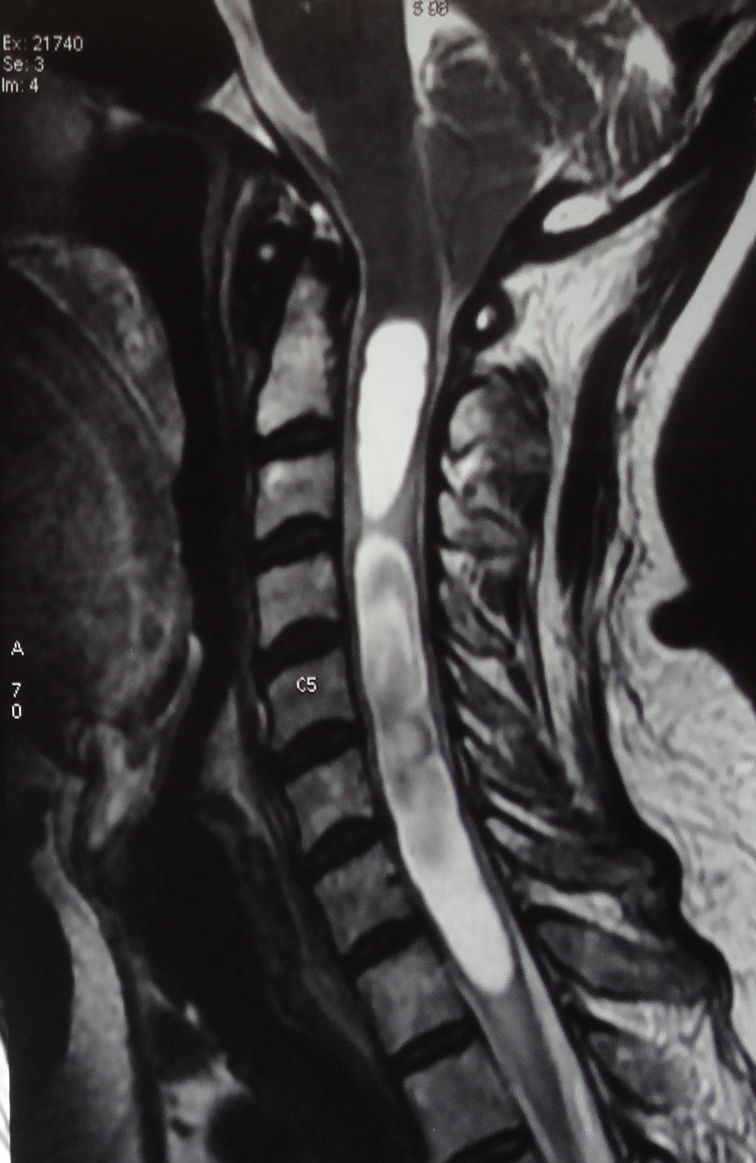
Magnetic resonance imaging (MRI) is the imaging of choice for the initial diagnosis of PTS. There is a lack of correlation between symptoms and cavity size. The cavity is usually found at the site of spinal column fracture or abnormal angulation. Computed tomographic scan myelography may be necessary for persons who cannot undergo MRI as it can delineate and obstruction to dye flow due to cord tethering/dural adhesions. Plain radiographs, including flexion-extension views, are used to detect spinal instability, spinal kyphosis, fractures, and dislocations. Electromyography may include findings of various forms of abnormal spontaneous activity. These are nonspecific, and electrodiagnosis is best used for the exclusion of other causes producing similar symptoms. Motor evoked potentials can be used to demonstrate and follow prolongation of central motor conduction time and should be used intraoperatively for incomplete patients, however, this technology is not widely available. MRI is used to follow up patients and evaluate the surgical treatment.[8]

Treatment / Management

Syringomyelia is difficult to treat.[9] Treatment of syrinx cavities is primarily surgical if they are producing symptoms and disability. Some authors feel that motor loss is infrequent or late and therefore conservative management is indicated.[2] The majority advocate early surgical as a means of reducing progressive delayed deficit. Conservative treatment is usually associated with the potential risk of neurologic worsening in up to 68% of the patients within a year. Some patients can have spontaneous regression of the cyst.[10][11] The shunting of the syrinx cavity (syringoperitoneal, syringopleural) is often attempted initially.[12][13][14] Shunts may become clogged with debris and require replacement. Shunt failure with cavity recurrence and shunt-related complications are very high in some series.[15] Other drainage procedures such as needle aspiration, myelotomy, and cord opening are less common. More recently, favored surgical approaches include those aimed at reestablishing normal CSF flow through areas of narrowing due to dural scar, adhesions, and spinal cord tethering. These include laminectomy with intradural exploration, lysis of adhesions, and widening of the CSF space via duraplasty.[2][4][14][2][16][17][18] Although this is a posterior surgical approach, care should be taken to evaluate and treat anterior tethering as well. In high cervical lesions or cases with extensive multilevel scar tissue, this approach may not be possible. Recurrence of syrinx formation can occur with all procedures. Some have reported that a change in syrinx size does not correlate with clinical outcomes[8], but others have found the opposite[4].

A promising therapy is being recently investigated in which autologous bone marrow-derived mesenchymal stromal cells are injected in the syrinx of posttraumatic syringomyelia.[19]

Serial neurologic examinations are critical for following patients with known syringomyelia. Handheld dynamometry of key muscle groups can provide a useful objective adjunct to manual muscle testing. The patient's report of changes in function, such as ambulation, wheelchair propulsion, or transfers, can be the most important factor to determine the progression of the condition. Patients with cervical syringomyelia need monitoring of pulmonary function to assess for worsening of vital capacity. Interdisciplinary evaluations by rehabilitation teams can assess the need for changes in mobility devices, seating/walking activity, and activities of daily living. The team will help to mitigate increasing risks of complications such as pressure ulcers, a decline in mobility, and falls. 

Differential Diagnosis

Spinal instability

Tethered spinal cord

Spinal hematoma

Glial scar formation with mass effect

Subacute progressive ascending myelopathy[20]

Apoptosis of spinal tissue

Prognosis

Conservative treatment usually leads to progressive neurological deterioration within a year in those patients who present with deficits.

Small cysts in asymptomatic or minimally symptomatic patients do not need initial surgery.

For those symptomatic patients who undergo a surgical procedure, most have good results for radicular symptoms but less encouraging results for autonomic symptoms or spasticity. Cessation of symptoms or improvement occurs in nearly 90% of the patients.[4]

Complications

Progressive numbness

Progressive weakness

Increased spasticity

Hyperhidrosis

Blood pressure instability

Postural hypotension

Pressure ulcers

Decline in mobility

Falls

Neuropathic arthropathies (Charcot's joint)

Burns to senseless areas

Progressive scoliosis

Loss of bowel and bladder function 

Sensory-motor deterioration if surgery is performed

Emotional instability

Consultations

Neurosurgical consultation is indicated when surgery is being considered

Electrodiagnostic consultation can be helpful to evaluate for other causes or concomitant radiculopathy

Spinal cord injury specialists

Physical and occupational therapists to address mobility, seating, self-care, neurogenic bowel

Pain specialists

Neuro-urologic specialists if a neurogenic bladder develops[21] 

Deterrence and Patient Education

Most of these patients have emotional and physical challenges due to their initial primary spinal cord injury. As syringomyelia causes new deficits, patients should be encouraged that it is a treatable condition, and outcomes are good in the majority of patients with pain symptoms. Patients may need an aggressive rehabilitation phase to improve motor and spastic symptoms.

Enhancing Healthcare Team Outcomes

Collaboration shared decision-making, and communication are key elements for a good outcome. The interprofessional care provided to the patient must use an integrated care pathway combined with an evidence-based approach to planning and evaluation of all joint activities. An interprofessional team that provides a holistic and integrated approach to postoperative care can help achieve the best possible outcomes. Posttraumatic syringomyelia is a difficult disease to evaluate and treat. It requires a coordinated team approach of multiple specialists, and supportive care of physical and occupational therapists, nurses, and clinicians. [Level V]


(Click Image to Enlarge)
<p>Syringomyelia Visualized on MRI in a Patient With Hindbrain Herniation.</p>

Syringomyelia Visualized on MRI in a Patient With Hindbrain Herniation.


Contributed by V Shenoy, MD, MBBS

Details

Editor:

Sean M. McAvoy

Updated:

8/23/2023 12:39:14 PM

References


[1]

Krebs J, Koch HG, Hartmann K, Frotzler A. The characteristics of posttraumatic syringomyelia. Spinal cord. 2016 Jun:54(6):463-6. doi: 10.1038/sc.2015.218. Epub 2015 Dec 1     [PubMed PMID: 26620880]


[2]

Bonfield CM, Levi AD, Arnold PM, Okonkwo DO. Surgical management of post-traumatic syringomyelia. Spine. 2010 Oct 1:35(21 Suppl):S245-58. doi: 10.1097/BRS.0b013e3181f32e9c. Epub     [PubMed PMID: 20881468]


[3]

Schurch B, Wichmann W, Rossier AB. Post-traumatic syringomyelia (cystic myelopathy): a prospective study of 449 patients with spinal cord injury. Journal of neurology, neurosurgery, and psychiatry. 1996 Jan:60(1):61-7     [PubMed PMID: 8558154]


[4]

Karam Y,Hitchon PW,Mhanna NE,He W,Noeller J, Post-traumatic syringomyelia: outcome predictors. Clinical neurology and neurosurgery. 2014 Sep     [PubMed PMID: 25016238]


[5]

Lam S, Batzdorf U, Bergsneider M. Thecal shunt placement for treatment of obstructive primary syringomyelia. Journal of neurosurgery. Spine. 2008 Dec:9(6):581-8. doi: 10.3171/SPI.2008.10.08638. Epub     [PubMed PMID: 19035753]


[6]

Kim HG, Oh HS, Kim TW, Park KH. Clinical Features of Post-Traumatic Syringomyelia. Korean journal of neurotrauma. 2014 Oct:10(2):66-9. doi: 10.13004/kjnt.2014.10.2.66. Epub 2014 Oct 31     [PubMed PMID: 27169036]


[7]

Goldstein B, Hammond MC, Stiens SA, Little JW. Posttraumatic syringomyelia: profound neuronal loss, yet preserved function. Archives of physical medicine and rehabilitation. 1998 Jan:79(1):107-12     [PubMed PMID: 9440427]


[8]

Li YD, Therasse C, Kesavabhotla K, Lamano JB, Ganju A. Radiographic assessment of surgical treatment of post-traumatic syringomyelia. The journal of spinal cord medicine. 2021 Nov:44(6):861-869. doi: 10.1080/10790268.2020.1743086. Epub 2020 Mar 30     [PubMed PMID: 32223591]


[9]

Lee TT, Alameda GJ, Gromelski EB, Green BA. Outcome after surgical treatment of progressive posttraumatic cystic myelopathy. Journal of neurosurgery. 2000 Apr:92(2 Suppl):149-54     [PubMed PMID: 10763684]


[10]

Killeen T, Rosner J, Jutzeler CR, Hupp M, Heilbronner R, Curt A. Spontaneous resolution of an extensive posttraumatic syrinx. Neurology. 2016 Sep 20:87(12):1299-301. doi: 10.1212/WNL.0000000000003130. Epub 2016 Aug 19     [PubMed PMID: 27543642]


[11]

Maharaj MM, Phan K, Mobbs R. Spontaneous regression of post-traumatic syringomyelia: A case report and literature review. Journal of clinical neuroscience : official journal of the Neurosurgical Society of Australasia. 2017 Oct:44():249-253. doi: 10.1016/j.jocn.2017.06.065. Epub 2017 Jul 14     [PubMed PMID: 28712735]

Level 3 (low-level) evidence

[12]

Srikantha U, Hari A, Lokanath YK, Varma RG. Syringo-Subarachnoid Shunt Placement: A Minimally Invasive Technique Using Fixed Tubular Retractors-Three Case Reports and Literature Review. International journal of spine surgery. 2020 Apr:14(2):133-139. doi: 10.14444/7020. Epub 2020 Apr 30     [PubMed PMID: 32355617]

Level 3 (low-level) evidence

[13]

Hussain I, Greenfield JP. Ultrasound-guided Syringosubarachnoid Shunt Insertion for Cervicothoracic Syringomyelia. Clinical spine surgery. 2020 Jun:33(5):185-191. doi: 10.1097/BSD.0000000000000835. Epub     [PubMed PMID: 31220040]


[14]

Aghakhani N, Baussart B, David P, Lacroix C, Benoudiba F, Tadie M, Parker F. Surgical treatment of posttraumatic syringomyelia. Neurosurgery. 2010 Jun:66(6):1120-7; discussion 1127. doi: 10.1227/01.NEU.0000369609.30695.AB. Epub     [PubMed PMID: 20495426]


[15]

Batzdorf U, Klekamp J, Johnson JP. A critical appraisal of syrinx cavity shunting procedures. Journal of neurosurgery. 1998 Sep:89(3):382-8     [PubMed PMID: 9724111]


[16]

Falci SP, Indeck C, Lammertse DP. Posttraumatic spinal cord tethering and syringomyelia: surgical treatment and long-term outcome. Journal of neurosurgery. Spine. 2009 Oct:11(4):445-60. doi: 10.3171/2009.4.SPINE09333. Epub     [PubMed PMID: 19929342]


[17]

Holly LT, Johnson JP, Masciopinto JE, Batzdorf U. Treatment of posttraumatic syringomyelia with extradural decompressive surgery. Neurosurgical focus. 2000 Mar 15:8(3):E8     [PubMed PMID: 16676931]


[18]

Klekamp J. Treatment of posttraumatic syringomyelia. Journal of neurosurgery. Spine. 2012 Sep:17(3):199-211. doi: 10.3171/2012.5.SPINE11904. Epub 2012 Jul 13     [PubMed PMID: 22794351]


[19]

Vaquero J, Hassan R, Fernández C, Rodríguez-Boto G, Zurita M. Cell Therapy as a New Approach to the Treatment of Posttraumatic Syringomyelia. World neurosurgery. 2017 Nov:107():1047.e5-1047.e8. doi: 10.1016/j.wneu.2017.08.019. Epub 2017 Aug 10     [PubMed PMID: 28804041]


[20]

Levi V, Franzini A, Di Cristofori A, Bertani G, Pluderi M. Subacute posttraumatic ascending myelopathy (SPAM): A potential complication of subarachnoid shunt for syringomyelia? The journal of spinal cord medicine. 2020 Sep:43(5):714-718. doi: 10.1080/10790268.2018.1512735. Epub 2018 Aug 29     [PubMed PMID: 30156977]


[21]

Caremel R, Hamel O, Gerardin E, Lenormand L, Parker F, Lefort M, Grise P, Perrouin-Verbe B. [Post-traumatic syringomyelia: What should know the urologist?]. Progres en urologie : journal de l'Association francaise d'urologie et de la Societe francaise d'urologie. 2013 Jan:23(1):8-14. doi: 10.1016/j.purol.2012.09.009. Epub 2012 Oct 15     [PubMed PMID: 23287478]